Archive for June, 2020
[Magazine] HOPE Magazine – Summer 2020
Posted by Kostas Pantremenos in TBI on June 30, 2020

[ARTICLE] Enhancing epilepsy self-management and quality of life for adults with epilepsy with varying social and educational backgrounds using PAUSE to Learn Your Epilepsy – Full Text
Posted by Kostas Pantremenos in Epilepsy on June 30, 2020
Highlights
•PAUSE is a personalized epilepsy self-management (SM) education program.
•PAUSE was implemented in diverse and mostly underserved adults with epilepsy.
•Self-efficacy, frequency of SM behaviors, and QOL significantly improved over time.
•Personal negative impact of epilepsy significantly reduced over time.
•Greater improvement was seen in those with lower scores at baseline.
Abstract
Purpose
People with epilepsy (PWE) come from a wide variety of social backgrounds and educational skillsets, making self-management (SM) education for improving their condition challenging. Here, we evaluated whether a mobile technology-based personalized epilepsy SM education intervention, PAUSE to Learn Your Epilepsy (PAUSE), improves SM measures such as self-efficacy, epilepsy SM behaviors, epilepsy outcome expectations, quality of life (QOL), and personal impact of epilepsy in adults with epilepsy.
Methods
Recruitment for the PAUSE study occurred from October 2015 to March 2019. Ninety-one PWE were educated using an Internet-enabled computer tablet application that downloads custom, patient-specific educational programs from Epilepsy.com. Validated self-reported questionnaires were used for outcome measures. Participants were assessed at baseline (T0), the first follow-up at completion of the PWE-paced 8–12-week SM education intervention (T1), and the second follow-up at least 3 months after the first follow-up (T2). Multiple linear regression was used to assess within-subject significant changes in outcome measures between these time points.
Results
The study population was diverse and included individuals with a wide variety of SM educational needs and abilities. The median time for the first follow-up assessment (T1) was approximately 4 months following the baseline (T0) and 8 months following baseline for the second follow-up assessment (T2). Participants showed significant improvement in all SM behaviors, self-efficacy, outcome expectancy, QOL, and personal impact of epilepsy measures from T0 to T1. Participants who scored lower at baseline tended to show greater improvement at T1. Similarly, results showed that participant improvement was sustained in the majority of SM measures from T1 to T2.
Conclusion
This study demonstrated that a mobile technology-based personalized SM intervention is feasible to implement. The results provide evidence that epilepsy SM behavior and practices, QOL, outcome expectation for epilepsy treatment and management, self-efficacy, and outcome expectation and impact of epilepsy significantly improve following a personalized SM education intervention. This underscores a greater need for a pragmatic trial to test the effectiveness of personalized SM education, such as PAUSE to Learn Your Epilepsy, in broader settings specifically for the unique needs of the hard-to-reach and hard-to-treat population of PWE.
1. Introduction
Epilepsy, characterized by spontaneous recurrent seizures with unpredictable frequency, is a common and complex neurological disorder that affects the health and quality of life (QOL) of people with epilepsy (PWE) [1]. It is the fourth most common chronic neurological disorder after migraines, Alzheimer’s disease, and Parkinson’s disease in terms of 1-year prevalence per 1000 in the general population [2]. In 2015, approximately 1.2% of American adults reported living with epilepsy; 68.5% had seen a neurologist or epilepsy specialist; 93% were taking antiseizure medication (ASM), and, among those taking medication to control seizures, only 42.4% were seizure-free in the past year [3]. Epilepsy, especially with uncontrolled seizures, poses an immense burden to the people who have it, caregivers, and the society due to a number of factors including associated developmental, cognitive, and psychiatric comorbidities; ASM side effects; higher injury and mortality rates; poorer QOL; and increased financial burden. An estimated 3.0% of global disability-adjusted life years (DALYs) were from neurological disorders in 2010, a quarter of which were from epilepsy; epilepsy was the second-most burdensome chronic neurologic disorder worldwide in terms of DALYs [4].
Self-management (SM) education has shown to improve SM skills & behaviors and QOL in many chronic diseases including heart disease, diabetes, asthma, and arthritis [5,6]. Barlow defines self-management as an individual’s ability to manage the symptoms, treatments, physical and psychological consequences, and life style changes inherent in living with a chronic condition [7]. However, successful SM requires sufficient knowledge of the condition, its treatment, and necessary skills to perform SM activities. Like other chronic conditions, day-to-day management of epilepsy shifts from healthcare professionals to PWE. Epilepsy care demands active involvement of PWE in keeping up with the health effects of epilepsy and coping with social (e.g., family/friends, stigma, hobbies), health (e.g., seizure response/tracking, comorbidities such as depression/anxiety, sleep, safety, health literacy), employment (e.g., transportation, disability, absenteeism), and economic (e.g., cost of healthcare and medication) challenges. One can only self-manage their disease if they have the tools to do so, including knowledge, access to information relevant to their specific healthcare needs, and the ability to carry out the SM tasks needed for their condition. Evidence shows that many PWE are not knowledgeable about their disorder or often not educated about the risks of epilepsy, injury, and mortality [1,8]. Education needs also vary between individuals and subgroups of PWE. Women, in particular, may seek information on bone health and the effect of ASM on pregnancy or contraception, while older adults’ priorities may relate to fall safety and interactions of ASM with other medications. Existing evidence also reveals that, while patients with chronic diseases are willing to receive SM education materials, perceived information overload (i.e., too much or complex information) negatively influences their usage willingness [9]. Patients with low health literacy are even more susceptible to information overload [10]. The Institute of Medicine recognized SM education gaps for PWE and recommended (Recommendation 9) in its 2012 report, “Epilepsy Across the Spectrum: Promoting Health and Understanding,” to improve and expand educational opportunities for PWE and their families, as well as to ensure that all PWE and their families have access to accurate, clearly communicated educational materials and information [1].
Several studies have reported contradictory results after examining the efficacy of SM education interventions in improving PWE’s knowledge and understanding of epilepsy and QOL. The Modular Service Package Epilepsy study (MOSES) reported significant improvements in ASM tolerability, epilepsy knowledge, coping with epilepsy, and seizure frequency after 6 months following a 2-day SM education program [11]. Self-management education for people with poorly controlled epilepsy [SMILE (UK)] adapted MOSES for use in the United Kingdom and did not find the 2-day course to be effective in improving QOL or secondary outcome measures (anxiety and depression), after 12 months [12]. Though both MOSES and SMILE were randomized control trials (RCTs), MOSES included all adults with epilepsy whereas SMILE included only adults with chronic epilepsy who had two or more seizures in the prior 12 months. Another RCT compared the effectiveness of a multicomponent SM intervention consisting of five weekly, 2-hour group sessions each followed by a 2-hour group session after three weeks with usual care; they found no difference in measures of self-efficacy, though did find improvements in some epilepsy QOL domains and decreases in measures of ASM side effects [13]. Other studies examining the efficacy of in-person, group-based, online or phone/internet SM interventions, including the Centers for Disease Control and Prevention-supported Managing Epilepsy Well (MEW) network programs, did show improvement in epilepsy SM and QOL [[14], [15], [16], [17], [18]].
In addition to existing group-based programs, which require permission to use and specialized training, there is a greater need for patient-centered and patient-specific individualized education interventions for epilepsy SM that are publicly available, cost-effective, and easily disseminated to clinics or in community. The PAUSE to Learn Your Epilepsy (hereafter referred to as “PAUSE”), a MEW network collaboration center, was developed and implemented to address the needs of all PWE, especially those in underserved populations. This program uses publicly available education information from the Epilepsy Foundation (EF) website, epilepsy.com, linked to a mobile technology-based PAUSE application to provide patient-centered personalized epilepsy SM lesson plan to PWE. Detailed information about PAUSE including study design, recruitment, intervention, and assessments has been published previously [19,20]. We reported significantly lower epilepsy SM practices and behaviors among PWE from an underserved population as compared to all PWE. In this paper, we sought to determine whether the PAUSE intervention significantly improves self-efficacy, SM behavior & skills, QOL, personal impact of epilepsy, and epilepsy outcome expectancies over time in adults with epilepsy. We also assessed whether perceived depression symptoms influence longitudinal changes in SM measures following the PAUSE intervention.[…]
[ARTICLE] The Promotoer, a brain-computer interface-assisted intervention to promote upper limb functional motor recovery after stroke: a study protocol for a randomized controlled trial to test early and long-term efficacy and to identify determinants of response – Full Text
Posted by Kostas Pantremenos in Neuroplasticity, Paretic Hand on June 29, 2020
Abstract
Background
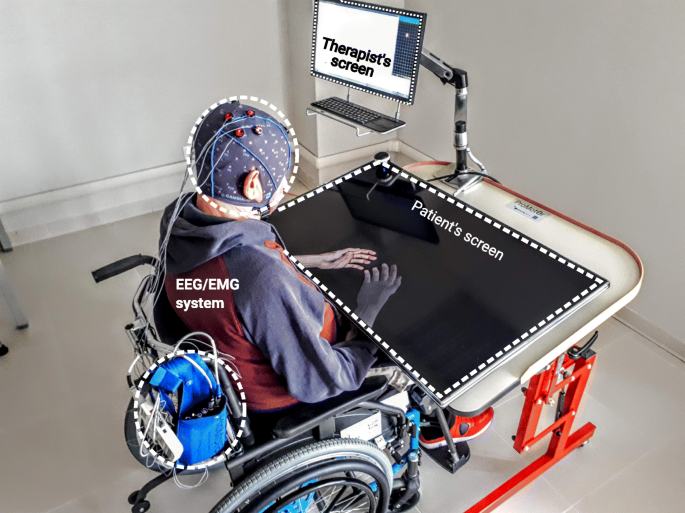
Stroke is a leading cause of long-term disability. Cost-effective post-stroke rehabilitation programs for upper limb are critically needed. Brain-Computer Interfaces (BCIs) which enable the modulation of Electroencephalography (EEG) sensorimotor rhythms are promising tools to promote post-stroke recovery of upper limb motor function. The “Promotoer” study intends to boost the application of the EEG-based BCIs in clinical practice providing evidence for a short/long-term efficacy in enhancing post-stroke hand functional motor recovery and quantifiable indices of the participants response to a BCI-based intervention. To these aims, a longitudinal study will be performed in which subacute stroke participants will undergo a hand motor imagery (MI) training assisted by the Promotoer system, an EEG-based BCI system fully compliant with rehabilitation requirements.
Methods
This longitudinal 2-arm randomized controlled superiority trial will include 48 first ever, unilateral, subacute stroke participants, randomly assigned to 2 intervention groups: the BCI-assisted hand MI training and a hand MI training not supported by BCI. Both interventions are delivered (3 weekly session; 6 weeks) as add-on regimen to standard intensive rehabilitation. A multidimensional assessment will be performed at: randomization/pre-intervention, 48 h post-intervention, and at 1, 3 and 6 month/s after end of intervention. Primary outcome measure is the Fugl-Meyer Assessment (FMA, upper extremity) at 48 h post-intervention. Secondary outcome measures include: the upper extremity FMA at follow-up, the Modified Ashworth Scale, the Numeric Rating Scale for pain, the Action Research Arm Test, the National Institute of Health Stroke Scale, the Manual Muscle Test, all collected at the different timepoints as well as neurophysiological and neuroimaging measures.
Discussion
We expect the BCI-based rewarding of hand MI practice to promote long-lasting retention of the early induced improvement in hand motor outcome and also, this clinical improvement to be sustained by a long-lasting neuroplasticity changes harnessed by the BCI-based intervention. Furthermore, the longitudinal multidimensional assessment will address the selection of those stroke participants who best benefit of a BCI-assisted therapy, consistently advancing the transfer of BCIs to a best clinical practice.
Trial registration
Name of registry: BCI-assisted MI Intervention in Subacute Stroke (Promotoer).
Trial registration number: NCT04353297; registration date on the ClinicalTrial.gov platform: April, 15/2020.
Background
Stroke is a major public health and social care concern worldwide [1]. The upper limb motor impairment commonly persists after stroke, and it represents the major contribution to long-term disability [2]. It has been estimated that the main clinical predictor of whether a patient would come back to work is the degree of upper extremity function [3]. Despite the intensive rehabilitation, the variability in the nature and extent of upper limb recovery remains a crucial factor affecting rehabilitation outcomes [4].
Electroencephalography (EEG)-based Brain-Computer Interface (BCI) is an emerging technology that enables a direct translation of brain activity into motor action [5]. Recently, EEG-based BCIs have been recognized as potential tools to promote functional motor recovery of upper limbs after stroke (for review see [6]). Several randomized controlled trials have shown that stroke patients can learn to modulate their EEG sensorimotor rhythms [7] to control external devices and this practice might facilitate neurological recovery both in subacute and chronic stroke phase [8,9,10].
We were previously successful in the design and validation of an EEG sensorimotor rhythms–based BCI combined with realistic visual feedback of upper limb to support hand motor imagery (MI) practice in stroke patients [11, 12]. Our previous pilot randomized controlled study [8] with the participation of 28 subacute stroke patients with severe motor deficit, suggested that 1 month BCI-assisted MI practice as an add-on intervention to the usual rehabilitation care was superior with respect to the add-on, 1 month MI training alone (ie., without BCI support) in improving hand functional motor outcomes (indicated by the significantly higher mean score at upper extremity Fugl-Meyer scale in the BCI with respect to control group). A greater involvement of the ipsilesional hemisphere, as reflected by a stronger motor-related EEG oscillatory activity and connectivity in response to MI of the paralyzed trained hand was also observed only in the BCI-assisted MI training condition. These promising findings corroborated the idea that a relatively low-cost technique (i.e. EEG-based BCI) can be exploited to deliver an efficacious rehabilitative intervention such as MI training and prompted us to undertake a translational effort by implementing an all-in-one BCI-supported MI training station– the Promotoer [13].
Yet, important questions remain to be addressed in order to improve the clinical viability of BCIs such as defining whether the expected early improvements in functional motor outcomes induced by the BCI-assisted MI training in subacute stroke [8] can be sustained in a long-term as it has been shown for other BCI-based approaches in chronic stroke patients [10, 14]. This requires advancements in the knowledge on brain functional re-organization early after stroke and on how this re-organization would correlate with the functional motor outcome (evidence-base medicine). Last but not least, the definition of the determinants of the patients response to treatment is paramount to optimize the process of personalized medicine in rehabilitation. We will address these questions by carrying out a randomized trial to eventually establish the fundamentals for a cost-effective use of EEG-based BCI technology to deliver a rehabilitative intervention such as the MI in hospitalized stroke patients.
Aim and hypotheses
The “Promotoer” study is a randomized controlled trial (RCT) designed to provide evidence for a significant early improvement of hand motor function induced by the BCI-assisted MI training operated via the Promotoer and for a persistency (up to 6 months) of such improvement. Task-specific training was reported to induce long-term improvements in arm motor function after stroke [15,16,17]. Thus, our hypothesis is that the BCI-based rewarding of hand MI tasks would promote long-lasting retention of early induced positive effect on motor performance with respect to MI tasks practiced in an open loop condition (ie, without BCI). Accordingly, the primary aim of the “Promotoer” RCT will be first to determine whether the BCI based intervention (MI-BCI) administered by means of a BCI system fully compatible with a clinical setting (the Promotoer), is superior to a non-BCI assisted MI training (MI Control) in improving hand motor function outcomes in sub-acute stroke patients admitted to the hospital for their standard rehabilitation care; secondly, we will test whether the efficacy of BCI-based intervention on hand motor function outcomes is sustained long-term after the end of intervention (6 months follow-up). A further hypothesis is that such clinical improvement would be sustained by a long-lasting neuroplasticity changes as harnessed by the BCI–based intervention. This hypothesis rises from current evidence for an early enhancement of post-stroke plastic changes enabled by BCI-based trainings [8,9,10]. To test this hypothesis, a longitudinal assessment of the brain network organization derived from advanced EEG signal processing (secondary objective) will be performed.
The heterogeneity of stroke makes prediction of treatment responders a great challenge [18]. The potential value of a combination of neurophysiological and neuroimaging biomarkers with the clinical assessment in predicting post-stroke motor recovery has been recently highlighted [19]. Our hypothesis is that the longitudinal combined functional, neurophysiological and neuroimaging assessment over 6 months from the intervention will allow for insights into biomarkers and potential predictors of patients response to the BCI-Promotoer training (secondary aim). To this purpose, well-recognized factors contributing to recovery after stroke such as the relation between clinical profile, lesion characteristics and patterns of post-stroke motor cortical re-organization (eg., ipsilesional/contralesional primary and non-primary motor areas, cortico-spinal tract integrity, severity of motor deficits at baseline; for review see [19]) will be taken into account.[…}
[WEB PAGE] When A Stroke Strikes
Posted by Kostas Pantremenos in REHABILITATION on June 28, 2020

Physiotherapy for stroke patients is not just about exercises and movement, but also incorporates technology such as functional electrical stimulation and virtual reality. — Photos: SUNWAY MEDICAL CENTRE VELOCITY
There you are, walking around the house, maybe on your way to get something to eat or to the living room to watch some TV.
All of a sudden, one side of your body goes numb and weak, and you lose your balance, causing you to collapse on the floor.
When you try to call for help, you find that you can’t speak properly with your speech sounding slurred.
You realise that you can’t see properly either, as your vision has become blurry.
And when you look at your face in the mirror, you realise that one side is drooping down, but not the other.
You are most likely experiencing a stroke, the third most common cause of death in Malaysia.
This emergency condition occurs when the brain is deprived of vital oxygen supply, either when a blood clot blocks off a blood vessel supplying the brain (ischaemic stroke) or when one of these blood vessels ruptures and starts bleeding (haemorrhagic stroke).
According to consultant neurologist Dr Kok Chin Yong. your brain cells will start to die within minutes after the stroke hits you – about 2,000,000 cells every minute.
Therefore, you need to get help and go to the nearest hospital’s emergency department as soon as possible.
The good news is that there are immediate treatments that can not only save your life, but also minimise any disability you might have from the stroke.
Ischaemic strokes are the most common type of stroke, comprising three-quarters of all cases.
For this type of stroke, the first and most immediate treatment is the administration of tissue plasminogen activators (tPAs), which help break down blood clots.
Says Dr Kok: “For an ischaemic stroke, the target is to unblock the blood clot as soon as possible.
“We can do this with an intravenous (IV) clot-busting agent called alteplase.
“The current guidelines state a cut-off point of 4.5 hours from the onset of symptoms in order to derive benefit from this treatment.
“Hence, time is brain.”
He notes that alteplase is the only US Food and Drug Administration-approved tPA for acute ischaemic stroke at the moment.
Another treatment available in certain hospitals, he adds, is a clot-removal procedure called mechanical thrombectomy, which is done by an interventional radiologist.
Meanwhile, haemorrhagic strokes are usually treated by the neurosurgeon or interventional radiologist.
Explains consultant neurosurgeon Dr Gerard Arvind Martin: “Choosing whether to take the patient to the operating room or not can depend on various factors, such as age and condition of the patient, the level of consciousness and extent of bleeding, all of which the surgeon takes into consideration before performing surgery.
“In those cases where a subarachnoid haemorrhage has occurred due to a suspected aneurysmal rupture, a further scan called an angiogram will be required to determine precisely the site of bleeding.
“Angiograms can be either via computed tomography (CT) scan or a catheter, which is typically carried out by a radiologist in an angiogram suite.
“Depending on the findings, the surgeon can then elect to operate and clip the ruptured aneurysm, or consider endovascular techniques, which are performed by an interventional radiologist.”
If you are lucky, you would have survived your stroke with no or minor complications and have a quick recovery.
However, many stroke survivors will face long-term disability, which can be physical or cognitive.
For example, patients may experience paralysis of the side affected by the stroke; weak coordination; difficulty in speaking, understanding, reading and writing; and difficulty concentrating.
This is where rehabilitation comes in. A stroke patient exercises his muscles with the help of a machine, supervised by a physiotherapist.
Says Dr Kok: “This is an area that is often given less attention, when in fact, to me, it is equally important as the acute treatment for stroke.
“A good rehabilitation programme improves disability and prevents complications.”
According to Sunway Medical Centre Velocity Rehabilitation Centre head Maxim Chea, stroke rehabilitation has to be individualised to the patient as it depends on the part of the body or type of ability affected by the stroke.
He explains that there are three main types of rehabilitation therapy: physical therapy, technology-assisted physical therapy (e.g. functional electrical stimulation and virtual reality), and cognitive and emotional therapy.
Visual rehabilitation is also available for those whose vision is affected by stroke.
For example, consultant neuro-ophthalmologist Dr Lakana Kumar shares that “Double vision and peripheral loss of vision can be treated with prisms incorporated into glasses for patients to wear.”
Rehabilitation is usually carried out by a team consisting of physiotherapists, occupational therapists, speech therapists, and dieticians/nutritionists, among others.
In addition to physical and cognitive problems, patients might easily become depressed, overly anxious and panic easily.
Says consultant psychiatrist Dr Lim Wai Jenn: “Stroke survivors are at significantly higher risk for neuropsychiatric conditions such as post-stroke depression (one in three patients), anxiety (one in four patients), and other changes in personality and behaviour.
“These conditions impede the rehabilitation process and degree of recovery in post-stroke patients.
“They also significantly impact the patients’ long-term functioning and quality of life, and can even lead to higher mortality rates.
“Early psychiatric assessment and intervention is essential.”
She notes that caregivers also need support and psychoeducation on how best to support patients in regaining function.
While being affected by a stroke is a frightening event, rest assured that there are treatments and therapies available to help you manage this condition – just remember that you need to seek medical help as soon as possible.
This article is courtesy of Velocity Neurocentre, Sunway Medical Centre Velocity.
[WEB PAGE] Mozart may reduce seizure frequency in people with epilepsy
Posted by Kostas Pantremenos in Epilepsy, Music/Music therapy on June 28, 2020
June 10, 2020. Source: University Health Network
Summary
A new clinical research study has found that a Mozart composition may reduce seizure frequency in patients with epilepsy.
A new clinical research study by Dr. Marjan Rafiee and Dr. Taufik Valiante of the Krembil Brain Institute at Toronto Western Hospital, part of University Health Network, has found that a Mozart composition may reduce seizure frequency in patients with epilepsy.
The results of the research study, “The Rhyme and Rhythm of Music in Epilepsy,” was recently published in the international journal Epilepsia Open. It looks at the effects of the Mozart melody, “Sonata for Two Pianos in D Major, K. 448” on reducing seizures, as compared to another auditory stimulus — a scrambled version of the original Mozart composition, with similar mathematical features, but shuffled randomly and lacking any rhythmicity.
“In the past 15 to 20 years, we have learned a lot about how listening to one of Mozart’s compositions in individuals with epilepsy appears to demonstrate a reduction in seizure frequency,” says Dr. Marjan Rafiee, lead author on the study. “But, one of the questions that still needed to be answered was whether individuals would show a similar reduction in seizure frequency by listening to another auditory stimulus — a control piece — as compared to Mozart.”
The researchers recruited 13 patients to participate in the novel, year-long study. After three months of a baseline period, half of the patients listened to Mozart’s Sonata once daily for three months, then switched to the scrambled version for three months. The others started the intervention by listening to the scrambled version for three months, then switched to daily listening of Mozart.
Patients kept “seizure diaries” to document their seizure frequency during the intervention. Their medications were kept unchanged during the course of the study.
“Our results showed daily listening to the first movement of Mozart K.448 was associated with reducing seizure frequency in adult individuals with epilepsy,” says Dr. Rafiee. “This suggests that daily Mozart listening may be considered as a supplemental therapeutic option to reduce seizures in individuals with epilepsy.”
Epilepsy is the most common serious neurological disorder in the world, affecting approximately 300,000 Canadians and 50 million people worldwide.
Many experience debilitating seizures. The treatment is often one or more anti-seizure medications. But for 30 per cent of patients, the medications are not effective in controlling their seizures.
“As a surgeon, I have the pleasure of seeing individuals benefit from surgery, however I also know well those individuals for whom surgery is not an option, or those who have not benefitted from surgery, so, we are always looking for ways to improve symptom control, and improve quality of life for those with epilepsy,” says Dr. Taufik Valiante, senior author of the study and the Director of the Surgical Epilepsy Program at Krembil Brain Institute at UHN and co-Director of CRANIA.
“Like all research, ours raises a lot of questions that we are excited to continue to answer with further research and support from the epilepsy community.”
While these results are promising, the next step is to conduct larger studies with more patients, over a longer period of time.
Story Source:
Materials provided by University Health Network. Note: Content may be edited for style and length.
Journal Reference:
- Marjan Rafiee, Kramay Patel, David M. Groppe, Danielle M. Andrade, Eduard Bercovici, Esther Bui, Peter L. Carlen, Aylin Reid, Peter Tai, Donald Weaver, Richard Wennberg, Taufik A. Valiante. Daily listening to Mozart reduces seizures in individuals with epilepsy: A randomized control study. Epilepsia Open, 2020; 5 (2): 285 DOI: 10.1002/epi4.12400
[WEB PAGE] FDA Clears Ekso Bionics’ EksoNR for Rehab Use with Acquired Brain Injury
Posted by Kostas Pantremenos in Gait Rehabilitation - Foot Drop, Rehabilitation robotics on June 26, 2020
Ekso Bionics Holdings Inc announces it has received 501(k) clearance from the U.S. Food and Drug Administration (FDA) to market its EksoNR robotic exoskeleton for use with patients with acquired brain injury (ABI).

EksoNR is reportedly the first exoskeleton device to receive FDA clearance for rehabilitation use with ABI. It was previously cleared by the FDA for stroke and spinal cord injury rehabilitation in 2016.
Related Content:
Ekso Bionics Unveils the EksoNR Neurorehabilitation Device
Post Acute Medical Expands Exoskeleton Rehab with New EksoNR Devices
EksoGT Users Have Walked Around the World Twice, Company Estimates
ABI is comprised of both traumatic (TBI) and non-traumatic (n-TBI) brain injury causes. TBI includes severe head injuries and concussions, while n-TBI includes a broader subset of conditions, such as stroke, aneurysms, brain tumors, anoxia, degenerative and metabolic conditions, infections, and surgical injuries, among others, according to a media release from Ekso Biokics.

“With the expanded indications to include the broad category of acquired brain injuries, the EksoNR has the potential to mobilize significantly more patients and improve patient recovery. Based on their experience with EksoNR, customers at leading rehabilitation centers have acknowledged the benefits our technology can offer during recovery from brain injuries. We are excited to see the device used more widely in neurorehabilitation.”
— Jack Peurach, CEO and president of Ekso Bionics
[Source(s): Ekso Bionics, Globe Newswire]
[Abstract] Impact of Telerehabilitation for Stroke-Related Deficits
Posted by Kostas Pantremenos in Tele/Home Rehabilitation on June 26, 2020
Abstract
Background: Stroke is the leading cause of serious long-term disability in the United States. Barriers to rehabilitation include cost, transportation, lack of trained personnel, and equipment. Telerehabilitation (TR) has emerged as a promising modality to reduce costs, improve accessibility, and retain patient independence. TR allows providers to remotely administer therapy, potentially increasing access to underserved regions.
Objectives: To describe types of stroke rehabilitation therapy delivered through TR and to evaluate whether TR is as effective as traditional in-person outpatient therapy in improving satisfaction and poststroke residual deficits such as motor function, speech, and disability.
Methods: A literature search of the term “telerehabilitation and stroke” was conducted across three databases. Full-text articles with results pertaining to TR interventions were reviewed. Articles were scored for methodological quality using the PEDro scale.
Results: Thirty-four articles with 1,025 patients were included. Types of TR included speech therapy, virtual reality (VR), robotic, community-based, goal setting, and motor training exercises. Frequently measured outcomes included motor function, speech, disability, and satisfaction. All 34 studies reported improvement from baseline after TR therapy. PEDro scores ranged from 2 to 8 with a mean of 4.59 ± 1.94 (on a scale of 0-10). Studies with control interventions, randomized allocation, and blinded assessment had significantly higher PEDro scores. All 15 studies that compared TR with traditional therapy showed equivalent or better functional outcomes. Home-based robotic therapy and VR were less costly than in-person therapy. Patient satisfaction with TR and in-person clinical therapy was similar.
Conclusions: TR is less costly and equally as effective as clinic-based rehabilitation at improving functional outcomes in stroke patients. TR produces similar patient satisfaction. TR can be combined with other therapies, including VR, speech, and robotic assistance, or used as an adjuvant to direct in-person care.
Similar articles
- Efficacy of Home-Based Telerehabilitation vs In-Clinic Therapy for Adults After Stroke: A Randomized Clinical Trial.Cramer SC, Dodakian L, Le V, See J, Augsburger R, McKenzie A, Zhou RJ, Chiu NL, Heckhausen J, Cassidy JM, Scacchi W, Smith MT, Barrett AM, Knutson J, Edwards D, Putrino D, Agrawal K, Ngo K, Roth EJ, Tirschwell DL, Woodbury ML, Zafonte R, Zhao W, Spilker J, Wolf SL, Broderick JP, Janis S; National Institutes of Health StrokeNet Telerehab Investigators.JAMA Neurol. 2019 Jun 24;76(9):1079-87. doi: 10.1001/jamaneurol.2019.1604. Online ahead of print.PMID: 31233135 Free PMC article.
- Maximizing post-stroke upper limb rehabilitation using a novel telerehabilitation interactive virtual reality system in the patient’s home: study protocol of a randomized clinical trial.Kairy D, Veras M, Archambault P, Hernandez A, Higgins J, Levin MF, Poissant L, Raz A, Kaizer F.Contemp Clin Trials. 2016 Mar;47:49-53. doi: 10.1016/j.cct.2015.12.006. Epub 2015 Dec 4.PMID: 26655433 Clinical Trial.
- Telerehabilitation services for stroke.Laver KE, Schoene D, Crotty M, George S, Lannin NA, Sherrington C.Cochrane Database Syst Rev. 2013 Dec 16;2013(12):CD010255. doi: 10.1002/14651858.CD010255.pub2.PMID: 24338496 Free PMC article. Updated. Review.
- Study protocol: home-based telehealth stroke care: a randomized trial for veterans.Chumbler NR, Rose DK, Griffiths P, Quigley P, McGee-Hernandez N, Carlson KA, Vandenberg P, Morey MC, Sanford J, Hoenig H.Trials. 2010 Jun 30;11:74. doi: 10.1186/1745-6215-11-74.PMID: 20591171 Free PMC article. Clinical Trial.
- Scoping review of outcome measures used in telerehabilitation and virtual reality for post-stroke rehabilitation.Veras M, Kairy D, Rogante M, Giacomozzi C, Saraiva S.J Telemed Telecare. 2017 Jul;23(6):567-587. doi: 10.1177/1357633X16656235. Epub 2016 Jun 24.PMID: 27342850 Review.
[Survey] Driving with an intracranial tumor
Posted by Kostas Pantremenos in Epilepsy, Uncategorized on June 25, 2020
EAN Scientific Panel Neuro-oncology invites you to take part in their survey
https://www.surveymonkey.com/r/38MG6YR?embedded=1
Meningeomas and brain tumors may interfere with the ability to drive a vehicle in a number of ways. Seizures, cognitive impairment, motor dysfunction and visual field defects may all impair safe driving. Intracranial tumors are highly heterogenous, ranging from benign meningeomas that nevertheless may cause seizures, to high grade gliomas and brain metastasis. The clinician always considers seizure frequency, compliance and focal deficits when assessing the ability to drive for neurological patients. However, oncological prognosis, risk of recurrence and effects of treatment are factors unique to patients with intracranial tumors. These factors must be evaluated when deciding if or when a patient with a brain tumor or a meningeoma may drive. In addition, different medical professions may differ in awareness of the driving dilemma as well as in practice policy concerning this issue.
Clinical studies and reviews that address driving ability in patients with brain tumors are sparse. Most countries do not have national guidelines concerning this issue, and general as well as specific driving legislations vary between countries. In the absence of guidelines or legislation, most clinicians probably prohibit or allow driving on a case-by-case basis, or by adhering to legislation concerning epilepsy or neoplastic disease in general. The use of neuro-psychological evaluation or practical testing is unknown.
The EAN Scientific Panel of neuro-oncology wants to address this issue by performing a survey of national legislations and practice patterns among European neurologists. As a start, we aim to do a survey among the members of the Scientific Panels of Neuro-Oncology and Epilepsy.
The answers will be a guidance for whether there are inconsistences in clinical practice and reason to do a more extensive survey.
References
Thomas S1, Mehta MP, Kuo JS, Ian Robins H, Khuntia D. Current practices of driving restriction implementation for patients with brain tumors.
J Neurooncol. 2011l;103(3):641-7. doi: 10.1007/s11060-010-0439-7.
Louie AV, D’Souza DP, Palma DA, Bauman GS, Lock M, Fisher B, Patil N, Rodrigues GB.
Curr Oncol. 2012;19(3):e117-22.
Chan E, Louie AV, Hanna M, Bauman GS, Fisher BJ, Palma DA, Rodrigues GB, Sathya A, D’Souza DP.
Curr Oncol. 2013;20(1):e4-e12. doi: 10.3747/co.20.1198
Louie AV, Chan E, Hanna M, Bauman GS, Fisher BJ, Palma DA, Rodrigues GB, Warner A, D’Souza DP. Assessing fitness to drive in brain tumour patients: a grey matter of law, ethics, and medicine. Curr Oncol. 2013;20(2):90-6.
Mansur A1,2, Desimone A2, Vaughan S2, Schweizer TA1,2,3, Das S. To drive or not to drive, that is still the question: current challenges in driving recommendations for patients with brain tumours. J Neurooncol. 2018;137(2):379-385. doi: 10.1007/s11060-017-2727-y.
[Case Report] Use of a myoelectric upper limb orthosis for rehabilitation of the upper limb in traumatic brain injury – Full Text
Posted by Kostas Pantremenos in Paretic Hand, Rehabilitation robotics, TBI on June 25, 2020
Abstract
Background
Upper limb motor deficits following traumatic brain injury are prevalent and effective therapies are needed. The purpose of this case report was to illustrate response to a novel therapy using a myoelectric orthosis in a person with TBI.
Case description: A 42-year-old female, 29.5 years post-traumatic brain injury with diminished motor control/coordination, and learned nonuse of the right arm. She also had cognitive deficits and did not spontaneously use her right arm functionally.
Intervention
Study included three phases: baseline data collection/device fabrication (five weeks); in-clinic training (2×/week for nine weeks); and home-use phase (nine weeks). The orthosis was incorporated into motor learning-based therapy.
Outcomes: During in-clinic training, active range of motion, tone, muscle power, Fugl-Meyer, box and blocks test, and Chedoke assessment score improved. During the home-use phase, decrease in tone was maintained and all other outcomes declined but were still better upon study completion than baseline. The participant trained with the orthosis 70.12 h, logging over 13,000 repetitions of elbow flexion/extension and hand open/close.
Discussion
Despite long-standing traumatic brain injury, meaningful improvements in motor function were observed and were likely the results of high repetition practice of functional movement delivered over a long duration. Further assessment in a larger cohort is warranted.
Introduction
Traumatic brain injury (TBI) affects 1.7 million people in the general US population annually1 and is one of the most common neurologic disorders causing disability.2 Motor deficits are present in 30% of TBI survivors with arm and hand problems occurring in about 17%,3 limiting the ability to perform activities of daily living (ADL). However, there is less research on motor recovery in patients with TBI compared with other neurologic diseases involving the brain, such as stroke.2
Activity-based interventions hope to maximize rehabilitation outcomes and enhance adaptive neural plasticity;3 however, optimal doses and schedules of training have not been adequately established. Repetition is one parameter important for activity-dependent neural plasticity. Studies assessing US rehabilitation found that stroke and TBI survivors receive an average of 32–50 repetitions of upper extremity active and passive movement per therapy session, significantly less than the 400–600 repetitions achieved in animal studies.3 Although persons with TBI benefit from traditional therapy,4 it is clear that more is needed to attain full recovery, especially with severely affected individuals.
Consistent with this idea, Krebs and Volpe5 argued that the basis of all assistive and therapeutic devices should be to induce the intent to move followed by that movement actually happening, referred to as “intent-driven rehabilitation”. One way this can be accomplished is through myoelectric control wherein a weak electromyography (EMG) signal from the muscle of an impaired limb is detected, processed, and used to activate a motor within the orthosis. The motor then assists the user in producing the desired movement. The patient-directed “intentional” action of the device promotes patient engagement as the orthosis will only reward the patient with movement when they use the correct muscles to complete a task. Previous studies of myoelectric driven lab-based robotic interventions6 showed improved Fugl-Meyer motor control scores of the upper extremity7 and reduced spasticity as assessed by the modified Ashworth scale (MAS).8 While demonstrating a benefit of “intent-driven rehabilitation”, in-lab robotic intervention restricted the amount of practice because training was restricted to the short lab sessions only and no home practice was possible.6
While myolectrically-driven orthotic technology has been in development for many years,9–11 recent advances have made it more accessible and clinically deployable for rehabilitation. However, initial research has focused on persons with stroke12–18 and not TBI. The ability of severely hemiplegic stroke survivors to activate a powered elbow orthosis using myoelectric control has been reported,13 along with increased elbow range of motion with orthosis use.18 Kim et al.15 reported that after a combined period of training and at home-use of an elbow-only myoelectric orthosis, a statistically significant three-point change in Fugl-Meyer motor control score was found in the upper extremity of nine persons post-stroke. We have recently reported a case series of chronic stroke survivors who used a myoelectric upper limb orthosis over a period of several months and achieved a 9.0 ± 4.8 point improvement in Fugl-Meyer.19 Since upper limb motor deficits in TBI are a problem that can lead to decreased independence in ADLs and given that there is a lack of effective therapies and supportive devices for upper limb impairment, the purpose of this case report was to illustrate the response to therapy combined with a myoelectric elbow–wrist–hand orthosis in a person with longstanding TBI.
Case description
The protocol described in this case report complies with standards of the Declaration of Helsinki and was approved by the Institutional Review Boards of participating institutions (IRB #16039-H29 and STU00203728) and met the Health Insurance Portability and Accountability Act (HIPAA) requirements for disclosure of protected health information. Written informed consent for participation was obtained from the patient’s legal guardian.
The participant was a 42-year-old female who sustained a TBI from being struck by a motor vehicle at age 12. At study entry, she was 29.5 years post injury, dependent on caregivers for most ADL/instrumental activities of daily living (IADLs), used a manual wheelchair for mobility, resided in a group home setting, and required assistance from caregivers to help her make decisions. She attended an adult workshop where she would perform general fitness/mobility activities along with interacting with peers socially, and had done so for several years. As a result of her injury, she had abnormal tone, weakness and dysmetria/ataxia leading to decreased motor control and coordination of the right upper limb. She has avoided using her right (dominant) arm, which has led to learned nonuse of the right arm and overuse of the left arm. Furthermore, she had cognitive, short-term memory, and perceptual deficits. Right visual processing deficits made it difficult for her to read and distinguish color. Her mini mental state exam (MMSE) score at baseline was 15 out of 30. Due to these impairments, she did not spontaneously use her right upper limb functionally. Over the years since her injury, interventions including traditional physical and occupational therapy (functional mobility training, upper limb task practice, aquatic therapy provided by licensed therapists) have been implemented with limited success to increase the use of her right upper limb.
Intervention
The participant underwent casting and a myoelectric elbow–wrist–hand orthosis (MyoPro Motion-G, Myomo Inc., Cambridge, MA) was custom fabricated by a certified and licensed orthotist. The orthosis is intended to help individuals with a weakened or paralyzed arm to complete patient-initiated movements and enhance function (Figure 1).

[WEB PAGE] The (Almost) Psychic Wheelchair – Rehab Managment
Posted by Kostas Pantremenos in Assistive Technology, Gait Rehabilitation - Foot Drop on June 24, 2020

What if a wheelchair could sense collisions and dangerous drop offs before its user knew there were there? The world is about to find out.
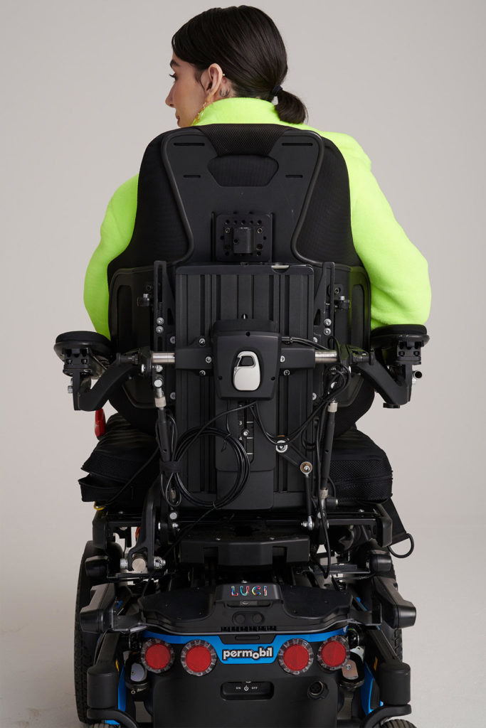
New to the marketplace is Nashville, Tenn-based LUCI, whose premiere product, also named LUCI, is a hardware and software platform that uses sensor-fusion technologies to allow a power wheelchair to “see” its environment.
Once mounted onto a power wheelchair between the power base and the seat, LUCI aims to help users avoid collisions and dangerous drop-offs while maintaining personalized driving control. Through cloud-based capabilities, LUCI can also monitor and alert users and caregivers of low battery, possible tipping scenarios, and other important updates regarding the chair and the user.
“Wheelchair users were left behind when it comes to most innovative technology,” says Barry Dean, CEO and Founder of LUCI. Dean is also a Grammy-nominated songwriter, and his daughter Katherine, 19, has cerebral palsy and has used a wheelchair her whole life.
“We realized no one else was working on this problem in a meaningful way, so my brother Jered [Dean, CTO of LUCI] and I set out to create a solution for Katherine,” he says, in a media release.
“What started as a labor of love among family members has ultimately created a safer, more stable way for people with disabilities to navigate their world and stay connected to loved ones. Today, we’re excited to launch LUCI and continue collaborating with researchers, universities and other companies using our open platform to move the industry forward together,” he adds.
The LUCI team spent the past two and half years collaborating with clinical professionals and logging over 25,000 hours of user testing to develop an invention to help people with physical disabilities drive safely, precisely and independently. LUCI’s R&D efforts have already resulted in a total of 16 patents (eight pending).
“When we started tinkering with my niece Katherine’s chair, we had no idea where this journey would lead,” says Jered Dean, CTO, who has spent 2 decades in design and systems engineering, most recently serving as director of the Colorado School of Mines’s Capstone Design@Mines program.
“From developing advancements in millimeter-wave radar technology to collaborating with engineering leaders from Intel RealSense Technology Group to maximize the application of some of the world’s smartest cameras, I’m incredibly proud of the unprecedented work our team has accomplished to solve the challenges our customers face,” he continues, in the release.
“LUCI leverages Intel RealSense to map the world in a low-power, cost-effective way to make drop-off protection and collision avoidance possible, and we’re excited to be a part of this inspirational effort to deliver innovation that improves lives,” says Joel Hagberg, head of product management and marketing, Intel RealSense Group
LUCI’s technology combines stereovision, infrared, ultrasonic and radar sensors to offer users these critical features, per the release:
- Collision avoidance: LUCI is designed to prevent wheelchair users from running into objects (walls, people, pets, furniture, etc) as they navigate their daily lives. It does this by smoothly helping to navigate the chair in coordination with user steering inputs based on obstacle detection in the driver’s surroundings.
- Drop-off protection: It doesn’t take a large drop-off to tip a wheelchair (less than 3 inches in some cases). LUCI helps users avoid tipping by recognizing steps or drop-offs and smoothly helping the chair continue on a safer path.
- Anti-tipping alert system: LUCI monitors the steepness of a ramp or the ground users are driving on and provides an audible alert if it becomes a tipping danger. In the event that a chair tips over, LUCI sounds an alarm and can be configured to quickly alert other individuals, such as a caregiver or loved one, of the exact location of the rider and the tipped chair.
- Cloud-based communications and alerts: The MyLUCI portal allows users to view their data and share it with loved ones or clinicians. LUCI can be set up to alert others of specific events, such as the user’s location if their battery gets dangerously low. LUCI also now works with Hey Google and Amazon Alexa so users can interact with MyLUCI using their voice. MyLUCI portal is available as a mobile app for both iOS and Android phones, as well as for desktop with the Web Portal.
- Secure health monitoring: LUCI users can choose to share their heart rate data with their team using either Google Fit or Apple HealthKit from day one.
[Source: LUCI]
-
You are currently browsing the archives for June, 2020

