Posts Tagged Neurofeedback

[Abstract + References] Interactive Brain Stimulation Neurotherapy Based on BOLD Signal in Stroke Rehabilitation

ABSTRACT

Interactive brain stimulation is a new generation of neurofeedback characterized by a radical change in the targets of cognitive (volitional, adaptive) influence. These targets are represented by specific cerebral structures and neural networks, the reconstruction of which leads to the brain functions’ restoration and behavioral metamorphoses.

Functional magnetic resonance imaging (fMRI) in the neurofeedback contour uses a natural intravascular tracer, a blood-oxygenation-level-dependent (BOLD) signal as feedback. The subject included into the “interactive brain contour” learns to modulate and modify his or her own cerebral networks, creating new ones or “awakening” pre-existing ones, in order to improve (or restore) mental, sensory, or motor functions.

In this review we focus on interactive brain stimulation based on BOLD signal and its role in the motor rehabilitation of stroke, briefly introducing the basic concepts of the so-called “network vocabulary” and general biophysical basis of the BOLD signal. We also discuss a bimodal fMRI-EEG neurofeedback platform and the prospects of fMRI technology in controlling functional connectivity, a numerical assessment of neuroplasticity.

REFERENCES

Alionte, C., Notte, C., & Strubakos, C. D. (2022). From symmetry to chaos and back: Understanding and imaging the mechanisms of neural repair after stroke. Life Sciences, 288, Article 120161. https://doi.org/10.1016/j.lfs.2021.120161

Almeida, S. R. M., Vicentini, J., Bonilha, L., De Campos, B. M., Casseb, R. F., & Min, L. L. (2017). Brain connectivity and functional recovery in patients with ischemic stroke. Journal of Neuroimaging, 27(1), 65–70. https://doi.org/10.1111/jon.12362

Alstott, J., Breakspear, M., Hagmann, P., Cammoun, L., & Sporns, O. (2009). Modeling the impact of lesions in the human brain. PLoS Computational Biology, 5(6), Article e1000408. https://doi.org/10.1371/journal.pcbi.1000408

Alves, R., Henriques, R. N., Kerkelä, L., Chavarrías, C., Jespersen, S. N., & Shemesh, N. (2022). Correlation tensor MRI deciphers underlying kurtosis sources in stroke. NeuroImage, 247, Article 118833. https://doi.org/10.1016/j.neuroimage.2021.118833

Bassett, D. S., Meyer-Lindenberg, A., Achard, S., Duke, T., & Bullmore, E. (2006). Adaptive reconfiguration of fractal small-world human brain functional networks. Proceedings of the National Academy of Sciences of the United States of America, 103(51), 19518–19523. https://doi.org/10.1073/pnas.0606005103

Bentley, W. J., Li, J. M., Snyder, A. Z., Raichle, M. E., & Snyder, L. H. (2016). Oxygen level and LFP in task-positive and task-negative areas: Bridging BOLD fMRI and electrophysiology. Cerebral Cortex, 26(1), 346–357. https://doi.org/10.1093/cercor/bhu260

Bezmaternykh, D. D., Kalgin, K. V., Maximova, P. E., Mel’nikov, M. Y., Petrovskii, E. D., Predtechenskaya, E. V., Savelov, A. A., Semenikhina, A. A., Tsaplina, T. N., Shtark, M. B., & Shurunova, A. V. (2021). Application of fMRI and simultaneous fMRI-EEG neurofeedback in post-stroke motor rehabilitation. Bulletin of Experimental Biology and Medicine, 171(3), 379–383. https://doi.org/10.1007/s10517-021-05232-1

Bezmaternykh, D. D., Mel’nikov, M. E., Petrovskii, E. D., Kozlova, L. I., Shtark, M. B., Savelov, A. A., Shubina, O. S., & Natarova, K. A. (2018). Spontaneous changes in functional connectivity of independent components of fMRI signal in healthy volunteers at rest and in subjects with mild depression. Bulletin of Experimental Biology and Medicine, 165(3), 325–330. https://doi.org/10.1007/s10517-018-4161-3

Birn, R. M., Bandettini, P. A., Cox, R. W., & Shaker, R. (1999). Event-related fMRI of tasks involving brief motion. Human Brain Mapping, 7(2), 106–114. https://doi.org/10.1002/(SICI)1097-0193(1999)7:2<106::AID-HBM4>3.0.CO;2-O

Biswal, B., Yetkin, F. Z., Haughton, V. M., & Hyde, J. S. (1995). Functional connectivity in the motor cortex of resting human brain using echo-planar MRI. Magnetic Resonance in Medicine, 34(4), 537–541. https://doi.org/10.1002/mrm.1910340409

Bonmassar, G., Anami, K., Ives, J., & Belliveau, J. W. (1999). Visual evoked potential (VEP) measured by simultaneous 64-channel EEG and 3T fMRI. NeuroReport, 10(9), 1893–1897. https://doi.org/10.1097/00001756-199906230-00018

Buckner, R. L. (1998). Event-related fMRI and the hemodynamic response. Human Brain Mapping, 6(5–6), 373–377 https://doi.org/10.1002/(SICI)1097-0193(1998)6:5/6<373::AID-HBM8>3.0.CO;2-P

Buckner, R. L., & DiNicola, L. M. (2019). The brain’s default network: Updated anatomy, physiology and evolving insights. Nature Reviews Neuroscience, 20(10), 593–608. https://doi.org/10.1038/s41583-019-0212-7

Carter, A. R., Astafiev, S. V., Lang, C. E., Connor, L. T., Rengachary, J., Strube, M. J., Pope, D. L. W., Shulman, G. L., & Corbetta, M. (2010). Resting interhemispheric functional magnetic resonance imaging connectivity predicts performance after stroke. Annals of Neurology, 67(3), 365–375. https://doi.org/10.1002/ana.21905

Carter, A. R., Shulman, G. L., & Corbetta, M. (2012). Why use a connectivity-based approach to study stroke and recovery of function? NeuroImage, 62(4), 2271–2280. https://doi.org/10.1016/j.neuroimage.2012.02.070

Cheng, H.-J., Ng, K. K., Qian, X., Ji, F., Lu, Z. K., Teo, W. P., Hong, X., Nasrallah, F. A., Ang, K. K., Chuang, K.-H., Guan, C., Yu, H., Chew, E., & Zhou, J. H. (2021). Task-related brain functional network reconfigurations relate to motor recovery in chronic subcortical stroke. Scientific Reports, 11(1), Article 8442. https://doi.org/10.1038/s41598-021-87789-5

Duncan, P. W., Lai, S. M., & Keighley, J. (2000). Defining post-stroke recovery: Implications for design and interpretation of drug trials. Neuropharmacology, 39(5), 835–841. https://doi.org/10.1016/s0028-3908(00)00003-4

Evans, J. R., Dellinger, M. B., & Russell, H. L. (Eds.). (2019). Neurofeedback: The first fifty years (1st ed.). Academic Press.

Feigin, V. L., Norrving, B., George, M. G., Foltz, J. L., Roth, G. A., & Mensah, G. A. (2016). Prevention of stroke: A strategic global imperative. Nature Reviews Neurology, 12(9), 501–512. https://doi.org/10.1038/nrneurol.2016.107

Feigin, V. L., Norrving, B., & Mensah, G. A. (2017). Global burden of stroke. Circulation Research, 120(3), 439–448. https://doi.org/10.1161/CIRCRESAHA.116.308413

Fornito, A., Zalesky, A., & Breakspear, M. (2015). The connectomics of brain disorders. Nature Reviews Neuroscience, 16(3), 159–172. https://doi.org/10.1038/nrn3901

Fovet, T., Jardri, R., & Linden, D. (2015). Current issues in the use of fMRI-based neurofeedback to relieve psychiatric symptoms. Current Pharmaceutical Design, 21(23), 3384–3394. https://doi.org/10.2174/1381612821666150619092540

Friston, K. J. (2011). Functional and effective connectivity: A review. Brain Connectivity, 1(1), 13–36. https://doi.org/10.1089/brain.2011.0008

Gauthier, C. J., & Fan, A. P. (2019). BOLD signal physiology: Models and applications. NeuroImage, 187, 116–127. https://doi.org/10.1016/j.neuroimage.2018.03.018

Golanov, E. V., Yamamoto, S., & Reis, D. J. (1994). Spontaneous waves of cerebral blood flow associated with a pattern of electrocortical activity. American Journal of Physiology, 266(1 Pt. 2), R204–R214. https://doi.org/10.1152/ajpregu.1994.266.1.R204

Guggisberg, A. G., Koch, P. J., Hummel, F. C., & Buetefisch, C. M. (2019). Brain networks and their relevance for stroke rehabilitation. Clinical Neurophysiology, 130(7), 1098–1124. https://doi.org/10.1016/j.clinph.2019.04.004

Herrmann, C. S., & Debener, S. (2008). Simultaneous recording of EEG and BOLD responses: A historical perspective. International Journal of Psychophysiology, 67(3), 161–168. https://doi.org/10.1016/j.ijpsycho.2007.06.006

Huster, R. J., Debener, S., Eichele, T., & Herrmann, C. S. (2012). Methods for simultaneous EEG-fMRI: An introductory review. The Journal of Neuroscience, 32(18), 6053–6060. https://doi.org/10.1523/JNEUROSCI.0447-12.2012

Ives, J. R., Warach, S., Schmitt, F., Edelman, R. R., & Schomer, D. L. (1993). Monitoring the patient’s EEG during echo planar MRI. Electroencephalography and Clinical Neurophysiology, 87(6), 417–420. https://doi.org/10.1016/0013-4694(93)90156-p

Keynan, J. N., Cohen, A., Jackont, G., Green, N., Goldway, N., Davidov, A., Meir-Hasson, Y., Raz, G., Intrator, N., Fruchter, E., Ginat, K., Laska, E., Cavazza, M., & Hendler, T. (2019). Electrical fingerprint of the amygdala guides neurofeedback training for stress resilience. Nature Human Behaviour, 3(1), 63–73. https://doi.org/10.1038/s41562-018-0484-3

Keynan, J. N., Meir-Hasson, Y., Gilam, G., Cohen, A., Jackont, G., Kinreich, S., Ikar, L., Or-Borichev, A., Etkin, A., Gyurak, A., Klovatch, I., Intrator, N., & Hendler, T. (2016). Limbic activity modulation guided by functional magnetic resonance imaging-inspired electroencephalography improves implicit emotion regulation. Biological Psychiatry, 80(6), 490–496. https://doi.org/10.1016/j.biopsych.2015.12.024

Kim, D. H., & Kang, H. (2022). Changes in bihemispheric structural connectivity following middle cerebral artery infarction. Journal of Personalized Medicine, 12(1), 81. https://doi.org/10.3390/jpm12010081

Koush, Y., Rosa, M. J., Robineau, F., Heinen, K., Rieger, S. W., Weiskopf, N., Vuilleumier, P., Van De Ville, D., & Scharnowski, F. (2013). Connectivity-based neurofeedback: Dynamic causal modeling for real-time fMRI. NeuroImage, 81, 422–430. https://doi.org/10.1016/j.neuroimage.2013.05.010

Kwakkel, G., & Kollen, B. J. (2013). Predicting activities after stroke: What is clinically relevant? International Journal of Stroke, 8(1), 25–32. https://doi.org/10.1111/j.1747-4949.2012.00967.x

Larivière, S., Ward, N. S., & Boudrias, M.-H. (2018). Disrupted functional network integrity and flexibility after stroke: Relation to motor impairments. NeuroImage: Clinical, 19, 883–891. https://doi.org/10.1016/j.nicl.2018.06.010

Lecrux, C., & Hamel, E. (2016). Neuronal networks and mediators of cortical neurovascular coupling responses in normal and altered brain states. Philosophical Transactions of the Royal Society of B, Biological Sciences, 371(1705), Article 20150350. https://doi.org/10.1098/rstb.2015.0350

Li, W., Li, Y., Zhu, W., & Chen, X. (2014). Changes in brain functional network connectivity after stroke. Neural Regeneration Research, 9(1), 51–60. https://doi.org/10.4103/1673-5374.125330

Liew, S.-L., Rana, M., Cornelsen, S., Fortunato De Barros Filho, M., Birbaumer, N., Sitaram, R., Cohen, L. G., & Soekadar, S. R. (2016). Improving motor corticothalamic communication after stroke using real-time fMRI connectivity-based neurofeedback. Neurorehabilitation and Neural Repair, 30(7), 671–675. https://doi.org/10.1177/1545968315619699

Lioi, G., Butet, S., Fleury, M., Bannier, E., Lécuyer, A., Bonan, I., & Barillot, C. (2020). A multi-target motor imagery training using bimodal EEG-fMRI neurofeedback: A pilot study in chronic stroke patients. Frontiers in Human Neuroscience, 14, Article 37. https://doi.org/10.3389/fnhum.2020.00037

Lioi, G., Fleury, M., Butet, S., Lécuyer, A., Barillot, C., & Bonan, I. (2018). Bimodal EEG-fMRI neurofeedback for stroke rehabilitation: A case report. Annals of Physical and Rehabilitation Medicine, 61(Suppl), e482–e483. https://doi.org/10.1016/j.rehab.2018.05.1127

Lopes da Silva, F. (2013). EEG and MEG: Relevance to neuroscience. Neuron, 80(5), 1112–1128. https://doi.org/10.1016/j.neuron.2013.10.017

Luu, P., Tucker, D. M., Englander, R., Lockfeld, A., Lutsep, H., & Oken, B. (2001). Localizing acute stroke-related EEG changes: Assessing the effects of spatial undersampling. Journal of Clinical Neurophysiology, 18(4), 302–317. https://doi.org/10.1097/00004691-200107000-00002

Mano, M., Lécuyer, A., Bannier, E., Perronnet, L., Noorzadeh, S., & Barillot, C. (2017). How to build a hybrid neurofeedback platform combining EEG and fMRI. Frontiers in Neuroscience, 11, Article 140. https://doi.org/10.3389/fnins.2017.00140

Marek, S., & Dosenbach, N. U. F. (2018). The frontoparietal network: Function, electrophysiology, and importance of individual precision mapping. Dialogues in Clinical Neuroscience, 20(2), 133–140. https://doi.org/10.31887/DCNS.2018.20.2/smarek

Matthews, P. M., & Jezzard, P. (2004). Functional magnetic resonance imaging. Journal of Neurology, Neurosurgery, and Psychiatry, 75(1), 6–12.

Mehler, D. M. A., Williams, A. N., Krause, F., Lührs, M., Wise, R. G., Turner, D. L., Linden, D. E. J., & Whittaker, J. R. (2019). The BOLD response in primary motor cortex and supplementary motor area during kinesthetic motor imagery based graded fMRI neurofeedback. NeuroImage, 184, 36–44. https://doi.org/10.1016/j.neuroimage.2018.09.007

Mehler, D. M. A., Williams, A. N., Whittaker, J. R., Krause, F., Lührs, M., Kunas, S., Wise, R. G., Shetty, H. G. M., Turner, D. L., & Linden, D. E. J. (2020). Graded fMRI neurofeedback training of motor imagery in middle cerebral artery stroke patients: A preregistered proof-of-concept study. Frontiers in Human Neuroscience, 14, Article 226. https://doi.org/10.3389/fnhum.2020.00226

Meir-Hasson, Y., Keynan, J. N., Kinreich, S., Jackont, G., Cohen, A., Podlipsky-Klovatch, I., Hendler, T., & Intrator, N. (2016). One-class FMRI-inspired EEG model for self-regulation training. PLoS ONE, 11(5), Article e0154968. https://doi.org/10.1371/journal.pone.0154968

Meir-Hasson, Y., Kinreich, S., Podlipsky, I., Hendler, T., & Intrator, N. (2014). An EEG finger-print of fMRI deep regional activation. NeuroImage, 102(Pt. 1), 128–141. https://doi.org/10.1016/j.neuroimage.2013.11.004

Mel’nikov, M. Y., Shtark, M. B., Savelov, A. A., & Bruhl, A. (2017). Real time fuctional magnetic resonance imaging biofeedback: A new generation of neurotherapy. Zhurnal Vysshei Nervnoi Deiatelnosti imeni I. P. Pavlova, 67(1), 3–32.

Menon, V. (2011). Large-scale brain networks and psychopathology: A unifying triple network model. Trends in Cognitive Sciences, 15(10), 483–506. https://doi.org/10.1016/j.tics.2011.08.003

Miller, K. L., Luh, W.-M., Liu, T. T., Martinez, A., Obata, T., Wong, E. C., Frank, L. R., & Buxton, R. B. (2001). Nonlinear temporal dynamics of the cerebral blood flow response. Human Brain Mapping, 13(1), 1–12. https://doi.org/10.1002/hbm.1020

Molnar-Szakacs, I., & Uddin, L. Q. (2013). Self-processing and the default mode network: Interactions with the mirror neuron system. Frontiers in Human Neuroscience, 7, Article 571. https://doi.org/10.3389/fnhum.2013.00571

Morgenroth, E., Saviola, F., Gilleen, J., Allen, B., Lührs, M., W Eysenck, M., & Allen, P. (2020). Using connectivity-based real-time fMRI neurofeedback to modulate attentional and resting state networks in people with high trait anxiety. NeuroImage: Clinical, 25, Article 102191. https://doi.org/10.1016/j.nicl.2020.102191

Nudo R. J. (2003). Functional and structural plasticity in motor cortex: Implications for stroke recovery. Physical Medicine and Rehabilitation Clinics of North America, 14(1 Suppl.), S57–S76. https://doi.org/10.1016/s1047-9651(02)00054-2

Nudo, R. J., Wise, B. M., SiFuentes, F., & Milliken, G. W. (1996). Neural substrates for the effects of rehabilitative training on motor recovery after ischemic infarct. Science, 272(5269), 1791–1794. https://doi.org/10.1126/science.272.5269.1791

Ogawa, S., & Lee, T.-M. (1990). Magnetic resonance imaging of blood vessels at high fields: In vivo and in vitro measurements and image simulation. Magnetic Resonance in Medicine, 16(1), 9–18. https://doi.org/10.1002/mrm.1910160103

Ogawa, S., Menon, R. S., Kim, S. G., & Ugurbil, K. (1998). On the characteristics of functional magnetic resonance imaging of the brain. Annual Review of Biophysics and Biomolecular Structure, 27, 447–474. https://doi.org/10.1146/annurev.biophys.27.1.447

Paret, C., Goldway, N., Zich, C., Keynan, J. N., Hendler, T., Linden, D., & Cohen Kadosh, K. (2019). Current progress in real-time functional magnetic resonance-based neurofeedback: Methodological challenges and achievements. NeuroImage, 202, 116107. https://doi.org/10.1016/j.neuroimage.2019.116107

Petersen, S. E., & Sporns, O. (2015). Brain networks and cognitive architectures. Neuron, 88(1), 207–219. https://doi.org/10.1016/j.neuron.2015.09.027

Philiastides, M. G., Tu, T., & Sajda, P. (2021). Inferring macroscale brain dynamics via fusion of simultaneous EEG-fMRI. Annual Review of Neuroscience, 44, 315–334. https://doi.org/10.1146/annurev-neuro-100220-093239

Raichle, M. E., MacLeod, A. M., Snyder, A. Z., Powers, W. J., Gusnard, D. A., & Shulman, G. L. (2001). A default mode of brain function. Proceedings of the National Academy of Sciences of the United States of America, 98(2), 676–682. https://doi.org/10.1073/pnas.98.2.676

Rance, M., Ruttorf, M., Nees, F., Schad, L. R., & Flor, H. (2014). Neurofeedback of the difference in activation of the anterior cingulate cortex and posterior insular cortex: Two functionally connected areas in the processing of pain. Frontiers in Behavioral Neuroscience, 8, Article 357. https://doi.org/10.3389/fnbeh.2014.00357

Ritter, P., & Villringer, A. (2006). Simultaneous EEG-fMRI. Neuroscience & Biobehavioral Reviews, 30(6), 823–838. https://doi.org/10.1016/j.neubiorev.2006.06.008

Rowe, J. B. (2010). Connectivity analysis is essential to understand neurological disorders. Frontiers in Systems Neuroscience, 4, Article 144. https://doi.org/10.3389/fnsys.2010.00144

Rudnev, V., Melnikov, M., Savelov, A., Shtark, M., & Sokhadze, E. M. (2021). fMRI-EEG fingerprint regression model for motor cortex. NeuroRegulation, 8(3), 162–172. https://doi.org/10.15540/nr.8.3.162

Savelov, A. A., Shtark, M. B., Kozlova, L. I., Verevkin, E. G., Petrovskii, E. D., Pokrovskii, M. A., Rudych, P. D., & Tsyrkin, G. M. (2019). Dynamics of interactions between cerebral networks derived from fMRI data and motor rehabilitation during stokes. Bulletin of Experimental Biology and Medicine, 166(3), 399–403. https://doi.org/10.1007/s10517-019-04359-6

Savelov, A. A., Shtark, M. B., Mel’nikov, M. E., Kozlova, L. I., Bezmaternykh, D. D., Verevkin, E. G., Petrovskii, E. D., Pokrovskii, M. A., Tsirkin, G. M., & Rudych, P. D. (2019a). Prospects of synchronous fMRI-EEG recording as the basis for neurofeedback (exemplified on patient with stroke sequelae). Bulletin of Experimental Biology and Medicine, 166(3), 390–393. https://doi.org/10.1007/s10517-019-04357-8

Savelov, A. A., Shtark, M. B., Mel’nikov, M. E., Kozlova, L. I., Bezmaternykh, D. D., Verevkin, E. G., Petrovskii, E. D., Pokrovskii, M. A., Tsirkin, G. M., & Rudych, P. D. (2019b). Dynamics of fMRI and EEG parameters in a stroke patient assessed during a neurofeedback course focused on Brodmann area 4 (M1). Bulletin of Experimental Biology and Medicine, 166(3), 394–398. https://doi.org/10.1007/s10517-019-04358-7

Schaechter, J. D., Moore, C. I., Connell, B. D., Rosen, B. R., & Dijkhuizen, R. M. (2006). Structural and functional plasticity in the somatosensory cortex of chronic stroke patients. Brain, 129(10), 2722–2733. https://doi.org/10.1093/brain/awl214

Scharnowski, F., Veit, R., Zopf, R., Studer, P., Bock, S., Diedrichsen, J., Goebel, R., Mathiak, K., Birbaumer, N., & Weiskopf, N. (2015). Manipulating motor performance and memory through real-time fMRI neurofeedback. Biological Psychology, 108, 85–97. https://doi.org/10.1016/j.biopsycho.2015.03.009

Shtark, M. B. (2019). Neurofeedback: A scarce resource at the mental market. In J. R. Evans, M. B. Dellinger, & H. L. Russell (Eds.), Neurofeedback: The first fifty years (pp. 353–358). Academic Press. https://doi.org/10.1016/B978-0-12-817659-7.00046-4

Shtark, M. B., Korostyshevskaia, A. M., Rezakova, M. V., & Savelov, A. A. (2012). Functional magnetic resonance imaging and neuroscience Uspekhi Fiziologicheskikh Nauk, 43(1), 3–29.

Shtark, M. B., Verevkin, E. G., Kozlova, L. I., Mazhirina, K. G., Pokrovskii, M. A., Petrovskii, E. D., Savelov, A. A., Starostin, A. S., & Yarosh, S. V. (2015). Synergetic fMRI-EEG brain mapping in alpha-rhythm voluntary control mode. Bulletin of Experimental Biology and Medicine, 158(5), 644–649. https://doi.org/10.1007/s10517-015-2827-7

Siegel, J. S., Ramsey, L. E., Snyder, A. Z., Metcalf, N. V., Chacko, R. V., Weinberger, K., Baldassarre, A., Hacker, C. D., Shulman, G. L., & Corbetta, M. (2016). Disruptions of network connectivity predict impairment in multiple behavioral domains after stroke. Proceedings of the National Academy of Sciences of the United States of America, 113(30), E4367–E4376. https://doi.org/10.1073/pnas.1521083113

Sitaram, R., Veit, R., Stevens, B., Caria, A., Gerloff, C., Birbaumer, N., & Hummel, F. (2012). Acquired control of ventral premotor cortex activity by feedback training: An exploratory real-time FMRI and TMS study. Neurorehabilitation and Neural Repair, 26(3), 256–265. https://doi.org/10.1177/1545968311418345

Sokolova, O. O., Shtark, M. B., & Lisachev, P. D. (2010). Neuronal plasticity and gene expression. Uspekhi Fiziologicheskikh Nauk, 41(1), 26–44.

Sorger, B., Kamp, T., Weiskopf, N., Peters, J. C., & Goebel, R. (2018). When the brain takes ‘BOLD’ steps: Real-time fMRI neurofeedback can further enhance the ability to gradually self-regulate regional brain activation. Neuroscience, 378, 71–88. https://doi.org/10.1016/j.neuroscience.2016.09.026

Spampinato, D. A., Block, H. J., & Celnik, P. A. (2017). Cerebellar–M1 connectivity changes associated with motor learning are somatotopic specific. Journal of Neuroscience, 37(9), 2377–2386. https://doi.org/10.1523/JNEUROSCI.2511-16.2017

Sporns, O., & Honey, C. J. (2006). Small worlds inside big brains. Proceedings of the National Academy of Sciences of the United States of America, 103(51), 19219–19220. https://doi.org/10.1073/pnas.0609523103

Stoeckel, L. E., Garrison, K. A., Ghosh, S., Wighton, P., Hanlon, C. A., Gilman, J. M., Greer, S., Turk-Browne, N. B., deBettencourt, M. T., Scheinost, D., Craddock, C., Thompson, T., Calderon, V., Bauer, C. C., George, M., Breiter, H. C., Whitfield-Gabrieli, S., Gabrieli, J. D., LaConte, S. M., Hirshberg, L., … Evins, A. E. (2014). Optimizing real time fMRI neurofeedback for therapeutic discovery and development. NeuroImage: Clinical, 5, 245–255. https://doi.org/10.1016/j.nicl.2014.07.002

Sulzer, J., Haller, S., Scharnowski, F., Weiskopf, N., Birbaumer, N., Blefari, M. L., Bruehl, A. B., Cohen, L. G., deCharms, R. C., Gassert, R., Goebel, R., Herwig, U., LaConte, S., Linden, D., Luft, A., Seifritz, E., & Sitaram, R. (2013). Real-time fMRI neurofeedback: Progress and challenges. NeuroImage, 76, 386–399. https://doi.org/10.1016/j.neuroimage.2013.03.033

Ullsperger, M., & Debener, S. (Eds.). (2010). Simultaneous EEG and fMRI: Recording, analysis, and application. Oxford University Press. https://doi.org/10.1093/acprof:oso/9780195372731.001.0001

Tang, C., Zhao, Z., Chen, C., Zheng, X., Sun, F., Zhang, X., Tian, J., Fan, M., Wu, Y., & Jia, J. (2016). Decreased functional connectivity of homotopic brain regions in chronic stroke patients: A resting state fMRI study. PLoS ONE, 11(4), Article e0152875. https://doi.org/10.1371/journal.pone.0152875

van Meer, M. P. A., van der Marel, K., Wang, K., Otte, W. M., el Bouazati, S., Roeling, T. A. P., Viergever, M. A., Berkelbach van der Sprenkel, J. W., & Dijkhuizen, R. M. (2010). Recovery of sensorimotor function after experimental stroke correlates with restoration of resting-state interhemispheric functional connectivity. The Journal of Neuroscience, 30(11), 3964–3972. https://doi.org/10.1523/JNEUROSCI.5709-09.2010

Wang, C., Stebbins, G. T., Nyenhuis, D. L., deToledo-Morrell, L., Freels, S., Gencheva, E., Pedelty, L., Sripathirathan, K., Moseley, M. E., Turner, D. A., Gabrieli, J. D. E., & Gorelick, P. B. (2006). Longitudinal changes in white matter following ischemic stroke: A three-year follow-up study. Neurobiology of Aging, 27(12), 1827–1833. https://doi.org/10.1016/j.neurobiolaging.2005.10.008

Wang, L., Yu, C., Chen, H., Qin, W., He, Y., Fan, F., Zhang, Y., Wang, M., Li, K., Zang, Y., Woodward, T. S., & Zhu, C. (2010). Dynamic functional reorganization of the motor execution network after stroke. Brain, 133(4), 1224–1238. https://doi.org/10.1093/brain/awq043

Wang, W., Collinger, J. L., Perez, M. A., Tyler-Kabara, E. C., Cohen, L. G., Birbaumer, N., Brose, S. W., Schwartz, A. B., Boninger, M. L., & Weber, D. J. (2010). Neural interface technology for rehabilitation: Exploiting and promoting neuroplasticity. Physical Medicine and Rehabilitation Clinics of North America, 21(1), 157–178. https://doi.org/10.1016/j.pmr.2009.07.003

Yoo, S.-S., & Jolesz, F. A. (2002). Functional MRI for neurofeedback: Feasibility study on a hand motor task. NeuroReport, 13(11), 1377–1381. https://doi.org/10.1097/00001756-200208070-00005

Yu, X., Jiaerken, Y., Wang, S., Hong, H., Jackson, A., Yuan, L., Lou, M., Jiang, Q., Zhang, M., & Huang, P. (2020). Changes in the corticospinal tract beyond the ischemic lesion following acute hemispheric stroke: A diffusion kurtosis imaging study. Journal of Magnetic Resonance Imaging, 52(2), 512–519. https://doi.org/10.1002/jmri.27066

Yuan, K., Chen, C., Wang, X., Chu, W. C.-W., & Tong, R. K.-Y. (2021). BCI training effects on chronic stroke correlate with functional reorganization in motor-related regions: A concurrent EEG and fMRI study. Brain Sciences, 11(1), 56. https://doi.org/10.3390/brainsci11010056

Zeiler, S. R., Gibson, E. M., Hoesch, R. E., Li, M. Y., Worley, P. F., O’Brien, R. J., & Krakauer, J. W. (2013). Medial premotor cortex shows a reduction in inhibitory markers and mediates recovery in a mouse model of focal stroke. Stroke, 44(2), 483–489. https://doi.org/10.1161/STROKEAHA.112.676940

Zhang, Y., Liu, H., Wang, L., Yang, J., Yan, R., Zhang, J., Sang, L., Li, P., Wang, J., & Qiu, M. (2016). Relationship between functional connectivity and motor function assessment in stroke patients with hemiplegia: A resting-state functional MRI study. Neuroradiology, 58(5), 503–511. https://doi.org/10.1007/s00234-016-1646-5

Zhuravleva, K. V., Savelov, A. A., Korostyshevskaya, A. M., & Shtark, M. B. (2022). Diffusional characteristics of brain matter after stroke. Bulletin of Experimental Biology and Medicine, 172(4), 402–406. https://doi.org/10.1007/s10517-022-05402-9

Zotev, V., Phillips, R., Yuan, H., Misaki, M., & Bodurka, J. (2014). Self-regulation of human brain activity using simultaneous real-time fMRI and EEG neurofeedback. NeuroImage, 85(Pt. 3), 985–995. https://doi.org/10.1016/j.neuroimage.2013.04.126

Source
, , , , , ,

Leave a comment

[Abstract] New treatments for induction of motor plasticity after stroke – Thesis

Abstract

Stroke is a major health issue. It is one of the main causes of acquired adult disability. A common deficit after stroke is the hemiparesis of the contralateral upper limb, with more than 80% of stroke inpatients experiencing this condition and in spite of intensive rehabilitation care, more than 40% of the patients still have this impairment chronically. The overall aim of this thesis is to understand how we reduce impairment in stroke patients with new treatment approaches. Previous studies have shown possible targets for new treatments. Patients with severe motor deficits show severe damage of the cortico-spinal tract (CST) and secondary degradation of white matter during the weeks after stroke. In these patients, inducing plasticity at the level of the CST or avoiding secondary degradation may enable the motor recovery processes, which are usually very limited or absent in these patients. Patients with mild or moderate motor deficits show high FC between the motor areas and the rest of the brain in the first weeks after stroke, which associates with better clinical recovery. In these patients, enhancing FC may improve the motor outcome above the proportional rule. In a first study, we recruited patients with mild and moderate motor deficits. We targeted the neural interactions of the perilesional cortical areas with the application of 4 different transcranial direct current stimulation (tDCS) montages: conventional anodal, high-definition (HD) anodal, bi-hemispheric, and SHAM. We aimed to find the optimal montage that would increase FC the most and lead to the best clinical outcome. After comparison, the HD tDCS montage had the most effect on motor outcome.The effect only appeared in the Action Research Arm Test and not in our primary outcome which was the Fugl-Meyer assessment of the upper extremity (FMA-UE). On a neurophysiological level, the MEP analyses pointed out a tendency of the HD montage to increase the MEP amplitudes and reduce the rest motor threshold in the contralesional hemisphere rather than having an effect in the ipsilesional hemisphere. The computation of FC confirmed previous findings where connectivity was an early marker of good motor recovery, while its late arrival was associated with worse recovery. In a second study, we recruited patients with severe motor deficits. Here, a closed-loop setup combining functional electrical stimulations (FES) contingent with a brain-computer interface (BCI) aimed to prevent the secondary degeneration of the CST integrity. The motor scores did not display significant differences between the BCI and a control group consisting of triggering the FES based on the contingency of patients undergoing the real BCI training. However, BCI-FES reduced spasticity of the patients as evaluated with the Ashworth score. On a group level, global FC between the primary motor cortex and the rest of the brain was significantly reduced after BCI-FES as compared to after SHAM treatment, and this reduction of global FC was correlated with greater FMA-UE improvement. Regarding CST integrity, the two groups evolved similarly. FA asymmetry showed that the CST of all patients did not deteriorate during the brief observation time window. No significant difference was observed in the MEPs of the ipsilesional or the contralesional side. In a third study, we explored the feasibility of developing a neurofeedback training targeting FC for clinical use. We aimed at finding an analysis pipeline that would allow to compute source FC of a region of interest based on a low-density EEG coverage and a template MRI. We compared 3 several algorithms that would allow an FC reconstruction from only 19 channels. We used numerical simulations of coherent sources as well as real datasets. Of these analyses, the use of beamformer inverse solution emerged as the best performing under the constraints imposed. We finally tested its performance in an independent dataset recorded from a low-density EEG using dry-gel electrodes. However, we could not reproduce the findings of the previous dataset. This may be due to the quality of the signal or to the design of the FC reconstruction, which was not yet efficient enough. In conclusion, this thesis allowed the observation of innovative treatments on the patients themselves in the first two projects, and as a proof-of-concept in the third project. Although promising, the three treatments still need more research to fine-tune their setup for more efficient interventions. Faced with motor deficits that are still too present, it is important to pursue research to perfect these interventions until the condition of the patients is improved. To that end, from our studies the next steps are to 1) go in the main phase to compare HD-tDCS against SHAM-tDCS, 2) add more BCI sessions to reach the usual amount of training, 3) perform complementary FC analyses to observe the neural interactions between motor areas, 4) implement a neurofeedback targeting FC with a high-density EEG using sponge electrodes.

Thesis (6.9 MB) – PDF file – Free access

Source

, , , , , , , , , ,

Leave a comment

[Abstract] Improving the acceptability to enhance the efficiency of stroke rehabilitation procedures based on brain-computer interfaces: General public results – Poster

Abstract : Stroke leaves around 40% of surviving patients dependent in their activities of daily living, notably due to severe motor disabilities [Inserm, 2019]. Brain-Computer Interfaces (BCIs) have been shown to be efficient for improving motor recovery after stroke [Cervera et al., 2018], but this efficiency is still far from the level required to achieve the clinical breakthrough expected by both clinicians and patients. While technical levers of improvement have been identified, they are insufficient: fully optimised BCIs are pointless if patients and clinicians do not want to use them [Blain-Moraes et al., 2012]. We hypothesise that improving BCI acceptability and acceptance, by better informing stakeholders about BCI functioning and by personalising the BCI-based rehabilitation procedures to each patient, respectively, will favour engagement in the rehabilitation process and result in an increased efficiency. Our first objective was to identify the factors influencing the intention to use (IU) BCIs [Davis, 1989]. Based on the literature, we constructed a model of BCI acceptability and adapted it in questionnaires addressed to the general population (n=753) and post-stroke patients (n=33). Videos were included, one about the general functioning of BCIs, the second about their relevance for rehabilitation. We used random forest algorithms to explain IU based on our model’s factors. After the first video, IU was mainly explained by subjective and personal factors, i.e., perceived usefulness (PU), perceived ease of use (PEOU) and BCI playfulness for the general population, and PU, autonomy and engagement in the rehabilitation for the patients. After the second video, the explanatory factors became more scientific/rational, with PU, cost-benefits ratio and scientific relevance for the general population, and PU, scientific relevance and ease of learning for patients. The shift of main explanatory factors (before/after second video) from subjective representations to scientific arguments highlights the impact of providing patients with clear information regarding BCIs.

Source

, , , , , , ,

Leave a comment

[WEB SITE] ‘Brain training’ may be an effective treatment for post-traumatic stress disorder

by Lawson Health Research Institute

'Brain training' may be an effective treatment for post-traumatic stress disorder
The study tested use of a neurofeedback loop in which a person’s brain activity is measured through sensors placed on the scalp and displayed back to them using a computer interface. Brain activity was visualized as either a still cartoon or a distorted picture that would move or become clearer when the alpha rhythm was successfully reduced. Credit: Lawson Health Research Institute

Neurofeedback, also called ‘brain training,’ consists of exercises where individuals regulate their own brain activity. In a new study from Lawson Health Research Institute and Western University, researchers have found that neurofeedback may be an effective treatment for individuals with post-traumatic stress disorder (PTSD). Published in NeuroImage: Clinical, the clinical trial found that neurofeedback was effective in reducing symptoms of PTSD.

“Brain connectivity involves different parts of the brain communicating with each other and helps to regulate states of consciousness, thought, mood and emotion,” explains Dr. Ruth Lanius, scientist at Lawson, professor at Western’s Schulich School of Medicine & Dentistry and psychiatrist at London Health Sciences Centre. “Individuals with PTSD tend to have disrupted patterns of brain connectivity, but our research suggests they can exercise their brains to restore patterns to a healthy balance.”

Neurofeedback uses a system called a neurofeedback loop in which a person’s brain activity is measured through sensors placed on the scalp and displayed back to them using a computer interface. This allows the individual to complete exercises and visually see the results.

The trial tested neurofeedback with a total of 72 participants, including 36 participants with PTSD and 36 healthy control participants. Of those with PTSD, 18 were randomized to participate in neurofeedback treatment while the other 18 acted as a comparison group.

The study found that the severity of PTSD symptoms decreased in participants randomized to receive neurofeedback treatment. After treatment, 61.1 per cent of participants no longer met the definition for PTSD. This remission rate is comparable to gold standard therapies like trauma-focused psychotherapy.

The research team also used functional magnetic resonance imaging (fMRI) at St. Joseph’s Health Care London to capture brain scans of participants both before and after participation in the trial. They found that individuals with PTSD experienced positive changes in brain connectivity in the salience network and the default mode network following neurofeedback treatment.

“The salience network is involved in detecting threat as part of the ‘fight or flight’ response. It is normally hyperactive in individuals with PTSD. Meanwhile, the default mode network is activated during rest and is involved in autobiographical memory. We often see that this network is less active during rest and functionally disrupted among individuals with PTSD,” says Dr. Andrew Nicholson, affiliated scientist at Lawson. “Neurofeedback helped restore the functional connectivity of both networks to healthier levels.” Dr. Nicholson is an assistant professor at McMaster University and was formerly a post-doctoral fellow at Schulich Medicine & Dentistry.

The study involved weekly sessions of neurofeedback over 20 weeks. Participants were asked to reduce the intensity of the brain’s dominant brain wave—the alpha rhythm. Brain activity was visualized as either a still cartoon or a distorted picture. If the alpha rhythm was successfully reduced, the cartoon started playing or the picture started becoming clearer.

“Participants were not instructed on how to reduce the alpha rhythm. Rather, each individual figured out their own way to do so,” notes Dr. Lanius. “For example, individuals reported letting their mind wander, thinking about positive things or concentrating their attention.”

The team notes the treatment could have a number of clinical implications following further validation.

“Neurofeedback could offer an accessible and effective treatment option for individuals with PTSD,” says Dr. Lanius. “The treatment is easily scalable for implementation in rural areas and even at home.”


Explore further Game study not playing around with PTSD relief


More information: Andrew A. Nicholson et al, A randomized, controlled trial of alpha-rhythm EEG neurofeedback in posttraumatic stress disorder: A preliminary investigation showing evidence of decreased PTSD symptoms and restored default mode and salience network connectivity using fMRI, NeuroImage: Clinical (2020). DOI: 10.1016/j.nicl.2020.102490

Journal information: NeuroImage: Clinical

Provided by Lawson Health Research Institute

, , ,

Leave a comment

[VIDEO] Woman with Traumatic Brain Injury (TBI) Improves with Neurofeedback — Even Over 9 Years Later – YouTube

http://www.CenterforBrain.com

April has suffered debilitating symptoms for over nine years since an illness left her with a severe traumatic brain injury (TBI). After just six weeks of neurofeedback, she has experienced significant improvement. This interview with April, her daughter, and Mike Cohen of the Center for Brain Training explores the power neurofeedback can have in people’s lives, even many years after a brain injury occurs.

Neurofeedback, or brain training, can help people suffering repercussions of traumatic brain injury, post-concussive syndrome, and stroke.

A transcript of the video is available below:

A: She is a walking miracle for sure.

Ap: Yes.

A: I mean, even now if you look at her actual MRI or anything, there is so much damage that people thought that she should be a vegetable or something like that by now. So after she got out of the hospital she couldn’t read, she couldn’t write, she had no depth perception. She was very out of it. She didn’t remember our names or anything like that.

M: What are you both seeing change since you’ve been training your brain with the neurofeedback?

Ap: My communication is lot better. My finding direction is a lot better.

A: I think she’s becoming more of herself again. She’s getting some of her personality back. She has always been pretty feisty. She keeps going no matter what happens. She seems to be getting a lot better. She can tell her right from left, which is a big deal. She is becoming a lot more sharp, I guess, mentally. She definitely has a ways to go, but this is improving her for sure. I think that a lot of things are possible with her because, before she got sick, she had so much drive and she was very inventive and creative and never let anything in life stop her. She’s still like that now, but she is really having a hard time putting her thoughts together and being organized, and the sharper her brain gets, I think that she could take that a long way.

M: When I met you, one of the things I noticed, April, was that you were almost like in a fog.

Ap: Yes. I’m much more alert.

M: So you are better able to communicate with other people now?

Ap: Yes, much better. Sometimes I have to hear what they said, and I can hear what they said but I couldn’t process it all. This is very encouraging. It’s amazing how I am seeing the brain come back around.

M: Did any of your doctors ever mention anything like this?

Ap: No.

A: No.

M: Did any of the other therapies ever help your brain like this?

Ap: No.

M: I am just excited for you that your brain seems to be waking up.

Ap: Yes.

M: Is that what it seems like?

Ap: Yes, in so many ways. I mean, I had other things; it’s very interesting, like the taste and the smell. You know, that didn’t really work.

A: Yes she had no taste, no smell for the most part. Even her vision changes all the time. Like when she goes to the doctor, everything is different all the time, so they can never give her glasses or contacts that actually work the whole time, which is really interesting, but it seems like since she has been doing the brain therapy, some of that has been coming back.

M: And you are interested in medical and going to a med school, is that right?

A: Yes, definitely. So the brain is definitely something that I am really interested in now, especially after seeing this, because doctors don’t seem to use this, like you had said, so I would definitely be interested in learning more and seeing how far you can go with it.

, , ,

Leave a comment

[VIDEO] Traumatic Brain Injury and Neurofeedback – YouTube

A video about Traumatic Brain Injury and Neurofeedback


, , ,

Leave a comment

[WEB SITE] What is neurohacking and can it actually rewire your brain?

Marc Bordons / Stocksy

What is neurohacking and can it actually rewire your brain?

Although at one point, “hack” referred to a creative solution to a tech problem, the term can apply to pretty much anything now. There are kitchen hacks, productivity hacks, personal finance hacks. Brain hacks, or neurohacks, are among the buzziest, though, thanks largely to the Silicon Valley techies who often swear by them as a way to boost their cognitive function, focus, and creativity. Mic asked a neuroscientist to explain neurohacking, which neurohacking methods are especially promising, which are mostly hype, and how to make neurohacking work for you.

First things first: Neurohacking, is a broad umbrella term that encompasses anything that involves “manipulating brain function or structure to improve one’s experience of the world,” says neuroscientist Don Vaughn of Santa Clara University and the University of California, Los Angeles. Like the other myriad forms of hacking, neurohacking uses an engineering approach, treating the brain as a piece of hardware that can be systematically modified and upgraded.

Neurohacking techniques can fall under a number of categories — here are a few of the most relevant ones, as well as the thinking behind them.

Brain stimulation

This involves applying an electric or magnetic field to certain regions of the brain in non-neurotypical people to make their activity more closely resemble that seen in a neurotypical brain. In 2008, the Food and Drug Administration approved transcranial magnetic stimulation (TMS) — a noninvasive form of brain stimulation which delivers magnetic pulses to the brain in a noninvasive manner — for major depression. Since then, the FDA has also approved TMS for pain associated with migraines with auras, as well as obsessive-compulsive disorder. Established brain stimulation techniques (such as TMS or electroconvulsive therapy) performed by an expert provider, such as a psychiatrist or neuroscientist, are generally safe, Vaughn says.

Neurofeedback

This one involves using a device that measures brain activity, such as an electroencephalogram (EEG) or a functional magnetic resonance imaging (fMRI) machine. People with neuropsychological disorders receive feedback on their own brain activity — often in the form of images or sound — and focus on trying to make it more closely resemble the brain activity in a healthy person, Vaughn says. This could happen through changing their thought patterns, Vaughn says. Another possibility is that the feedback itself, or the person’s thoughts about the feedback, may somehow lead to a change in their brain’s wiring.

Reducing cognitive load

This means minimizing how much apps, devices, and other tech compete for your attention. Doing so can sharpen and sustain your focus, or what Vaughn refers to as your attention quotient (AQ). To boost his AQ, Vaughn listens to brown noise, which he likens to “white noise, but deeper.” (Think the low rush of a waterfall versus pure static.) He also chews gum, which he says provides an outlet for his restless “monkey mind” while still allowing him to focus on the task at hand.

Reducing cognitive load can also deepen your connection with others. Vaughn uses Voicea, an app based on an AI assistant that takes and store notes of meetings, whether over the phone or in-person, allowing him to focus solely on the conversation, not on recording it. “If we can quell those disruptions that occur because of the way work is done these days, it will allow us to focus and be more empathic with each other,” he says.

Monitoring sleep

Tracking your sleep patterns and adjusting them accordingly. Every night, you go through around five or so stages of sleep, each one deeper than the last. “People are less groggy and make fewer errors when they wake up in a lighter stage of sleep,” Vaughn says. He uses Sleep Cycle, an app that tracks your sleep patterns based on your movements in bed to rouse you during your lightest sleep stage.

Andrey Popov / Shutterstock

Microdosing

Microdosing is the routinely consumption of teensy doses of psychedelics like LSD, ecstasy, or magic mushrooms. Many who practice microdosing follow the regimen recommended by James Fadiman, psychologist and author of The Psychedelic Explorer’s Guide: Safe, Therapeutic, and Sacred Journeys: a twentieth to a tenth of a regular dose, once every three days for about a month. While a regular dose may make you trip, a microdose has subtler effects, with some users reporting, for instance, enhanced energy and focus, per The Cut.

Nootropics

These are OTC supplements or drugs taken to enhance cognitive function. They range from everyday caffeine and vitamin B12 (B12 deficiency has been associated with cognitive decline) to prescription drugs like Ritalin and Adderall, used to treat ADHD and narcolepsy, as well as Provigil (modafinil), used to treat extreme drowsiness resulting from narcolepsy and other sleep disorder. (All three of these drugs promote wakefulness.) The science behind nootropic supplements in particular remains rather murky, though.

Does neurohacking work, though?

Vaughn finds microdosing, neurostimulation, and neurofeedback especially promising for neuropsychological disorders. Although studies suggest that larger doses of psychedelics could help with disorders such as PTSD and treatment-resistant major depression, there are few studies on microdosing psychedelics. “The little science that has been done…is mixed—perhaps slightly positive,” Vaughn says. “Microdosing is promising mainly because of anecdotal evidence.” Meanwhile, neurostimulation can be used noninvasively in some cases, and TMS has already received FDA approval for a handful of conditions. Neurofeedback is not only non-invasive, but offers immediate feedback, and studies suggest it could be effective for PTSD and addiction.

But it’s important to note that just because these methods could positively alter brain function in people with neuropsychological disorders, that “doesn’t mean it’s going to take a normal system and make it superhuman,” Vaughn says. “I think there are lots of small hacks to be done that could add up to something big,” rather than huge hacks that can vastly upgrade cognitive function, a la Limitless. Thanks to millions of years of evolution, the human brain is already pretty damn optimized. “I just don’t know how much more we can tweak it to make it better,” Vaughn says.

As far as enhancements for neurotypical brains, he says that “you’ll probably see a much greater improvement” from removing distractions in your environment to reduce cognitive load than say, increasing your B12 intake — which brings us to an important disclaimer about nootropic supplements in particular. As with all supplements, they aren’t FDA-regulated, meaning that companies that sell them don’t need to provide evidence that they’re safe or effective. Vaughn recommends trying nootropics that research has shown to be safe and effective, like B12 or caffeine.

How can I start neurohacking?

As tempting as it is, adopting every neurohack under the sun is “not the answer,” Vaughn says. Remember, everyone is different. While your best friend may gush about how much her mood has improved since she began microdosing shrooms, your brain might not respond to microdosing—or maybe taking psychedelics just doesn’t align with your ethics.

Start by exploring different neurohacks, and of course, be skeptical of any product that makes outrageous claims. Since neurofeedback isn’t a common medical treatment, talk to your doctor about enrolling in academic studies on neurofeedback, or companies that offer it if you’re interested, Vaughn says. You should also talk to your doctor if you want to try brain stimulation. A doctor can prescribe you Adderall, Ritalin, or Provigil but only for their indicated medical uses, not for cognitive enhancement.

Ultimately, neurohacks are tools, Vaughn says. “You have to find the one that works for you.” If anything, taking this DIY approach to improving your brain function will leave you feeling empowered, a benefit that probably rivals anything a supplement or sleep tracking app could offer.


via What is neurohacking and can it actually rewire your brain?

, , , , , , , , , ,

Leave a comment

[Abstract + References] Multimodal Head-Mounted Virtual-Reality Brain-Computer Interface for Stroke Rehabilitation – Conference paper

Abstract

Rehabilitation after stroke requires the exploitation of active movement by the patient in order to efficiently re-train the affected side. Individuals with severe stroke cannot benefit from many training solutions since they have paresis and/or spasticity, limiting volitional movement. Nonetheless, research has shown that individuals with severe stroke may have modest benefits from action observation, virtual reality, and neurofeedback from brain-computer interfaces (BCIs). In this study, we combined the principles of action observation in VR together with BCI neurofeedback for stroke rehabilitation to try to elicit optimal rehabilitation gains. Here, we illustrate the development of the REINVENT platform, which takes post-stroke brain signals indicating an attempt to move and drives a virtual avatar arm, providing patient-driven action observation in head-mounted VR. We also present a longitudinal case study with a single individual to demonstrate the feasibility and potentially efficacy of the REINVENT system.

References

  1. 1.
    Mozaffarian, D., et al.: American heart association statistics committee and stroke statistics subcommittee: heart disease and stroke statistics–2015 update: a report from the American heart association. Circulation 131, e29–e322 (2015)Google Scholar
  2. 2.
    Miller, E.L., et al.: American heart association council on cardiovascular nursing and the stroke council: comprehensive overview of nursing and interdisciplinary rehabilitation care of the stroke patient: a scientific statement from the American heart association. Stroke 41, 2402–2448 (2010)CrossRefGoogle Scholar
  3. 3.
    Celnik, P., Webster, B., Glasser, D., Cohen, L.: Effects of action observation on physical training after stroke. Stroke J. Cereb. Circ. 39, 1814–1820 (2008)CrossRefGoogle Scholar
  4. 4.
    Ertelt, D., et al.: Action observation has a positive impact on rehabilitation of motor deficits after stroke. NeuroImage 36(Suppl 2), T164–T173 (2007)CrossRefGoogle Scholar
  5. 5.
    Garrison, K.A., Aziz-Zadeh, L., Wong, S.W., Liew, S.-L., Winstein, C.J.: Modulating the motor system by action observation after stroke. Stroke 44, 2247–2253 (2013)CrossRefGoogle Scholar
  6. 6.
    Ballester, B.R., et al.: The visual amplification of goal-oriented movements counteracts acquired non-use in hemiparetic stroke patients. J. Neuroeng. Rehabil. 12, 50 (2015)CrossRefGoogle Scholar
  7. 7.
    Vourvopoulos, A., Bermúdez i Badia, S.: Motor priming in virtual reality can augment motor-imagery training efficacy in restorative brain-computer interaction: a within-subject analysis. J. Neuroeng. Rehabil. 13, 69 (2016)CrossRefGoogle Scholar
  8. 8.
    Maclean, N., Pound, P., Wolfe, C., Rudd, A.: Qualitative analysis of stroke patients’ motivation for rehabilitation. BMJ 321, 1051–1054 (2000)CrossRefGoogle Scholar
  9. 9.
    Paraskevopoulos, I., Tsekleves, E., Warland, A., Kilbride, C.: Virtual reality-based holistic framework: a tool for participatory development of customised playful therapy sessions for motor rehabilitation. In: 2016 8th International Conference on Games and Virtual Worlds for Serious Applications (VS-Games), September (2016)Google Scholar
  10. 10.
    Wolpaw, J.R.: Brain-Computer Interfaces: Principles and Practice. Oxford University Press, Oxford (2012)CrossRefGoogle Scholar
  11. 11.
    Vourvopoulos, A., Bermudez i Badia, S.: Usability and cost-effectiveness in brain-computer interaction: is it user throughput or technology related? In: Proceedings of the 7th Augmented Human International Conference. ACM, Geneva, Switzerland (2016)Google Scholar
  12. 12.
    Schomer, D.L., Lopes da Silva, F.H.: Niedermeyer’s Electroencephalography: Basic Principles, Clinical Applications, and Related Fields. Lippincott Williams & Wilkins, Philadelphia (2011)Google Scholar
  13. 13.
    Kropotov, J.D.: Chapter 2.2 – Alpha rhythms. In: Kropotov, J.D. (ed.) Functional Neuromarkers for Psychiatry, pp. 89–105. Academic Press, San Diego (2016)CrossRefGoogle Scholar
  14. 14.
    Pfurtscheller, G., Lopes da Silva, F.H.: Event-related EEG/MEG synchronization and desynchronization: basic principles. Clin. Neurophysiol. Off. J. Int. Fed. Clin. Neurophysiol. 110, 1842–1857 (1999)CrossRefGoogle Scholar
  15. 15.
    Wu, J., et al.: Connectivity measures are robust biomarkers of cortical function and plasticity after stroke. Brain 138, 2359–2369 (2015)CrossRefGoogle Scholar
  16. 16.
    Zhou, R.J., et al.: Predicting gains with visuospatial training after stroke using an EEG measure of frontoparietal circuit function. Front. Neurol. 9, 597 (2018)CrossRefGoogle Scholar
  17. 17.
    Soekadar, S.R., Birbaumer, N., Slutzky, M.W., Cohen, L.G.: Brain–machine interfaces in neurorehabilitation of stroke. Neurobiol. Dis. 83, 172–179 (2015)CrossRefGoogle Scholar
  18. 18.
    Friedman, D.: Brain-computer interfacing and virtual reality. In: Nakatsu, R., Rauterberg, M., Ciancarini, P. (eds.) Handbook of Digital Games and Entertainment Technologies, pp. 151–171. Springer, Singapore (2017).  https://doi.org/10.1007/978-981-4560-50-4_2CrossRefGoogle Scholar
  19. 19.
    Vourvopoulos, A., Ferreira, A., Bermúdez i Badia, S.: NeuRow: an immersive VR environment for motor-imagery training with the use of brain-computer interfaces and vibrotactile feedback. In: 3rd International Conference on Physiological Computing Systems, Lisbon (2016)Google Scholar
  20. 20.
    Slater, M.: Place illusion and plausibility can lead to realistic behaviour in immersive virtual environments. Philos. Trans. R. Soc. B Biol. Sci. 364, 3549–3557 (2009)CrossRefGoogle Scholar
  21. 21.
    Spicer, R., Anglin, J., Krum, D.M., Liew, S.L.: REINVENT: a low-cost, virtual reality brain-computer interface for severe stroke upper limb motor recovery. In: 2017 IEEE Virtual Reality (VR), pp. 385–386 (2017)Google Scholar
  22. 22.
    Klem, G.H., Luders, H.O., Jasper, H.H., Elger, C.: The ten-twenty electrode system of the international federation. Electroencephalogr. Clin. Neurophysiol. 52, 3–6 (1999). The International Federation of Clinical NeurophysiologyGoogle Scholar
  23. 23.
    Kothe, C.: Lab streaming layer (LSL). https://github.com/sccn/labstreaminglayer. Accessed 26 Oct 2015 (2014)
  24. 24.
    Fugl-Meyer, A.R., Jääskö, L., Leyman, I., Olsson, S., Steglind, S.: The post-stroke hemiplegic patient. 1. a method for evaluation of physical performance. Scand. J. Rehabil. Med. 7, 13–31 (1975)Google Scholar
  25. 25.
    Duncan, P.W., Wallace, D., Lai, S.M., Johnson, D., Embretson, S., Laster, L.J.: The stroke impact scale version 2.0: evaluation of reliability, validity, and sensitivity to change. Stroke 30, 2131–2140 (1999)CrossRefGoogle Scholar
  26. 26.
    Kennedy, R.S., Lane, N.E., Berbaum, K.S., Lilienthal, M.G.: Simulator sickness questionnaire: an enhanced method for quantifying simulator sickness. Int. J. Aviat. Psychol. 3, 203–220 (1993)CrossRefGoogle Scholar
  27. 27.
    Bailey, J.O., Bailenson, J.N., Casasanto, D.: When does virtual embodiment change our minds? Presence Teleoperators Virtual Environ. 25, 222–233 (2016)CrossRefGoogle Scholar
  28. 28.
    Witmer, B.G., Singer, M.J.: Measuring presence in virtual environments: a presence questionnaire. Presence Teleoperator Virtual Environ. 7, 225–240 (1998)CrossRefGoogle Scholar
  29. 29.
    Bouchard, S., Robillard, G., Renaud, P., Bernier, F.: Exploring new dimensions in the assessment of virtual reality induced side effects. J. Comput. Inf. Technol. 1, 20–32 (2011)Google Scholar
  30. 30.
    Delorme, A., Makeig, S.: EEGLAB: an open source toolbox for analysis of single-trial EEG dynamics including independent component analysis. J. Neurosci. Methods 134, 9–21 (2004)CrossRefGoogle Scholar
  31. 31.
    Makeig, S.: Auditory event-related dynamics of the EEG spectrum and effects of exposure to tones. Electroencephalogr. Clin. Neurophysiol. 86, 283–293 (1993)CrossRefGoogle Scholar
  32. 32.
    Neuper, C., Wörtz, M., Pfurtscheller, G.: ERD/ERS patterns reflecting sensorimotor activation and deactivation. Prog. Brain Res. 159, 211–222 (2006)CrossRefGoogle Scholar
  33. 33.
    Pfurtscheller, G., Aranibar, A.: Evaluation of event-related desynchronization (ERD) preceding and following voluntary self-paced movement. Electroencephalogr. Clin. Neurophysiol. 46, 138–146 (1979)CrossRefGoogle Scholar
  34. 34.
    Pfurtscheller, G., Brunner, C., Schlögl, A., Lopes da Silva, F.H.: Mu rhythm (de)synchronization and EEG single-trial classification of different motor imagery tasks. NeuroImage 31, 153–159 (2006)CrossRefGoogle Scholar
  35. 35.
    Liew, S.-L., et al.: Laterality of poststroke cortical motor activity during action observation is related to hemispheric dominance. Neural Plast. 2018, 14 (2018)CrossRefGoogle Scholar
  36. 36.
    Ritter, P., Moosmann, M., Villringer, A.: Rolandic alpha and beta EEG rhythms’ strengths are inversely related to fMRI-BOLD signal in primary somatosensory and motor cortex. Hum. Brain Mapp. 30, 1168–1187 (2009)CrossRefGoogle Scholar
  37. 37.
    Westlake, K.P., et al.: Resting state alpha-band functional connectivity and recovery after stroke. Exp. Neurol. 237, 160–169 (2012)CrossRefGoogle Scholar
  38. 38.
    Dubovik, S., et al.: EEG alpha band synchrony predicts cognitive and motor performance in patients with ischemic stroke. https://www.hindawi.com/journals/bn/2013/109764/abs/

via Multimodal Head-Mounted Virtual-Reality Brain-Computer Interface for Stroke Rehabilitation | SpringerLink

, , , , , , , , , , , , ,

Leave a comment

[ARTICLE] Effects of a Brain-Computer Interface With Virtual Reality (VR) Neurofeedback: A Pilot Study in Chronic Stroke Patients – Full Text

Rehabilitation for stroke patients with severe motor impairments (e.g., inability to perform wrist or finger extension on the affected side) is burdensome and difficult because most current rehabilitation options require some volitional movement to retrain the affected side. However, although these patients participate in therapy requiring volitional movement, previous research has shown that they may receive modest benefits from action observation, virtual reality (VR), and brain-computer interfaces (BCIs). These approaches have shown some success in strengthening key motor pathways thought to support motor recovery after stroke, in the absence of volitional movement. The purpose of this study was to combine the principles of VR and BCI in a platform called REINVENT and assess its effects on four chronic stroke patients across different levels of motor impairment. REINVENT acquires post-stroke EEG signals that indicate an attempt to move and drives the movement of a virtual avatar arm, allowing patient-driven action observation neurofeedback in VR. In addition, synchronous electromyography (EMG) data were also captured to monitor overt muscle activity. Here we tested four chronic stroke survivors and show that this EEG-based BCI can be safely used over repeated sessions by stroke survivors across a wide range of motor disabilities. Finally, individual results suggest that patients with more severe motor impairments may benefit the most from EEG-based neurofeedback, while patients with more mild impairments may benefit more from EMG-based feedback, harnessing existing sensorimotor pathways. We note that although this work is promising, due to the small sample size, these results are preliminary. Future research is needed to confirm these findings in a larger and more diverse population.

Introduction

Stroke is a leading cause of adult long-term disability worldwide (Mozaffarian et al., 2015), and an increasing number of stroke survivors suffer from severe cognitive and motor impairments each year. This results in a loss of independence in their daily life, such as decreased ability to perform self-care tasks and decreased participation in social activities (Miller et al., 2010). Rehabilitation following stroke focuses on maximizing restoration of lost motor and cognitive functions and on relearning skills to better perform activities of daily living (ADLs). There is increasing evidence that the brain remains plastic at later stages after stroke, suggesting additional recovery remains possible (Page et al., 2004Butler and Page, 2006). To maximize brain plasticity, several rehabilitation strategies have been exploited, including the use of intensive rehabilitation (Wittenberg et al., 2016), repetitive motor training (Thomas et al., 2017), mirror therapy (Pérez-Cruzado et al., 2017), motor-imagery (Kho et al., 2014), and action observation (Celnik et al., 2008), amongst others.

Recently, growing evidence of the positive impact of virtual reality (VR) techniques on recovery following stroke has accumulated (Bermúdez i Badia et al., 2016). When combined with conventional therapy, VR is able to effectively incorporate rehabilitation strategies such as intensity, frequency, and duration of therapy in a novel and low-cost approach in the stroke population (Laver et al., 2017). However, patients with low levels of motor control cannot benefit from current VR tools due to their low volitional motor control, range of motion, pain, and fatigue. Rehabilitation for these individuals is challenging because most current training options require some volitional movement to train the affected side, however, research has shown that individuals with severe stroke may receive modest benefits from action observation and brain-computer interfaces (BCIs) (Silvoni et al., 2011).

Merging BCIs with VR allows for a wide range of experiences in which patients can feel immersed in various aspects of their environment. This allows patients to control their experiences in VR using only brain activity, either directly (e.g., movement in VR through explicit control) or indirectly (e.g., modulating task difficulty level based on workload as implicit control) (Vourvopoulos et al., 2016Friedman, 2017). This direct brain-to-VR communication can induce a sensorimotor contingency between the patient’s internal intentions and the environment’s responsive actions, increasing the patient’s sense of embodiment of their virtual avatar (Slater, 2009Ramos-Murguialday et al., 2013).

After a stroke resulting in severe motor impairments (e.g., inability to perform wrist or finger extension on the affected side), research shows that action observation combined with physical training enhances the effects of motor training (Celnik et al., 2008), eliciting motor-related brain activity in the lesioned hemisphere, leading to modest motor improvements (Ertelt et al., 2007Garrison et al., 2013). Moreover, action observation in a head-mounted VR increases motor activity in both healthy and the post-stroke brains (Ballester et al., 2015Vourvopoulos and Bermúdez i Badia, 2016a).

In addition, neurofeedback through BCIs has been proposed for individuals with severe stroke because BCIs do not require active motor control. Research on BCIs for rehabilitation has shown that motor-related brain signals are reinforced by rewarding feedback so they can be used to strengthen key motor pathways that are thought to support motor recovery after stroke (Wolpaw, 2012). Such feedback has previously shown modest success in motor rehabilitation for severe stroke patients (Soekadar et al., 2015).

The most common brain signal acquisition technology used with BCIs in stroke patients is non-invasive electroencephalography (EEG) (Wolpaw, 2012), which provide a cost-effective BCI platform (Vourvopoulos and Bermúdez i Badia, 2016b). In BCI paradigms for motor rehabilitation, EEG signals related to motor planning and execution are utilized. During a motor attempt, the temporal pattern of the Alpha rhythm (8–12 Hz) desynchronizes. The Alpha rhythm is also termed Rolandic mu or the sensorimotor rhythm (SMR) when it is localized over the sensorimotor cortices of the brain. Mu rhythms (8–12 Hz) are considered indirect indications of the action observation network (Kropotov, 2016) and reflect general sensorimotor activity. Mu rhythms are often detected with changes in the Beta rhythm (12–30 Hz) in the form of event-related desynchronization (ERD), in which a motor action is executed (Pfurtscheller and Lopes da Silva, 1999). These EEG rhythms, or motor-related EEG signatures, are primarily detected during task-based EEG (i.e., when the patient is actively moving or imagining movement) and used for neurofeedback.

Further, neurofeedback-induced changes in brain activity have also been linked to changes in brain activity at rest. That is, after training one’s brain activity using neurofeedback, the intrinsic, resting brain activity (i.e., EEG activity in the absence of a task) may also show changes. This resting brain activity can be used to assess more generalized brain changes, and baseline resting-state signatures may be used to predict recovery (Wu et al., 2015) or response to treatments (Zhou et al., 2018). When combined with neural injury information, resting EEG parameters can also help predict the efficacy of stroke therapy.

In this study, our goal was to combine the principles of virtual reality and BCIs to elicit optimal rehabilitation gains for stroke survivors. We hypothesized that merging BCIs with VR should induce illusions of movement and a strong feeling of embodiment within a virtual body via the action observation network, activating brain areas that overlap with those controlling actual movement, which is important for mobilizing neuroplastic changes (Dobkin, 2007). Using a VR-based BCI, those with severe stroke impairments can trigger voluntary movements of the virtual arm in a closed neurofeedback loop. This helps to increase the illusion of one’s own movements through the coordination between one’s intention and the observed first-person virtual action. Therefore, we developed a training platform called REINVENT, which uses post-stroke brain signals that indicate an attempt to move and then drives the movement of a virtual avatar arm, providing patient-driven action observation in head-mounted VR (Spicer et al., 2017). Our previous work using REINVENT with healthy individuals indeed showed that the combination of VR integrated into a BCI encouraged greater embodiment, and greater embodiment was related to greater neurofeedback performance (Anglin et al., 2019).

For this study, we recruited four chronic stroke survivors to undergo a longitudinal BCI-VR intervention using REINVENT to provide EEG-based neurofeedback with simultaneous EMG acquisition. We assessed intervention results using clinical measures, Transcranial Magnetic Stimulation (TMS) and Magnetic Resonance Imaging (MRI) and compared these measures before and after the intervention. The purpose of this study was twofold. First, we sought to determine whether REINVENT is feasible for stroke patients to use across repeated sessions and second, whether REINVENT might be able to strengthen motor-related brain signals in individuals with differing levels of motor impairment after stroke.[…]


Continue —>  Frontiers | Effects of a Brain-Computer Interface With Virtual Reality (VR) Neurofeedback: A Pilot Study in Chronic Stroke Patients | Frontiers in Human Neuroscience

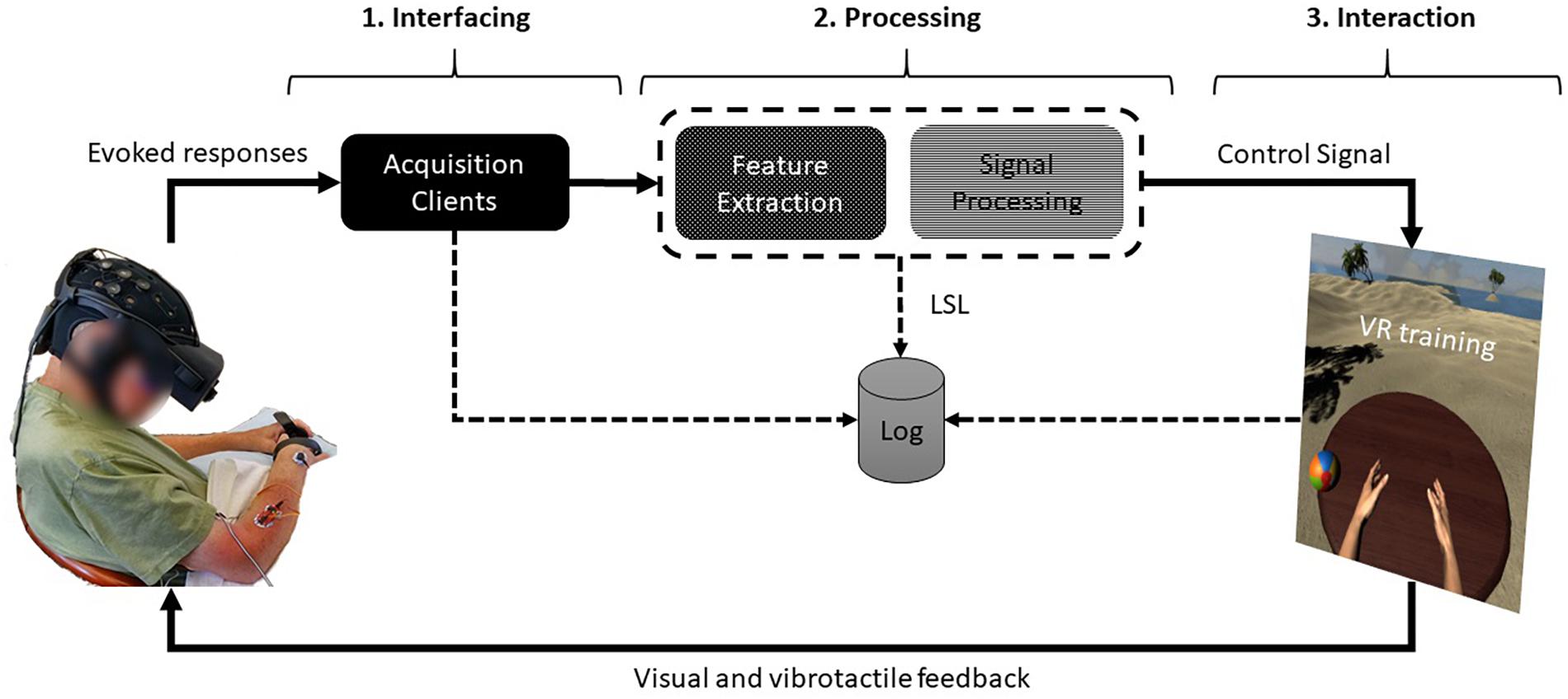
Figure 1. System architecture of a closed neurofeedback loop. From left, (1) the evoked physiological responses are captured at the interfacing layer through the data acquisition clients, (2) sent to the processing layer where the signals are filtered and logged, and then, (3) the extracted features (e.g., EEG bands) are sent to the interaction layer where VR training occurs. Written permission to use this photo was obtained from the individual.

, , , , , , ,

Leave a comment

[Abstract] Brain-machine interface of upper limb recovery in stroke patients rehabilitation: A systematic review

Abstract

BACKGROUND:

Technologies such as brain-computer interfaces are able to guide mental practice, in particular motor imagery performance, to promote recovery in stroke patients, as a combined approach to conventional therapy.

OBJECTIVE:

The aim of this systematic review was to provide a status report regarding advances in brain-computer interface, focusing in particular in upper limb motor recovery.

METHODS:

The databases PubMed, Scopus, and PEDro were systematically searched for articles published between January 2010 and December 2017. The selected studies were randomized controlled trials involving brain-computer interface interventions in stroke patients, with upper limb assessment as primary outcome measures. Reviewers independently extracted data and assessed the methodological quality of the trials, using the PEDro methodologic rating scale.

RESULTS:

From 309 titles, we included nine studies with high quality (PEDro ≥ 6). We found that the most common interface used was non-invasive electroencephalography, and the main neurofeedback, in stroke rehabilitation, was usually visual abstract or a combination with the control of an orthosis/robotic limb. Moreover, the Fugl-Meyer Assessment Scale was a major outcome measure in eight out of nine studies. In addition, the benefits of functional electric stimulation associated to an interface were found in three studies.

CONCLUSIONS:

Neurofeedback training with brain-computer interface systems seem to promote clinical and neurophysiologic changes in stroke patients, in particular those with long-term efficacy.

via: https://www.ncbi.nlm.nih.gov/pubmed/30609208

, , , , , , , ,

Leave a comment