Archive for category Educational
[Slideshow] Stroke: What You Need to Know
Posted by Kostas Pantremenos in Educational on December 17, 2023
Medically Reviewed by Carol DerSarkissian, MD on January 18, 2022

What Is a Stroke?
1/13
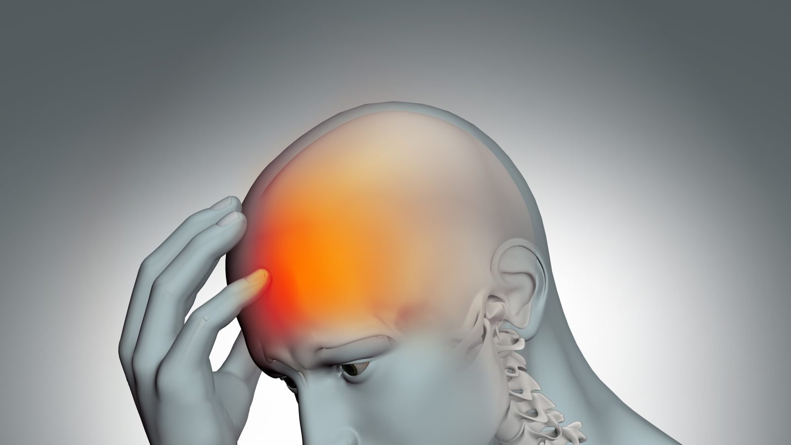
A stroke is a medical emergency. It happens when a blood vessel in the brain bursts or, more commonly, when a blockage happens. Without treatment, cells in the brain quickly begin to die. This can cause serious disability or death. If a loved one is having stroke symptoms, call 911 right away.

Stroke Symptoms
2/13
Call 911 right away about signs of a stroke, which may include sudden:
- Numbness or weakness of the body, especially on one side
- Vision changes in one or both eyes, or trouble swallowing
- Severe headache with an unknown cause
- Problems with dizziness, walking, or balance
- Confusion, trouble speaking or understanding others

Think FAST
3/13
The FAST test helps spot symptoms. It stands for:
Face drooping. Ask for a smile. Does one side droop?
Arm weakness or numbness.
Speech. Can the person repeat a simple sentence? Do they have trouble or slur words?
Time to call 911. Don’t delay.

Time = Brain Damage
4/13
Every second counts. Without oxygen, brain cells begin dying within minutes. Once brain tissue has died, the body parts controlled by that area won’t work right. This makes stroke a top cause of long-term disability. There are clot-busting drugs that can curb brain damage, and they must be given in a short time — usually within 3 hours of when symptoms start.

Diagnosis
5/13
Tests may start when you’re still in the ambulance. Once you get to the ER, you’ll get imaging tests such as a CT scan, MRI, or ultrasound. You may get other types of tests, such as an EKG (checks your heart’s electrical activity) and an EEG (checks your brain’s electrical activity).

Ischemic Stroke
6/13
This is the most common type of stroke: Nearly nine out of 10 fall into this category. An ischemic stroke happens when a blood clot blocks the supply of blood to or in the brain. The clot may start in that spot or travel through the blood from elsewhere in the body. Clogged arteries are a top cause.

Hemorrhagic Stroke
7/13
Hemorrhagic strokes happen when a weakened blood vessel in the brain bursts. The result is bleeding inside the brain that can be hard to stop. The most common cause is high blood pressure. Other causes include aneurysms and AVMs (arteriovenous malformations), which weaken blood vessels in the brain.
")
‘Mini-Stroke’ (TIA)
8/13
Transient ischemic attacks, often called “mini-strokes,” are also an emergency. When they happen, blood flow is temporarily hampered in part of the brain, causing stroke-like symptoms. When the blood flows again, the symptoms stop. You can’t tell at the time if it’s a stroke or TIA. So call 911. Having a TIA is also a warning sign, so see your doctor if you think you’ve had one.

Emergency Treatment
9/13
Ischemic strokes: The goal is to restore blood flow. A clot-busting medication called tPA is very good at dissolving clots and cutting the chance of long-term damage, but it must be given in time — usually within 3 hours. Hemorrhagic strokes: These are harder to manage. Treatment usually involves trying to control high blood pressure, bleeding, and brain swelling.

Causes
10/13
Ischemic strokes: Clogged arteries are a top cause. Plaque made of fat, cholesterol, and other things builds up in the arteries, leaving less space for blood to flow. A blood clot may lodge in this narrowed space and cause an ischemic stroke. All that plaque makes it easier for a clot to form and can also rupture, blocking blood flow.
Hemorrhagic strokes: These can happen if uncontrolled high blood pressure bursts a weakened artery.

Risk Factors
11/13
Your chance of having a stroke rises with age and if you have:
- Had a stroke or TIA before
- Heart disease
- High blood pressure
- High cholesterol
- Diabetes
- Obesity
- Sickle cell disease
Smoking, heavy drinking, and not being active also raise your risk.

What’s On Your Plate?
12/13
Eating too much fat and cholesterol can cause plaque to narrow arteries. Too much salt may lead to high blood pressure. Eating plenty of fruits, vegetables, whole grains, and fish may help lower your stroke risk.

Lowering Your Risk
13/13
Find out if you have any conditions that you need to treat to help prevent a stroke. That may mean taking medicine and also boosting healthy habits, from the foods you eat to being active and not smoking. It’s never too late to start.
[Textbook] Orthopedic Rehabilitation – Principles and Practice
Posted by Kostas Pantremenos in Books, Educational, REHABILITATION on August 12, 2023
Editors: Tony K. George, S. Ali Mostoufi, Alfred J. Tria Jr.
- A concise, practical guide to the principles and practice of orthopedic rehabilitation for residents and fellows
- Utilizes a consistent chapter format, arranged anatomically and covering each joint
- Written and edited by experts in both the orthopedic and physical medicine field
Sections
- Table of contents
- About this book
- Keywords
- Editors and Affiliations
- About the editors
- Bibliographic Information
This is a preview of subscription content, access via your institution.
Table of contents (11 chapters)
- Rehabilitation Principles for Interventional Orthopedics and Orthobiologics
- Walter I. Sussman, Marc P. Gruner, David R. Bakal, Kenneth R. Mautner
- Rehabilitation of Cervical Spine Disorders
- Laurent Delaveaux, Matthew Thomas, Brielle Hansen, Tony K. George
- Rehabilitation of Thoracic Spine Disorders
- Tony K. George, Sneha Varghese, Mindy Chu, Brittney Tout, Hemant Kalia
- Rehabilitation of Lumbar Spine Disorders
- Tony K. George, Matthew Thomas, Sruthi Nanduri, Liya Thomas, Wayne Bonkowski, Bobby Oommen
- Rehabilitation of Shoulder Disorders
- William Micheo, Anthony Lombardi, Claudia Jimenez
- Rehabilitation of Elbow Disorders
- Robert Bowers, Joshua M. Romero, Robert Pagan-Rosado, Dennis A. Colón
- Rehabilitation of Hand Disorders
- Remy V. Rabinovich, Robert M. Zbeda, Steven Beldner, Daniel B. Polatsch
- Rehabilitation of Wrist Disorders
- Robert M. Zbeda, Remy V. Rabinovich, Steven Beldner, Daniel B. Polatsch
- Rehabilitation of Hip Disorders
- David A. Harwood, Anna H. Green, John P. Stelmach, Alfred J. Tria Jr.
- Rehabilitation of Knee Disorders
- Giles R. Scuderi, Matt H. Nasra, Jeremy Silver, Kara L. Sarrel, Alfred J. Tria Jr.
- Rehabilitation of Foot and Ankle Disorders
- Seyed Behrooz Mostofi, Naveen Joseph Mathai
About this book
This pocket-sized guide provides a practical and comprehensive resource for orthopedic, PM&R, and musculoskeletal specialists, as well as primary care physicians who work in the community outpatient clinic setting. Its consistent chapter format covers each area with anatomy, physical examination, preoperative management, and postoperative rehabilitation sections for the spine and extremities.
The book presents treatment protocols for various injuries, including physical therapy measures such as weight bearing status, PRE, closed or open chain exercises, and timing for returning to routine or sport activities. Its concise presentation of rehabilitation for the upper and lower extremities, the hip and pelvis, and the spine enables quick reference and clinical decision-making.
Furthermore, the book includes a chapter on rehabilitation following the use of orthobiologics, making it a valuable resource for healthcare professionals involved in orthopedic rehabilitation after regenerative intervention.
[WEB] Qigong for the Physical Therapist
Posted by Kostas Pantremenos in Educational on April 23, 2023

The goal of this article is to highlight the elements of qigong that fit well with physical therapy – qigong for the physical therapist. A PT does not need to become an expert in qigong to start using qigong principles and exercises with patients.
By Karen Danchalski, PT, DPT
Disclaimer: I am not a master of qigong, I have never led a tai chi class, and I have let my yoga class card expire more times than I can remember. I am writing to you as a physical therapist and qigong practitioner who has found joy and purpose fusing the two modalities. I teach as I learn, my practice and teaching reinforce each other, and I often practice alongside my patients as I lead them through the exercises. A physical therapist does not need to become an expert in a particular type of qigong, have hundreds of hours of practice, or hold a certification to start using qigong principles and exercises with patients.
The goal of this article is to highlight the elements of qigong that fit well with physical therapy. It is not my intention to delve into all the principles of traditional Chinese medicine (TCM) or describe qigong in absolute detail. How can qigong help physical therapy patients? Why is a physical therapist qualified to teach qigong? How can learning qigong add to a therapist’s toolbox and enhance a patient’s therapeutic experience? These are some of the questions I intend to answer.
As discussed in my recent article The Benefits of Mindful Movement in Physical Therapy, qigong is an ancient Chinese form of exercise that combines breath and intention with mindful movement. Qi is the Chinese word for energy and gong means work. Qigong therefore means “energy work” and refers to the practice of moving energy throughout the body for health and well-being. In TCM, illness results from a disruption or blockage in the flow of qi. Although western medicine does not recognize qi as it is described in TCM, scientific evidence supports the practice of qigong in reducing pain, promoting healing, and improving physical, mental, and emotional health.
Listen to more about using mindful movement: Podcast: Mindful Movement with Karen Danchalski, PT, DPT
Physical therapists have strong foundations in body mechanics, posture, and movement. Qigong exercises practiced purely on the physical level can be thought of as light to moderate exercise. Therapists, if they choose, can focus their teaching on this physical level and introduce patients to a number of new routines for stretching, strengthening, and balance. Qigong, when taught as mindful movement, however, requires a broader understanding of TCM concepts and cues. Beyond the physical practice, there are five additional levels which differ in intention but not necessarily difficulty. Which levels are most relevant to physical therapy, and what are some specific qigong cues that can be helpful to patients? Let’s dive in.
Six Levels of Qigong Practice
Physical level (Li) is the movement or choreography of the exercise and is often taught first. The patient learns the placement of the hand, which knee to bend, or the direction of a weight shift, for example. Qigong movements are repetitive. This reinforces the motor pattern, allowing the patient to then focus on the feeling of the exercise and enter into a flow state.
Elastic level (Jing) refers to the feeling of stretch or elasticity in the soft tissues of the body. Some exercises such as “pushing the pillars – pressing the hands out to the side” or “turning the hands – alternating pronation/supination with the arms extended” create and release neural tension and look and feel quite similar to traditional physical therapy neural flossing or gliding techniques. There is a method of qigong practice called YiJin Jing or “muscle tendon changing,” which focuses on transforming and developing the elasticity and strength of the soft tissues.
Mental level (Yi) focuses on visualization and imagery to guide the movement. Imagery in qigong is usually linked to nature. Patients are cued to feel strong and powerful like they are pushing a rock, rooting their feet like trees into the ground, or floating the arms upward like ashes from a fire. Exercises typically have names like the “crane,” “emerging tiger,” or “rolling like a wave,” which characterizes the feeling and quality of the movement.
Energy level (Qi) focuses on moving stagnant Qi and improving circulation and flow of energy throughout the body. In TCM, there is the concept of yin and yang in which energy is either turned inward and restored (Yin) or moved outward and released (Yang). In TCM, there should be a balance of yin and yang energy for optimal health and well-being. Patients learn to feel sensations in the body such as warmth, tingling, changes in the breath, or release of muscle tension, which can be signs of increased Qi flow.
Spirit level (Shen) is the intention of practicing qigong at the level of one’s spirit. The shen, or “spirit,” emanates from the heart center which houses our emotions and instincts. Qigong exercises named for different animals, such as the dragon, monkey, or bear can be practiced at the shen level where the patient feels and expresses the nature of the animal. One can also practice at the Shen level when connecting to a “universal or divine” source of energy which can depend on a patient’s religious or spiritual beliefs.
Emptiness (Kong) is the intention to enter into a meditative state on the feeling of emptiness while practicing both static and moving postures. This type of practice can be highly restorative and healing but is generally more difficult to achieve. It can feel like time is standing still as patients enter into a flow state. Our busy minds and lives make it challenging to practice at this level.
I have explored Li, Jing, Yi, and Qi with my patients, as I feel these levels are most relevant to physical therapy. Below I have given a few examples of patients who I have treated successfully at various levels of practice.
Patient Examples of Qigong for the Physical Therapist
A 65-year-old man with a history of degenerative disc disease and stenosis wants to learn some new ways to stretch and loosen up his back. He also complains of stiffness in his hips and weakness in his legs. I select a hip circle and back circle warm-up in standing and the “wheel” with emphasis on pushing and pulling through the legs in a staggered stance. The focus is on the physical aspects of the exercises.
A 40-year-old female with a history of cervical radiculopathy feels tightness in her neck and in her right arm down to her hand. She complains of feeling restricted in reaching and using her right arm for doing housework and caring for her toddler. She asks if there are any exercises to loosen up the muscles in her arm. I select “pushing the pillars” with focus on good cervical alignment and “drawing the bow” for stretching through the pec and arm with gentle rotation of the neck. The focus is on the elasticity felt during the exercises.
A 70-year-old man with Parkinson’s disease complains of impaired movement, difficulty getting up from a chair, balance problems, and difficulty in turning direction while walking. He wants to learn some exercises that focus on his balance and walking. I select the “buddha walk” which is a slow side step emphasizing pressing the foot on/off the floor, and “cloud hands” which requires coordination of the hands with trunk rotation while weight shifting side to side. The focus is on the visual imagery of rooting the feet into the ground and moving the hands like they are parting the clouds.
A 55-year-old woman with a diagnosis of dermatomyositis complains of pain and restriction in her chest and neck muscles. She states she has had increased anxiety since caring for her elderly parents and sometimes feels restricted in taking a deep breath. She wants to feel like her body can move more freely. I select a routine of three different movements that release tension in the neck and shoulders named the “bear,” “parting the horse’s mane,” and emerging tiger” and end with a standing meditation with a focus on the breath and release of muscle tension. The focus is on moving energy through the body.
Directions of Movement
As therapists, we know that quality of movement matters. We talk to our patients about slowing down repetitions and controlling movements. Qigong uses specific language that can help describe the direction or quality of movement. There are seven directions of movement in qigong: upward, downward, outward, inward, turning, circling, and spiraling.
| Upward movements | Lifting, floating, pressing or pushing upward. |
| Downward movements | Grounding, settling, heavy or sinking, can be passive and gravity assisted, or active as in pressing or pushing downward. |
| Outward movements | Growing, expanding, or the feeling of “letting go” of excess energy or pain, moving from one’s center. |
| Inward movements | Pulling in, gathering, or compressing toward one’s center. |
| Turning | Rotating around the spine. Turning is performed within a patient’s comfortable range of motion. Twisting moves further into a restriction and can be used to increase mobility. |
| Circling | Making the shape of a circle with the arms, hands, hips, etc. |
| Spiraling | Non-linear, combining energies moving in different directions while feeling grounded and lifted simultaneously. Spiraling is a more complex motion to feel. |
Additional Concepts and Cues
In addition to direction and quality of movement, therapists can cue breathing and mindfulness, and impart insightful nuggets of wisdom to their patients during routines. Breathing can be slow and diaphragmatic, coordinated with movement, or cued to “just be natural.” Therapists can encourage meditation or mindfulness while working on posture. A home program could include standing for 3-5 minutes in “ready stance” or “horse’s stance.” Patients learn to focus their mind on their pelvic and spinal alignment, staying lifted through the head and neck, softening their knees, keeping their shoulders down and breathing deeply. Patients learn to feel just enough tone to maintain good posture while letting go of unnecessary muscle tension. Simultaneously they learn the benefit of taking a break from their busy day and being mindful.
Qigong can be part of a patient’s home exercise program and support the patient’s therapeutic goals. The number of repetitions, the speed of the exercise, and the level of effort will be dependent on how the patient feels at the moment and can change from one session to the next. Since the movements are gentle, they can be performed as few as 5 times or for as long as several minutes. Patients are taught the concept of “effortless effort,” which means they are fully present and focused but use about 70% physical effort to allow space for self-discovery and conserving energy.
The goal of qigong for the physical therapist or for anyone is to practice with a calm and focused mind, to become aware of the sensations of the body, and ultimately enter into a flow state. Patients are unique and will bring their former experiences and injuries to their practice. Patients should learn not to compare themselves to others or be preoccupied with making their movements look perfect. Therapists can adapt any qigong exercise to the needs of the patient by changing the position to sitting or lying down, reducing the number of components, or building one cue at a time.
I have discovered tremendous versatility in qigong and have used it with a wide variety of patients with differing diagnoses, physical and cognitive abilities, and even with patients who are non-ambulatory or bedbound. Not all patients will be receptive to learning qigong. They may not think of it as conventional exercise or may have preconceived notions about its origins. They may be uncomfortable with the stillness and patience required to move slowly and to feel rather than to just do the exercise.
The best way to learn and teach qigong to your patients is to find a teacher and start practicing yourself. In the final article of this series, I will introduce some different forms of qigong and suggest some ways to get started on your qigong journey as both a practitioner and a teacher. Stay tuned!
Do you have any questions for the author or commentary about how you would use these techniques with patients or do already? Sound off in the comments!

Karen Danchalski, PT, DPT, has been practicing physical therapy for 24 years. She currently practices in an outpatient orthopedic clinic and provides outpatient services in the home setting. Karen has a special interest in mindful movement, has been a certified Stott Pilates instructor for the past 10 years, and is a member of the National Qigong Institute. She integrates Pilates and qigong exercises into traditional physical therapy treatments whenever it can benefit the patient. She is the author of several articles written for therapists on the topics of exercise for seniors, pain, and personalized healthcare.
Main Photo: Photo 159817417 © Robert Kneschke | Dreamstime.com
Read Karen Danchalski’s other posts in this series:
The Case for Mindful Movement in PT
[BLOG POST] Right Hemisphere Brain Damage: therapy exercises that may help
Posted by Kostas Pantremenos in Educational, TBI on March 28, 2023

Have you recently been told that you or someone close to you has experienced damage to the right side of the brain? If so, you are not alone. However, you may be eager to learn more about the condition and have questions, such as:
- How does the right brain participate in daily functioning?
- What causes damage to the right brain?
- What impairments are common after a right hemisphere brain injury?
- What resources can someone with RHD turn to rebuilding skills that their neurological event or injury may have impacted?
In parallel with our recent post discussing left brain injury, this BrainWire article will demystify right brain injury and answer the above questions. Our hope is that with this information as a starting point, you can begin Brain Injury Awareness Month this March fully empowered with the knowledge you need to optimize your or your loved one’s recovery journey.
What are the key functions of the right brain?
The human brain is divided into two sides (called cerebral hemispheres)–AKA the “left brain” and “right brain”–that work together to control most aspects of the human experience, from emotion to cognition to movement. The left and right hemispheres overlap in some tasks, such as sensory processing and motor regulation., They can work in tandem due to the corpus callosum, a thick bundle of nerves that allows brain signals to travel between the two sides of the brain. However, each brain hemisphere does have specialized functions that differentiate it from the other.
The right side of the brain is especially important for:
- Visuospatial awareness
- Emotion processing
- Deciphering social and nonverbal cues
- Facial recognition
- Creative, artistic, and musical awareness
- Left-side motor control
- Abstract reasoning

This infographic describes some of the critical functions of the right side of the brain and common symptoms of right hemisphere brain damage.
How does right hemisphere brain damage occur?
Right hemisphere brain damage, also commonly known as right hemisphere disorder or RHD, can result from any number of neurological injuries or events. Common causes include strokes, which interrupt the supply of oxygenated blood to the brain; traumatic brain injuries, which are caused by an external blow to the head; tumors; and infections.
What happens when the right side of the brain is damaged?
While no two instances of right hemisphere brain damage are exactly alike, the disorder can exhibit a pattern of similar symptoms spanning the domains of emotion regulation, social interaction, cognition, and movement. For example, a person with RHD may experience:
- Left-side motor impairment: Weakness, paralysis, or difficulty with left-side motor control. (While counterintuitive, the right side of the brain controls movement on the left side of the body.)
- Visuospatial perceptual changes, such as failure to notice or react to stimuli that fall within the left visual field (also known as left-side or left neglect).
- Difficulty orienting and maintaining attention and remembering information.
- Struggles with facial recognition.
- Challenges with complex and abstract reasoning.
It is important to remember that right hemisphere brain damage, regardless of its cause, can manifest differently across individuals. You may be confronting all or none of the above symptoms following damage to the right brain, and both of those experiences—as well as everything in between—are normal for an individual with RHD.
Which Constant Therapy exercises might help with recovery from right hemisphere disorder?
Fortunately, Constant Therapy has a vast library of evidence-based speech, language, and cognitive therapy activities that can help you build back skills which may be more challenging after a right hemisphere brain disorder diagnosis. Below are suggestions for Constant Therapy exercises in skill areas frequently impacted by RHD.
Attention, processing speed, and impulse control Constant Therapy exercises:
- Find the same symbols
- Remember the right card
- Choose which direction
- Remember pictures in order (N-back)
- Remember and say numbers

“Remember and say numbers” Constant Therapy exercise (attention)
Visual memory Constant Therapy exercises:

“Match faces” Constant Therapy exercise (visual memory)
Visuospatial processing Constant Therapy exercises:

“Read a calendar” Constant Therapy exercise (visuospatial processing)
Auditory memory Constant Therapy exercises:

“Understand stories you hear” Constant Therapy exercise (auditory memory)
Analytical reasoning Constant Therapy exercises:

“Put steps in order” Constant Therapy exercise (analytical reasoning)
Conclusion
It can be overwhelming to think about where to start as you recover from right hemisphere disorder. At Constant Therapy, we are excited to partner with you along your journey, and we gladly offer a 2-week free trial to ensure that our program is a good match for your individual needs.
Above all, it is crucial to remember to take care of and be kind to yourself. Some days will be more difficult than others, and when things get especially challenging, don’t feel afraid to reach out for help from a friend, loved one, or trained health professional. Beyond that, remember that neuroplasticity means that your brain has an innate ability to adapt, including after experiencing right hemisphere brain damage. That means that growth is possible, so long as you stay as optimistic as possible through the inevitable ups and downs of recovery.
Curious how left-side brain damage affects daily functions? Read our Left Brain Damage: effects + therapy activities that can help post.
[BLOG POST] All You Should Know About Brain Stroke
Posted by Kostas Pantremenos in Educational on March 23, 2023
All You Should Know About Brain Stroke
A stroke, sometimes called a brain attack, occurs when blood vessel supplying ay part of brain gets blocked or bursts. In either case, parts of the brain become damaged or die. A stroke can cause lasting brain damage, long-term disability, or even death. Brain stroke is 3rd most common killer disease in the world.
What are the types of strokes?
Ischemic stroke: It occurs when blood vessel gets blocked leading to death of part of brain supplied by the blood vessel.
Hemorrhagic stroke: It occurs when blood vessel bursts leading to bleeding within brain tissue and damage of tissue.
One more entity called transient ischemic attack (TIA), as the name suggests stroke symptoms start and resolve on own due t restoration of blood supply to brain. Many times TIAs occur days or weeks before actual stroke, hence acting as warning sign for future stroke. Recognizing and treating TIAs can avert major strokes.
What happens during stroke:
Brain needs continues supply of oxygen and nutrients. Due to blocked or burst blood vessel energy supply gets affected leading to death of neurons within minutes. Each half of brain roughly controls opposite half of body. Right Brain stroke leads to problems in left half of body and vice versa.
What are the risk factors for stroke?
Risk factors for stroke can be classified as modifiable (that can be changed) and non-modifiable(that can’t be changed).
Modifiable risk factors: High blood pressure( > 140/90), Diabetes mellitus, Lack of sleep, Stress, Obesity, Smoking, High plasma lipids, Lack of exercise, Oral contraceptive pills, heart disease, abnormal heart rhythm.
Non modifiable risk factors are: older age, male gender, ethnicity- African Americans, history of prior strokes, genetic risk factors for strokes.
What are the symptoms of stroke:
Stroke symptoms happen suddenly, symptoms vary and can occur in combination of any of the following:
- Weakness or numbness of the face, arm, or leg, usually on one side of the body
- Having trouble speaking or understanding
- Problems with vision, such as dimness or loss of vision in one or both eyes
- Dizziness or problems with balance or coordination, vomiting
- Problems with movement or walking
- Fainting (loss of consciousness) or seizure
- Severe headaches with no known cause, especially if they happen suddenly
Stroke symptoms can be easily identified with acronym BEFAST: Balance, Eye problems, Facial droop, Arm weakness, Speech change, Time since onset of symptoms. If any of these symptoms occur, time of onset should be noted and immediate medical attention should be sought.
Treatment of stroke: Treatment is immediate evaluation, medical, surgical, rehabilitative.
As soon as Stroke is suspected neuroimaging in the form of CT or MRI brain is needed. Type of stroke need to be identified- Ischemic (due to blocked blood vessel) or hemorrhagic (burst blood vessel)
Medical: If patient reaches hospital within 4.5hrs of symptoms onset, Injection called tissue plasminogen activator can be given in eligible patients to dissolve clot and restore blood supply. This is used only in ischemic stroke.
Surgical or interventional: If a large blood vessel is blocked, a procedure called mechanical thrombectomy is done where in clot is removed with endovascular techniques to restore blood vessel. This can be done up to 24hr from symptom onset in eligible patients.
Rehabilitation: patients who have persistent neurological deficits need rehabilitation in the form of physiotherapy, balance therapy, speech therapy. Rehabilitation can take months in major strokes.
Stroke can strike anyone, anytime and can change one’s life forever. Prevention with risk factor modification, good lifestyle, early detection of warning signs, seeking medical attention on time can prevent major disability and death.
[WEB] Helping Stroke Recovery with Stem Cells
Posted by Kostas Pantremenos in Educational on March 17, 2023
As a stroke occurs, some parts of the brain may be temporarily deprived of oxygen due to the interruption of blood flow or a hemorrhage. This causes the death of millions of neurons. According to the CDC report, almost 87% of all strokes are ischemic strokes, i.e., they are caused by a blockage. The two available treatments for stroke include mechanical thrombectomy and stem cell therapy. In mechanical thrombectomy, the blood clots from the brain are removed by administering anti-clotting drugs and using a catheter-based system. This method is effective within six hours of a stroke.
In cell-based therapy, the mesenchymal stem cells are injected or infused into the patient to fuel the repair process within the site of a stroke. This therapy can be given to a patient with either of two forms of stroke and any time after the cardiovascular accident (CVA). While this method is not common yet, the stem cell stroke clinical trial has proven that it aids in expediting repair mechanisms after a stroke. This article will teach you how this research and development can help stroke patients recover.

Use of stem cells in the medical world
Stem cells can seek out damaged tissues in the body and trigger regenerative and anti-inflammatory effects there. Many scientists worldwide are turning towards stem cells for clinical trials. They are trying to figure out how they can help in stroke recovery. In a research conducted at Stanford University School of Medicine, stem cell transplant therapy was used on the brains of 18 individuals who suffered a stroke (DOI: 10.1161/STROKEAHA.116.012995).
Their brains were injected with SB623 cells (mesenchymal stem cells). These were derived from the bone marrow of two donors. Interestingly, the trial participants showed an average increase of 11.4 points on the Fugl-Meyer Assessment, which is a stroke-specific impairment test. This test proved that they are indeed effective in stroke recovery years after its occurrence, regardless of the person’s age.
The trial also showed motor-function improvements in patients with no immune rejection of cells within and after the therapy.
Treatment of stroke conditions with stem cells
They belong to a class of undifferentiated cells. What makes them special is that they can differentiate into specialized types. In other words, they have the ability to become nerve, cardiac, or blood cells, depending on their location within the body. Another factor that makes them special is that they can divide infinitely.
This makes them ideal for replacing damaged cells due to neurological injuries such as stroke. As per a study by Michael Levy and his colleagues, intravenous injection of allogeneic mesenchymal stem cells is an effective treatment for long-term post-stroke recovery (DOI: 10.1161/STROKEAHA.119.026318). Once they are injected into the brain, it helps promote regeneration and repair the damaged tissue.
| “These [stem] cells don’t actually integrate into the brain long term and become neurons to reconstitute circuits. What they do is pump out very powerful growth factors, molecules and proteins that enhance native mechanisms of recovery, such as new synapses of neurons that are there, new blood vessels, and they have a very profound effect on modulating the immune system. And in that way, what we believe they do is to turn the adult brain into a neonatal or infant brain, which has a lot of ability to recover after injury.” Gary Steinberg, MD, PhD, practices at Stanford Health CareStanford Medicine Children’s Health. |
Results of stem cell treatment
Contrary to popular belief, the injected stem cells by themselves don’t replace the neurons. Instead, they enhance the native mechanisms of recovery in the brain and turn it into a cell-regenerating machine. This treatment also helps stimulate neuroplasticity, which reorganizes the circuits in the brain. Here is the evidence:
- Some participants in the study conducted by Michael Levy and his colleagues regained their ability to walk.
- In Dr Steinberg’s clinical study, a substantial motor improvement was reported by participants.
It was proved that this treatment increases functional recovery regardless of the person’s age or the type of stroke that they suffered.
| “Stem cell research is the key to developing cures for degenerative conditions like Parkinson’s and motor neuron disease from which I and many others suffer.” Stephen Hawking, professor, scientist, and astrophysicist. |
Key takeaways
Stem cell therapy involves injecting separated and cultivated adult stem cells into the bloodstream where they reach the site of injury, or directly into the brain. What is important, there are no serious side effects from this therapy. Once the treatment is complete, it improves the natural capacity of the brain to regrow neurons.
The chronic stroke patients who were provided with this therapy demonstrated substantial recovery even long after the occurrence. Hence, this treatment has great potential to restore physical functions after a period of immobility. This gives hope to those who have not been able to achieve progress in recovery after a stroke using conventional rehabilitation methods.
[BLOG POST] A guide to left hemisphere brain damage
Posted by Kostas Pantremenos in Educational, TBI on March 2, 2023

A guide to left hemisphere brain damage
Marissa Russell, MS, CF-SLP
Have you ever heard someone talk about being “left-brained” or “right-brained”? People who consider themselves to be “right-brained” are often thought to be artistic, creative, and intuitive. “Left-brained” individuals, on the other hand, may be more logical, analytical, and detail-oriented.
While it may not be entirely accurate to view personality in this way, it makes sense that people think of the brain as being separated into two parts. The brain is divided into the right side (or hemisphere) and the left side. These hemispheres are connected in the middle by a bundle of nerve fibers known as the corpus callosum. Because of this, the two sides of the brain work together in some ways. However, they control different functions and abilities that we use in our day-to-day lives. That is why people who have damage to the left side of their brain often experience different challenges compared to those who have right hemisphere brain damage.
What is controlled by the left side of the brain?
Some people call the left hemisphere the “language hub” of the brain. It is not hard to see where this name came from, because this part of the brain plays a significant role in our ability to use and understand language, including reading and writing. We also rely on the left hemisphere to help us speak, solve problems, make computations, and move the right side of our body, since each hemisphere of the brain controls movement on the opposite side of the body.
How left hemisphere brain damage impacts daily life
People who have been impacted by left hemisphere brain damage often experience communication, cognition, and movement-related problems.
Left hemisphere brain damage can lead to:
- Difficulty expressing and understanding language at the word, sentence, or conversational level
- Trouble reading and writing
- Changes in speech
- Deficits in planning, organization, and memory as those skills relate to language
- Weakness or lack of movement on the right side of the body
If you have damage to this part of your brain, you may find it hard to retrieve the words you want and to then put those words together following grammatical rules when you are talking to someone. Additionally, it may be hard to understand what others are saying to you. These difficulties are characteristic of aphasia. You may have trouble coordinating the movements of your mouth to say words (apraxia), experience weakness in the muscles you need for speech (dysarthria), or have trouble moving the right side of your body altogether. It can also be more difficult to reason through problems, remember things you hear, and put ideas in order. Any of these impairments can create challenges that impact quality of life.
How is left hemisphere brain damage Treated?
Individuals with brain damage have been shown to benefit from rehabilitation, including (but not limited to) speech-language therapy, physical therapy, and occupational therapy. Clinicians can employ evidence-based therapeutic methods to improve and support speech, language, cognitive, and physical abilities that may have been impacted post-injury. These therapies can enable patients to live more functionally and independently within their communities.
The other great news is that the brain has amazing abilities to heal and compensate for damage. Neuroimaging studies have proven that our brains are “plastic”, meaning that they can change the way they work. We’ve seen evidence of different parts of the brain taking over for damaged parts – even areas of the brain in the opposite hemisphere! Check out our blog post on the 10 principles of neuroplasticity to learn more.
Tips for caring for someone with left hemisphere brain damage
There are many ways that caregivers can support a loved one as they navigate these newfound challenges. While the areas in which someone may need help will vary, here are a few suggestions:
- Change how you communicate – There are many strategies that can both help your loved one understand you, as well as assist them in conveying their own message. A few helpful actions may include:
- Speaking slowly
- Using simple language and short phrases
- Writing down key words
- Making sure you have their attention before speaking
- Limiting interruptions
- Giving your loved one extra time to formulate their message and then speak
- Utilizing alternative ways of communicating (e.g., writing, drawing, and using gestures)
- Asking “yes/no” questions during communication breakdowns
- Eliminate background noise – excess noise can be distracting and make it hard to focus, communicate, and implement strategies.
- Help with planning and problem-solving – this can involve helping your loved one write out the steps to a task before completing it or using tools such as planners and calendars.
- Lead with empathy – Your loved one will be struggling with certain skills that were previously second nature, and empathy can foster patience when situations become difficult. Remember, your mental and emotional health is important throughout this process, and empathy can help.
- Take care of yourself – just like emergency protocols before a flight, “put on your own oxygen mask before assisting others”. In order to provide the best care for your loved one, you must make sure you are taking proper care of yourself. Seek support, take time to participate in the activities you enjoy, and be mindful of your own health.

Marissa is a practicing speech-language pathologist clinical fellow serving English and Spanish-speaking patients at Northeast Rehabilitation Hospital. She is also a Clinical and Scientific Consultant for Constant Therapy Health, where she is involved in content development, advisement on product features, and other app-related clinical support.
References and further reading
Brain Injury Association of America. (2020, March 18). Functions of the Brain. Retrieved from https://www.biausa.org/brain-injury/about-brain-injury/basics/function-of-the-brain
Cleveland Clinic. How to Better Communicate with Stroke Patients. Retrieved from https://my.clevelandclinic.org/health/articles/10408-right–and-left-brain-strokes-tips-for-the-caregiver
Josse, G., & Tzourio-Mazoyer, N. (2004). Hemispheric specialization for language. Brain Research Reviews, 44(1), 1-12.
[BLOG POST] Left Brain Damage: effects + therapy activities that can help
Posted by Kostas Pantremenos in Educational, TBI on February 3, 2023

Kate Ying
If you or your loved one has recently sustained a left hemisphere brain injury — due to a stroke or other brain injury — you may be overwhelmed with questions like:
- What role does the left brain play in everyday life?
- How does left-sided brain damage impact cognitive processing capabilities?
- What differentiates a left side brain injury?
- What tools might be available to help regain valuable skills during the recovery journey?
In anticipation of Brain Injury Awareness Month in March, this post will answer those questions and provide an overview of specific Constant Therapy exercises that may help in recovery from a left brain injury.
What is the left side of the brain responsible for?
The brain is divided into two hemispheres, left and right, which are bridged by the corpus callosum, a bundle of nerves that functions like a highway to connect the two sides of the brain. While the left and right sides of the brain do communicate with one other, each hemisphere has primary control over particular functions.
Counterintuitively, each side of the brain controls the opposite side of the body. One of the key functions the left brain is responsible for, therefore, is right side body mobility.
Beyond that, the left hemisphere of the brain is especially important for language expression and comprehension. This is in part because several brain regions especially critical for language, such as Broca’s area and Wernicke’s area, are located in the left hemisphere of the brain. The left brain is therefore largely responsible for speaking, listening, reading, writing, and grammatical processing.
The left hemisphere is also crucial for logical reasoning skills—including numeracy, computation, and information processing—and executive functions like time awareness.

What is a left-side brain injury?
Due to the relative specialization of brain function, injuries to a particular hemisphere of the brain often assume common characteristics. Such damage can result from a traumatic brain injury, which typically follows a sudden, strong physical impact to the brain, or a non-traumatic brain injury, which can be caused by oxygen deprivation (during a stroke, for example) or chemical exposure.
A left brain injury occurs when the left cerebral hemisphere sustains damage that impacts its functioning. It is important to keep in mind, however, that many brain injuries ultimately affect both sides of the brain.
What would be impaired if the left side of your brain were injured?
Every brain, and every brain injury, is unique. It is impossible to predict how an injury to the left cerebral hemisphere might manifest in a specific individual. However, there are patterns of brain damage that have been broadly observed by researchers and clinicians.
When the left side of the brain is damaged from a neurological injury (like a TBI) or event (like a stroke), language difficulties are one of the most common side effects. People who experience left brain injuries might have difficulty producing and comprehending spoken and/or written language (aphasia); coordinating the motor aspects of speech (apraxia); or with slurring their speech (dysarthria).
Left side brain damage may also result in challenges with numerical computations (such as counting or math operations) and processing and solving complex problems.
Movement on the right side of the body is also commonly affected with left side brain damage, and it may result in weakness and decreased sensitivity on the right side as well.
Which Constant Therapy exercises can help with my brain injury recovery?
As you or your loved one is recovering from a left side brain damage, Constant Therapy’s customizable program can be a crucial partner in recovery success. The program’s extensive body of evidence-based exercises, which target specific skill areas, can be leveraged by you and/or your clinician to best address high-priority recovery targets.
Here are some Constant Therapy exercises that might be especially helpful for someone with left cerebral hemisphere damage:
Word retrieval, naming, and repetition speaking brain exercises

Name Pictures exercise (Word Retrieval) in Constant Therapy
Language comprehension reading therapy activities:
- Understand Written Words
- Read Multiple Paragraphs
- Complete Active Sentences
- Read and Identify Related Words

Read and Identify Related Words exercise (Reading Comprehension) in Constant Therapy
Spelling and writing brain exercises:

Spell What You Hear exercise (Spelling) in Constant Therapy
Auditory comprehension listening therapy activities:

Understand Voicemail exercise (Auditory Comprehension) in Constant Therapy
Numerical skill therapy activities:

Solve Number Patterns exercise (Quantitative Reasoning) in Constant Therapy
Conclusion
The above exercises are just a small sampling of the many exercises Constant Therapy offers through its program that might aid in recovery from a left side brain injury. If you or a loved one might benefit from such therapy exercises during their recovery journey, Constant Therapy offers a 2-week free trial so that prospective users can try it out and experience the benefits for themselves. Most of all, it is important to remember that with time, dedication, and hope, improvement is possible. Therapy exercises that might seem impossible today can eventually become seamless with hard work and determination.
[BLOG POST] Hemiplegia vs. Hemiparesis
Posted by Kostas Pantremenos in Educational on January 19, 2023
Do you ever get mixed up trying to distinguish hemiplegia from hemiparesis? With all of the confusing stroke terminology out there, it’s hard to tell the difference.

Hemiplegia and hemiparesis may seem very similar because hemiplegia and hemiparesis have the same underlying symptoms. However, these terms have slightly different meanings.
In this article, we’ll break down the difference between hemiplegia and hemiparesis, describe how to manage hemiplegic or hemiparetic arm weakness, and review the long-term outcomes of each condition.
Hemiplegia and Hemiparesis
Definitions of Hemiplegia and Hemiparesis
How can one tell the differences between hemiparesis and hemiplegia? Both conditions may be accompanied by symptoms including loss of movement, loss of sensation, abnormal muscle tone (hyper or hypotonicity), and loss of postural control. One-sided weakness from hemiparesis or hemiplegia may span from the head (including facial muscles), through the trunk and the arm, and down to the legs. These can conditions even cause weakness on muscles inside of the body!
Ultimately, there is a distinction between the two terms:
Hemiparesis
Hemiparesis indicates a mild to moderate degree of muscular weakness on one side of the body.
Hemiplegia
Hemiplegia indicates a severe or complete loss of movement function on one side of the body.
Although these terms are similar and often used synonymously, hemiparesis relates to a milder form of one-sided weakness than hemiplegia. Hopefully that begins to clear things up!
Causes
Hemiplegia and hemiparesis often have the same causes; however, use of one term or the other depends on the severity of muscle weakness and dysfunction resulting from central nervous system (brain or spinal cord) injury.
Brain damage can disrupt many functions, including movement control, sensation, coordination, and thinking skills. Damage may alter or destroy the brain’s underlying neural pathways, the structures responsible for sending messages related to controlling the rest of the body. Abnormalities in brain areas responsible for controlling movement and muscle tone are primarily responsible for causing hemiparesis or hemiplegia.
Hemiparesis or hemiplegia may occur as a result of the following diagnoses:
- Stroke: Stroke is one of the most common causes of one-sided bodily weakness. The severity of muscle weakness that you experience can depend on the size and location damage within the brain
- Traumatic brain injury (TBI): A TBI can cause permanent brain damage. If the trauma only affects one side of the brain, hemiplegia can result on the opposite side of the body. Common causes of trauma include car accidents, falls, or blows to the head.
- Spinal cord injury (SCI): SCI has many presentations. Depending on where the spinal cord was damaged, resultant weakness may affect only one side of the body.
- Brain Tumors: Brain tumors can lead to a variety of physical problems including hemiplegia. Symptoms of hemiplegia may get worse as the tumor grows and invades brain areas that control movement.
- Additional Causes: Multiple sclerosis, muscular dystrophy, cerebral palsy, amyotrophic lateral sclerosis (ALS)
Symptoms
Hemiplegia or hemiparesis may present differently from person to person. Common symptoms of these conditions include:
- Muscle weakness wasting, or atrophy
- Loss of coordination lack of gross or fine motor skills resulting in difficulties sitting, standing, walking, or reaching
- Loss of normal muscle tone resulting in stiffness, spasticity, flaccidity
- Loss of sensation difficulty detecting texture, temperature, touch, or pain
Management
There are many ways to manage symptoms of one-sided weakness from hemiplegia or hemiparesis. Common treatment approaches include remediating physical function by strengthening muscles and restoring lost skills or compensating for independent function using supportive devices.
Rehabilitation Treatment
The concept of neuroplasticity, which states that the body can adapt to the environment and change even after the brain is damaged, suggests that it is possible to improve hemiplegic symptoms through rehabilitation. High-frequency and high repetition strengthening exercise and functional task training may help reduce weakness.
Equipment/devices
Assistive devices are used to enable independent functioning by supplementing skills that the user cannot perform independently.
Common assistive devices used to make up for one-sided weakness include:
- Mobility aids: wheelchair, walker, cane
- Meal aids: universal cuff, built-up utensils, suction cup plates, high wall plates, no-slip mats
- Dressing aids: reacher, sock aid, elastic shoelaces, long shoehorn
- Bath accessories: shower chair, tub bench, raised toilet seat, grab bars, long-handled sponge
- Bowel and bladder aids: catheter equipment, bowel program equipment
- Splints and braces for the arm or leg
Mental health management
A significant medical condition associated with reduced physical functioning, daily routine changes, or increased dependency on others can contribute to feelings of hopelesness or depression. It is recommended that survivors partake in psychological counseling to provide support, promote appropriate adjustment to disability, and instill coping mechanisms and hope. Always ask your primary doctor about mental health resources or a referral to a counselor in the event that you are struggling.
Prognosis
What is the prognosis for hemiparesis or hemiplegia?
The prognoses of hemiplegia and hemiparesis are difficult to predict because symptoms may change over time. Each person is different in how they recover from brain, spinal cord, or nerve damage due to variables like age, current health, and other underlying factors. Some will recover quickly with no treatment while others will only see minor improvements even with extensive therapy.
However, even after damage, nerve cells in the brain and spinal cord show capability to rewire and adapt to regain lost functions. It is possible for older nerve pathways to heal and resume their original function or for different, undamaged areas to perform the functions of the damaged area instead.
In order to promote neuroplastic changes for improved physical functioning, health management, rehabilitation, and therapeutic activities and exercise are vital. It helps to have a supportive medical team to guide you throughout the process.
WRITTEN BY
- Ashley Gatewood, OTR/LAshley is an occupational therapist and health writer based out of Richmond, VA. Ashley has seven years of experience across multiple practice settings and is currently working in adult home health.
- June LeeClinical Manager / Physical Therapist
[WEB] Conditions Related to Epilepsy
Posted by Kostas Pantremenos in Educational, Epilepsy on January 10, 2023
Medically reviewed by Amit M. Shelat, D.O.
Article written by Brooke Dulka, Ph.D.
Epilepsy includes a spectrum of seizure disorders caused by abnormal electrical activity in the brain. Approximately 1 percent of the general population has epilepsy, but having a related condition may put you at greater risk. When two or more conditions occur at the same time — such as epilepsy and depression — the conditions are said to be comorbid.
Sometimes, a comorbid disorder occurs alongside epilepsy as a consequence of epilepsy. Other times, a preexisting condition can increase the risk of later developing epilepsy. Epilepsy and comorbid conditions often share underlying biological mechanisms that can increase the risk of either condition.
Managing two conditions at the same time can affect your quality of life. If you have epilepsy, it may help to understand your risk of related conditions so you can work with your doctor or health care team to help prevent or treat the other conditions. Here’s what to know about the comorbidities of epilepsy.
Comorbid Neurological Disorders
Neurological disorders are brain-related conditions. Abnormalities in sodium channels, which help regulate electrical activity in the brain, may be a shared mechanism in many neurological disorders that occur alongside epilepsy.
Alzheimer’s Disease
Alzheimer’s disease is a disease of cognitive decline, brain atrophy, and dementia. Research has found that people who have Alzheimer’s disease are at an increased risk of seizures or epilepsy. Approximately 10 percent to 22 percent of people with Alzheimer’s will experience at least one seizure, typically beginning in the later stages of Alzheimer’s disease.
Research in mice has shown that high levels of beta-amyloid protein in the brain (a characteristic trait of Alzheimer’s disease) can cause seizure activity. This finding has led experts to believe that Alzheimer’s disease may be a risk factor for epilepsy development.
Migraine
Migraines cause intense headaches accompanied by sensitivity to light, nausea, and other symptoms. Headaches (including migraines) appear at a higher rate in people with epilepsy (52 percent of men and 57 percent of women) compared to the general population (16 percent of men and 28 percent of women). Migraines may share common genetic roots with epilepsy.
Brain Tumors
Brain tumors, particularly gliomas, can cause epilepsy to develop. Approximately 40 percent to 60 percent of people with brain tumors will experience seizures.
Encephalitis
Viral encephalitis is an infection of the brain that causes inflammation. Like brain tumors, encephalitis can cause epilepsy. The frequency of seizures in cases of encephalitis ranges from 7 percent to 46 percent.
Cerebrovascular Disease
Cerebrovascular disease refers to a variety of conditions that disrupt blood flow or damage blood vessels within the brain. Problems with blood flow can lead to stroke. According to the Epilepsy Foundation, strokes are the most common cause of epilepsy in the elderly and account for 22 percent to 69 percent of seizures in this age group.
Comorbid Cognitive and Psychiatric Disorders
Several psychiatric and cognitive disorders are related to epilepsy, including attention deficit hyperactivity disorder (ADHD), depression, anxiety, autism, and psychosis.
Attention Deficit Hyperactivity Disorder
ADHD is a disorder of attention and impulsiveness that affects children and adults. ADHD has been shown to occur at a higher rate in children with epilepsy than in the general population. Among all children, rates of ADHD are approximately 9 percent. Research has shown that the rate of ADHD in children with epilepsy, however, is between 27 percent and 40 percent, making ADHD one of the most common psychiatric comorbidities of childhood epilepsy.
Depression
Depression is a mood disorder related to feelings of sadness. Overall, people with epilepsy tend to have depression at higher rates than people without epilepsy. Changes in the brain chemical messengers (neurotransmitters) norepinephrine and serotonin may underlie features of both depression and epilepsy.
Anxiety Disorder
Epilepsy may result in the development of anxiety, a psychiatric condition that causes excessive worry and fear. Anxiety has been reported to occur in approximately 23 percent of people with epilepsy, compared to 11 percent in the general population. Because anxiety medications can be used to treat epilepsy and antiepileptic drugs can be used for anxiety, scientists believe that the conditions also share similar brain biology underpinnings.
Autism
Autism, or autism spectrum disorder, is a developmental disorder of communication. Neurotypical children (children without autism) experience epilepsy rates around 1 percent to 2 percent. However, in children with autism, the rate of epilepsy is around 30 percent. This relationship is likely due to shared genetic risk factors, as research shows that autism and epilepsy share 20 genes.
Psychosis
Psychosis, or when a person loses contact from reality and experiences delusions, can be a symptom of epilepsy. Research shows that people with epilepsy are at eight times the risk of psychosis than the general population.
Comorbid Sleep Disorders
Insomnia and sleep apnea are two sleep disorders that are associated with epilepsy.
Insomnia
Insomnia is a chronic problem of not being able to fall asleep or stay asleep. Insomnia can occur as a symptom of epilepsy. In one study, 55 percent of people with epilepsy experienced insomnia.
Sleep Apnea
Sleep apnea, or obstructive sleep apnea, is a serious condition in which a person repeatedly stops breathing while sleeping. According to one study, about 30 percent of individuals with epilepsy also experienced sleep apnea. The typical treatment for sleep apnea is continuous positive airway pressure (CPAP). CPAP use has also been shown to reduce seizure activity.
Comorbid Autoimmune Disorders
Autoimmune disorders also frequently occur in the context of epilepsy. The International League Against Epilepsy classification system suggests that there may be an immune component of epilepsy that could explain the association between epilepsy and some autoimmune disorders.
Type 1 Diabetes Mellitus
Doctors and researchers have noticed a relationship between type 1 diabetes mellitus (a condition in which the pancreas does not produce enough insulin) and epilepsy. Studies show that approximately 25 percent of people with diabetes also experience epilepsy, whereas epilepsy occurs in approximately 1 percent of the general population. Some scientists believe that changes in brain metabolism related to low blood sugar levels may be the link between these two conditions.
Thyroid Disorders
A thyroid disorder is a condition involving the dysregulation of thyroid hormones, which contribute to many functions in the body. Thyroid conditions and epilepsy have both been linked to mitochondrial dysfunction and oxidative stress. Some research suggests that thyroid disorders may lead to the development of epilepsy.
Myasthenia Gravis
A chronic weakness of skeletal muscles known as myasthenia gravis has also been observed in people with epilepsy. One study shows that about 3 percent of people with epilepsy also experience myasthenia gravis, a condition that affects less than 1 percent of the general population.
Multiple Sclerosis
Multiple sclerosis (MS) is an autoimmune disease that attacks the brain and spinal cord. This condition occurs more frequently than by chance in people with epilepsy. Some researchers suspect that MS lesions in the brain can act as a focal point for an epileptic seizure.
Lupus
Lupus is an autoimmune disease involving inflammation and pain in connective tissues. Research shows that people with lupus are at nearly a sixfold risk of developing epilepsy compared to people without lupus.
Other Medical Comorbidities
Other medical conditions have also been shown to be comorbid with epilepsy. These disorders related to epilepsy include:
- Musculoskeletal system disorders
- Gastrointestinal and digestive disorders
- Respiratory system disorders
- Chronic pain disorders
- Neoplasia
- Arthritis
- Obesity
- Fractures
- Allergies
Talk To Your Doctor About Related Conditions
Your doctor can help you understand your risk factors for developing related conditions. The doctor can also recommend steps to manage or lower your risk. Always make sure your health care provider is aware of every medication you are taking for any condition, whether the medication is available over the counter or by prescription, including any vitamins or herbal supplements. Some medications can cause dangerous interactions. Open communication with your doctor ensures you can be partners in decision-making about your health.
Condition Guide
- Epilepsy — An Overview
- Epilepsy — The Path to Diagnosis
- Causes of Seizures and Epilepsy
- Seizure Types and Symptoms of Epilepsy
- Treatments for Epilepsy
- Types of Epilepsy
- Epilepsy — Mayo Clinic
- Characteristic Distribution of Interictal Brain Electrical Activity in Idiopathic Generalized Epilepsy — Epilepsia
- Defining Comorbidity: Implications for Understanding Health and Health Services — Annals of Family Medicine
- Depression in Epilepsy — Neurology
- Association of Epilepsy and Comorbid Conditions — Future Neurology
- Sodium Channel Mutations in Epilepsy and Other Neurological Disorders — The Journal of Clinical Investigation
- Epilepsy and Cognitive Impairments in Alzheimer Disease — Archives of Neurology
- What Is Alzheimer’s Disease? — Alzheimer’s Association
- Aberrant Excitatory Neuronal Activity and Compensatory Remodeling of Inhibitory Hippocampal Circuits in Mouse Models of Alzheimer’s Disease — Neuron
- Amyloid Cascade Hypothesis: Pathogenesis and Therapeutic Strategies in Alzheimer’s Disease — Neuropeptides
- Seizures in Elderly Patients With Dementia: Epidemiology and Management — Drugs & Aging
- Evidence of Shared Genetic Risk Factors for Migraine and Rolandic Epilepsy — Epilepsia
- Migraines — Mayo Clinic
- Evidence for a Shared Genetic Susceptibility to Migraine and Epilepsy — Epilepsia
- High Prevalence of Headaches in Patients With Epilepsy — The Journal of Headache and Pain
- Epilepsy-Related Brain Tumors — Seizure
- Seizure Prognosis in Brain Tumors: New Insights and Evidence-Based Management — The Oncologist
- Viral Encephalitis and Epilepsy — Epilepsia
- Cerebrovascular Disease — University of Michigan Health
- Cerebrovascular Disease — Epilepsy Foundation
- What Is ADHD? — Centers for Disease Control and Prevention
- Prevalence of Epilepsy and Attention-Deficit Hyperactivity (ADHD) Disorder: A Population-Based Study — Journal of Child Neurology
- Childhood Epilepsy, Attention Problems, and ADHD: Review and Practical Considerations — Seminars in Pediatric Neurology
- Measures of Psychopathology in Children With Complex Partial Seizures and Primary Generalized Epilepsy With Absence — Journal of the American Academy of Child and Adolescent Psychiatry
- Depression — National Institute of Mental Health
- Depression in Epilepsy: A Neurobiologic Perspective — Epilepsy Currents
- Anxiety — Epilepsy Foundation
- Psychiatric Comorbidity in Epilepsy: A Population-Based Analysis — Epilepsia
- Using Anxiolytics in Epilepsy: Neurobiological, Neuropharmacological and Clinical Aspects — Epileptic Disorders
- What Is Autism? — Autism Speaks
- Epilepsy in Autism — The Lancet Neurology
- Multiplex Gene and Phenotype Network to Characterize Shared Genetic Pathways of Epilepsy and Autism — Scientific Reports
- What Is Psychosis? — National Institute of Mental Health
- Psychosis — Epilepsy Foundation
- The Prevalence of Psychosis in Epilepsy; A Systematic Review and Meta-Analysis — BMC Psychiatry
- Comorbidity Between Epilepsy and Sleep Disorders — Epilepsy Research
- Insomnia — Mayo Clinic
- Insomnia and Epilepsy: A Questionnaire-Based Study — Journal of Clinical Sleep Medicine
- Obstructive Sleep Apnea — Mayo Clinic
- Effect of Continuous Positive Airway Pressure Treatment on Seizure Control in Patients With Obstructive Sleep Apnea and Epilepsy — Epilepsia
- Epilepsy and Autoimmune Diseases: Comorbidity in a National Patient Cohort — Seizure
- ILAE Classification of the Epilepsies: Position Paper of the ILAE Commission for Classification and Terminology — Epilepsia
- Type 1 Diabetes — Mayo Clinic
- Association Between Seizures and Diabetes Mellitus: A Comprehensive Review of Literature — Current Diabetes Reviews
- Metabolic Brain Adaptations to Recurrent Hypoglycaemia May Explain the Link Between Type 1 Diabetes Mellitus and Epilepsy and Point Towards Future Study and Treatment Options — Diabetologia
- Thyroid Disease — Cleveland Clinic
- Thyroid Hormones: Possible Roles in Epilepsy Pathology — Seizure
- Myasthenia Gravis Fact Sheet — National Institute of Neurological Disorders and Stroke
- Treatment of Epilepsy in Patients With Myasthenia Gravis: Is Really Harder Than It Looks? — Journal of Clinical Neuroscience
- Multiple Sclerosis — Mayo Clinic
- Association Between Multiple Sclerosis and Epilepsy: Large Population-Based Record-Linkage Studies — BMC Neurology
- What Is Lupus? — Lupus Foundation of America
- Comorbidities of Pediatric Systemic Lupus Erythematosus: A 6-Year Nationwide Population-Based Study — Journal of Microbiology, Immunology and Infection
- Data and Statistics About ADHD — Centers for Disease Control and Prevention
- Epilepsy — World Health Organization
- Myasthenia Gravis — National Organization for Rare Disorders
-
You are currently browsing the archives for the Educational category.

