Posts Tagged grasping
[Abstract] Robustness of Hand Motor Function Evaluation System on Home Rehabilitation Device
Posted by Kostas Pantremenos in Paretic Hand, Tele/Home Rehabilitation on April 20, 2022
Abstract:
Continuous hand rehabilitation at home is important for hemiplegic patient because the hand is difficult to recover. Nevertheless, a patient cannot sufficiently receive rehabilitation compared with in clinic and it may lead to worsening condition. In this study, we try to solve problems by developing hand rehabilitation device which enables patient to do self-rehabilitation. To realize the device, evaluation system for feedback is important. We have also proposed the evaluation system by calculating dissimilarity with healthy subject’s finger movement which is measured by pressure sensors. Although our system shows good relationship with patients who has various condition, it is unclear whether the system has same tendency even hardware is modified. Therefore, we experiment the robustness of evaluation system by using 2 devices. As a result, evaluation system does not depend on hardware modification.
[PhD Thesis] ADDRESSING THE GAPS IN ASSESSMENT AND TREATMENT OF THE UPPER EXTREMITY IN REHABILITATION OF INDIVIDUALS WITH NEUROLOGICAL CONDITIONS – Full Text PDF
Posted by Kostas Pantremenos in Functional Electrical Stimulation (FES), Paretic Hand, REHABILITATION on December 4, 2021
Regaining upper extremity function is critical following stroke and spinal cord injury
(SCI). The objectives of this thesis were to develop and assess the psychometric
properties of a 3D printed version of an upper extremity outcome assessment tool called the Toronto Rehabilitation
Institute-Hand Function Test (TRI-HFT) and to assess the feasibility of stimulating interscapular
muscles using transcutaneous functional electrical stimulation (FES).
The first study explored the feasibility of 3D printing the original TRI-HFT objects and
assessed its inter and intra-rater reliability and convergent validity in chronic stroke. The second
study focused on assessing its psychometric properties in the sub-acute and chronic SCI
populations. We hypothesized that the TRI-HFT could be 3D printed and that the 3D printed test
would have high reliability and validity in stroke and SCI populations. In the third study I
explored the feasibility and benefits of stimulating the Lower Trapezius (LT), Serratus Anterior
(SA) and Upper Trapezius (UT) along with Anterior Deltoid during forward flexion and along
with Middle Deltoid during abduction in able-bodied individuals. The underlying hypothesis was
that it would result in an increased range and more natural reaching movement.
In the first and second study we found that all objects of the TRI-HFT could be
successfully 3D printed with an error margin of less than 10% except for the Paper and the
Sponge objects. The 3D TRI-HFT showed high inter and intra-rater reliability in stroke and SCI.
The 3D TRI-HFT showed strong criterion validity when compared to the Graded Redefined
Assessment of Strength, Sensibility and Prehension test in the SCI population. The 3D TRIHFT showed moderate to strong construct validity when compared to the Chedoke-McMaster
Stroke Assessment-Arm and Hand and the Fugl Meyer Assessment-Hand in chronic stroke. In
the third study, the LT, SA and UT could be successfully stimulated using surface FES. The
maximum reach in abduction for FES of middle deltoid along with the interscapular muscles
was 51.77°±17.54° compared to FES for middle deltoid alone which was 43.76°±15.32°.
This work essentially builds on the current state of assessment and FES treatment of the
upper extremity in the rehabilitation domain.[…]
[Abstract] Exoskeletal Development of a Hand Complex for Rehabilitation Activities
Posted by Kostas Pantremenos in Paretic Hand, Rehabilitation robotics on August 21, 2021
Abstract
Hand exoskeletons are becoming inevitable in the journey of rehabilitation robotics. The practical considerations of human interactions are always constraining the developments in hand exoskeletons. In this paper, the design, kinematic modeling, and static structural analysis are carried out to synthesis the mechanism for a hand exoskeleton. The minimum grasping capacity for the index finger is analyzed and found to be in the range of the literature. This paper also finally presents the prototype fabrication of a lightweight and robust hand exoskeleton.
[Doctoral Thesis] Fully Wearable Powered Wrist Exoskeleton for Rehabilitation Training after Stroke – Abstract
Posted by Kostas Pantremenos in Paretic Hand, Rehabilitation robotics, Tele/Home Rehabilitation on April 18, 2021
Abstract
The human wrist is to the hand, as what the pen is to the poet. The latter cannot work without the former. The wrist not only serves to orientate the hand before a grasp, but also to stabilize it during a grasp. These two functions of the upper limb are inseparable, and a disorder of the wrist joint negatively affects dexterity and grip strength of the hand. Upper limb paresis is the most common impairment following neurological disorders such as stroke, and affects more than 3’700 individuals in Switzerland each year. Stroke survivors often suffer from abnormal muscle tone such as spasticity, tremors, and pain, which affects wrist function and negatively impacts independence and quality of life.
The rehabilitation of these functions is possible during conventional therapy and can be enhanced through high dose and intensive movement-based training delivered by robotic systems. Robot-assisted therapy promotes active participation combined with proprioceptive feedback that reinforces motor learning and somatosensory recovery. By quantitatively assessing the recovery and providing a motivating environment, robot-assisted therapy stands as an adequate candidate to supplement conventional therapy. Nevertheless, therapy administered via robotic devices remains in the minority of treatments and many patients after discharge from the hospital suffer from persistent wrist and hand impairments. Therefore, novel and accessible technologies that empower the patient to self-initiate and continue rehabilitation training must be developed and made commonplace.
Home-based rehabilitation using robotic technologies is a promising and growing field that has triggered the development of many devices. In addition to promoting independent rehabilitation training, powered wearable devices have the potential to provide assistance during functional everyday tasks. However, besides meeting the requirements for supporting a given motor function, the development of such solutions must strike a balance between functionality, usability and wearability. In particular, the ease to mount and unmount (don and doff) is an essential aspect that has so far been rarely addressed in many projects targeting home-based therapy.
The aim of this thesis was to develop, characterize and evaluate a fully wearable wrist exoskeleton – the eWrist – that actively supports extension and flexion movements. Envisioned as a tool for assistance during daily tasks, the development focused on usability and wearability of the device. Furthermore, this thesis aimed at implementing a robust and intuitive control scheme on a wearable exoskeleton that promotes voluntary effort using physiological signals.
To achieve this goal, existing technologies targeting the upper limb were reviewed and requirements for a wearable wrist exoskeleton were determined. Weight, size, actuation torque, angular velocity, range of motion, and most importantly ease of implementation were aspects considered when choosing an appropriate transmission type that meets the requirements. With the goal of building a device for independent use, a mechanism to don the eWrist with a single hand was implemented. Moreover, the development of the first prototype and subsequent iterations prioritized the selection of widespread and affordable components, and the use of 3D printing techniques and open-source software that would facilitate potential integration into maker communities. The non-backdrivability of the transmission imposed the implementation of an admittance control scheme that allowed smooth and stable interactions between the user and the robot. To investigate the feasibility of intuitive control promoting voluntary effort, an sEMG-based controller was implemented and evaluated on a single healthy subject. The results showed that the fastening system enabled quick and easy donning and doffing, and a firm attachment to the forearm and hand. Moreover, the sEMG controller proved to drive the assistance support in accordance with the intention of the user.
To further improve functionality and wearability, a new iteration of the eWrist was characterized and evaluated in fifteen healthy participants and two stroke survivors. Shortcomings of the previous iteration were addressed by: reducing weight and physical profile, increasing durability, improving interaction with the device, and further improving the donning procedure. A novel fastening system including electronics and battery was developed that enabled donning of the entire exoskeleton using one hand. Standardized human-robot interaction metrics and impedance planes were used to characterize and evaluate the various behaviours that can render the device. Based on the established requirements, the developed solution fulfilled or even outperformed expectations. The time required to mount the eWrist revealed that after a few practice trials participants could don it independently in about 1 min. In addition, standardized usability questionnaires completed by the participants showed that they all embraced the device and found its attachment system efficient and simple to use.
The non-backdrivability of the transmission combined with a stiffening of the wrist joint generates instabilities in the physical human-robot interaction (pHRI) that were assessed in a goal-directed visuomotor task. A variable admittance control scheme was implemented to detect and dampen these disturbances, and was evaluated in ten healthy participants and six stroke survivors performing the task. In addition, an improved sEMG-based controller, together with a gravity compensation controller were implemented to promote voluntary effort and support wrist weakness. The stability and transparency of the pHRI, characterized by metrics such as jerk, interaction force, and angular velocity/acceleration, was used to assess the effectiveness of the variable admittance scheme. In the context of the visuomotor task, the variable admittance controller proved to significantly reduce instabilities in the human-robot interaction with healthy participants. Additionally, both controllers could enhance wrist functionality of stroke survivors, especially in the most extreme angular positions and more impaired patients.
After many iterations, the latest version of the eWrist exoskeleton has resulted in a solution that combines lightweight, low physical profile, ease of donning, and intuitive control to support extension and flexion wrist function in patients with neuromotor impairment. Furthermore, the portability of the eWrist makes it suitable for deployment in various environments whether in a clinic or at an individual’s home. Finally, thanks to a focus on accessibility and simplicity throughout the design process, the eWrist meets an optimal trade-off between complexity and functionality to increase access to affordable orthoses for stroke rehabilitation.
[ARTICLE] Functional Electrical Stimulation Therapy for Retraining Reaching and Grasping After Spinal Cord Injury and Stroke – Full Text
Posted by Kostas Pantremenos in Functional Electrical Stimulation (FES), Paretic Hand on November 3, 2020
Neurological conditions like hemiplegia following stroke or tetraplegia following spinal cord injury, result in a massive compromise in motor function. Each of the two conditions can leave individuals dependent on caregivers for the rest of their lives. Once medically stable, rehabilitation is the main stay of treatment. This article will address rehabilitation of upper extremity function. It is long known that moving the affected limb is crucial to recovery following any kind of injury. Overtime, it has also been established that just moving the affected extremities does not suffice, and that the movements have to involve patient’s participation, be as close to physiologic movements as possible, and should ideally stimulate the entire neuromuscular circuitry involved in producing the desired movement. For over four decades now, functional electrical stimulation (FES) is being used to either replace or retrain function. The FES therapy discussed in this article has been used to retrain upper extremity function for over 15 years. Published data of pilot studies and randomized control trials show that FES therapy produces significant changes in arm and hand function. There are specific principles of the FES therapy as applied in our studies: (i) stimulation is applied using surface stimulation electrodes, (ii) there is minimum to virtually no pain during application, (iii) each session lasts no more than 45–60 min, (iv) the technology is quite robust and can make up for specificity to a certain extent, and (v) fine motor function like two finger precision grip can be trained (i.e., thumb and index finger tip to tip pinch). The FES therapy protocols can be successfully applied to individuals with paralysis resulting from stroke or spinal cord injury.
Introduction
Application of functional electrical stimulation (FES) for therapeutic purposes in rehabilitation settings dates back to the 1960’s when Liberson et al. (1961) used an FES system to stimulate the peroneal nerve to correct foot drop by triggering a foot switch, a single-channel electrical stimulation device stimulated the common peroneal nerve via a surface electrode, producing ankle dorsiflexion during the swing phase of gait (Liberson et al., 1961). This led to the first commercially available FES system with surface stimulation electrodes. Since then FES technology has been researched extensively to evaluate its benefits in diverse neurological conditions, and using an array of application techniques (Baldi et al., 1998; Field-Fote, 2001; Popovic et al., 2005, 2011, 2012, 2016; Yan et al., 2005; Frotzler et al., 2008; Griffin et al., 2009; Daly et al., 2011; Kapadia et al., 2011, 2013, 2014a; Giangregorio et al., 2012; Malešević et al., 2012; Martin et al., 2012; Kawashima et al., 2013; Lee et al., 2013; Sadowsky et al., 2013; Ho et al., 2014; Kapadia N. et al., 2014; Popović, 2014; Sharif et al., 2014; Bauer et al., 2015; Howlett et al., 2015; Vafadar et al., 2015; Buick et al., 2016; Cuesta-Gómez et al., 2017; Fu et al., 2019; Straudi et al., 2020). The two common uses of FES are to replace function (i.e., as an orthotic device) and to retrain function (i.e., as a therapeutic device). In this article we will limit ourselves to the therapeutic application of FES.
In the therapeutic application (FES therapy), FES is used as a short-term treatment modality. The expectation is that, after training with the FES system, the patients will be able to voluntarily perform the trained activities without FES (i.e., patients are expected to regain voluntary function). To date, a few high-quality randomized controlled trials have been performed, proving the efficacy of FES therapy over other rehabilitation techniques (Sharififar et al., 2018; Yen et al., 2019). This paucity in multicenter randomized controlled trials and the limited access to systems that can properly deliver FES therapy might have affected its uptake in clinical settings (Ho et al., 2014; Auchstaetter et al., 2016). Fortunately, both these issues are being addressed as new FES systems that are specifically developed for FES therapy are being introduced, as well as large scale multicenter randomized controlled trials are being planned to further confirm the efficacy of this rehabilitation modality. This article will provide readers with the details on how transcutaneous multichannel FES therapy for the upper extremity can be applied in clinical trials and as such the same methodology can be used in clinical practice by physiotherapists and occupational therapists.
The FES methodology discussed here has been developed with the intent to be user friendly, robust and to be able to produce better functional gains than the presently available best-practice rehabilitation techniques. The FES system used in our laboratory is a surface stimulation system with up to 4 stimulation channels that can produce gross motor function as well as precision grips such as two finger pinch grip. However, the methodology of FES application discussed here is pertinent to any multichannel transcutaneous FES device. We have used transcutaneous FES to retrain reaching and grasping in individuals with both spinal cord injury and stroke (Thrasher et al., 2008; Kapadia and Popovic, 2011; Kapadia et al., 2011, 2013; Popovic et al., 2012; Hebert et al., 2017). The results obtained in both patient populations indicate functional improvements after 8–14 weeks of therapy (20–48 h of stimulation). Patients showed reduced dependency on caregivers, and some even became independent in their activities of daily living.
This article will extensively detail how FES was applied in these previously successful clinical trials to retrain reaching and grasping functions in individuals who sustained a spinal cord injury or a stroke.[…]
[Abstract] Restoring Activities of Daily Living Using an EEG/EOG-Controlled Semiautonomous and Mobile Whole-Arm Exoskeleton in Chronic Stroke
Posted by Kostas Pantremenos in Paretic Hand, Rehabilitation robotics on October 6, 2020
Abstract
Stroke survivors with chronic paralysis often have difficulties to perform various activities of daily living (ADLs), such as preparing a meal or eating and drinking independently. Recently, it was shown that a brain/neural hand exoskeleton can restore hand and finger function, but many stroke survivors suffer from motor deficits affecting their whole upper limb.
Therefore, novel hybrid electroencephalography/electrooculography (EEG/EOG)-based brain/neural control paradigms were developed for guiding a whole-arm exoskeleton. It was unclear, however, whether hemiplegic stroke survivors are able to reliably use such brain/neural-controlled device.
Here, we tested feasibility, safety, and user-friendliness of EEG/EOG-based brain/neural robotic control across five hemiplegic stroke survivors engaging in a drinking task that consisted of several subtasks (e.g., reaching, grasping, manipulating, and drinking). Reliability was assumed when at least 75% of subtasks were initialized within 3 s. Fluent control was assumed if average “time to initialize” each subtask ranged below 3 s. System’s safety and user-friendliness were rated using Likert-scales.
All chronic stroke patients were able to operate the system reliably and fluently. No undesired side effects were reported. Four participants rated the system as very user-friendly. These results show that chronic stroke survivors are capable of using an EEG/EOG-controlled semiautonomous whole-arm exoskeleton restoring ADLs.
Source: https://ieeexplore.ieee.org/abstract/document/9199380
[Abstract] Automated design of a custom-made hand rehabilitation robot – Full Text PDF
Posted by Kostas Pantremenos in Paretic Hand, REHABILITATION, Rehabilitation robotics on November 26, 2019
Abstract
A frequent consequence of stroke is a limited hand function. Numerous studies have shown, that repetitive passive training enhances the rehabilitation process. As there are high anthropometric variances in hand and finger anatomy, this contribution presents a custom-made hand rehabilitation robot. The individual design is proposed to ensure an ergonomic interface which allows long-time wearing. To provide a cost-effective production, we present an automated design process. The individual fingers are manufactured monolithically using the selective laser sintering of polyamide. The presented device is portable and can be used for training as well as for grasping things.
[Abstract + References] Novel Assessment Measures of Upper-Limb Function in Pre and Poststroke Rehabilitation: A Pilot Study – IEEE Conference Publication
Posted by Kostas Pantremenos in Paretic Hand, Uncategorized on October 19, 2018
Abstract
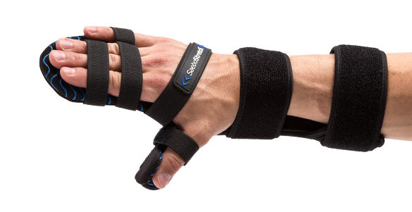
[BLOG POST] Why?: Dynamic Hand Splint Rehab Gloves For Stroke Recovery Patients – Saebo
Posted by Kostas Pantremenos in Paretic Hand on July 30, 2018

Stroke is among the top three causes of death in the United States, but nothing comes close to stroke as the leading cause of long-term disability. After patients survive a stroke, their risk of having another stroke increases, along with their likelihood of suffering a serious disability as a result. However, medical and technological advances have made it easier to help patients cope and recover. Occupational therapy is an effective way to restore mobility and reduce future risks for stroke survivors.
Therapy for stroke survivors often involves “re-training” or reprogramming the brain after neurological damage. As we learn more about the relationship between the brain, muscles, and connective tissue, one stimulating innovation is emerging as a top tool for recovery. Today, many patients are relying on a stroke rehabilitation gloves & dynamic splints to reverse damage, restore mobility, and reduce pain after a stroke.
But how, exactly, does wearing these orthoses treat symptoms of stroke survivors? Truth is, there are many benefits for patients who incorporate a glove or a dynamic splint into their recovery process.
Problems Stroke Can Cause
Especially with strokes, survivors can suffer from impaired function, weakness and spasticity. Spasticity causes involuntary muscle contractions in the arms and can even cause even short-term or long-term paralysis as the tendons and tissues around the muscles get tighter.
Strokes can really affect upper arm movements too. Survivors only use their affected upper limb approximately 3 hours per day.
Individuals who have not suffered a neurological injury use their dominant hand for an average of 9 hours per day. Patientsless than 14 days following stroke use their affected upper limb only 38 minutes out of a 9-hour day.
Shortening of muscles and connective tissue can start occurring within hours/days. Maintaining a shortened position for a prolonged period of time leads to fibrous adhesion formation, loss of sarcomeres and a loss of tissue extensibility.
There Is Hope After A Stroke
Fortunately, we can respond to spasticity, and lessened arm movements and muscle tone by harnessing the brain’s own plasticity. Cortical Plasticity, also known as neuroplasticity, is the brain’s remarkable ability to reorganize itself by forming new neural connections based on individual experiences, lifestyle and environment. It essentially is the brain’s ability to “re-program” itself through mass practice, task-oriented arm training.
To get these neuroplastic changes, patents participate in skill-dependent rather than simply use-dependent activities. Skill-dependent activities are specific and progressively challenging tasks whereas use-dependent activities are repetition tasks in the absence of a meaningful challenge or an activity that requires problem solving strategies.
With these skill-dependent activities cortical maps are continuously remodeled throughout life and after injury by experiences and learning in response to activity and behavior from the stroke survivor. Stimulated through this task training, the brain has the ability to reorganize and form new connections between the intact neurons. The healthy surrounding tissue takes over some of the functions of the damaged area of the brain.
This Is Where Stroke Rehabilitation Gloves and Dynamic Splints Come Into Place
Task-specific training with rehabilitation gloves and dynamic splits improve upper extremity function in individuals suffering from neurological injuries. Treatment options are limited for neurological clients who cannot effectively incorporate their hand for functional grasp and release activities. This is where dynamic splints can really help rehabilitation.
Dynamic Splints Help Train the Brain
If the hand and arm muscles are no longer functional, it’s especially important to re-learn basic functions first, such asgrasping and releasing objects. A stroke rehabilitation device like the SaeboFlex can make this process easier for some patients and possible for those who otherwise would have no function left.

For a vast majority of stroke survivors, especially ones with incomplete spinal cord injury, patients do not exhibit sufficient active wrist and/or finger extension to allow the hand to be functional. Stroke recovery gloves like SaeboFlex has the biomechanical advantage in allowing prehension grasp and release activities for individuals with moderate to severe hemiparesis.
The SaeboFlex and other rehabilitative dynamic splints actually step in to compensate for some of the patient’s biomechanical disadvantages.
The majority of patients with neurological or spinal cord damage cannot extend their fingers or move their wrists, but this orthosis imitates the hand’s natural functions and makes it possible to grasp and release objects. The goal is to make the hand functional again, but it also minimizes joint damage and pain.
Dynamic Splints Help Fight Contracture
When stroke survivors lose function in their upper limbs after a stroke, sometimes hard static splints are used to keep the arm and wrist in a “neutral” position and avoid muscle contracture. Unfortunately, some studies have shown that static splinting is ineffective against muscle contracture, and others have actually linked the practice to joint damage and contracture. Contracture is a loss of motion over time due to abnormal shortening of the soft tissue structures spanning one or more joints. These include skin, ligaments, tendon, muscles and joint capsules.
The ideal splint is dynamic, moveable and helps stretch out muscles, tendons and ligaments like the SaeboStretch.
The splint’s energy-storing technology allows individuals suffering from spasticity to stretch comfortably and safely resulting in increased motivation and compliance. It allows the fingers to move through flexion caused by associated reactions and increased tone.
A dynamic splint may prevent contracture after a stroke, as well as:
- Reduce joint pain
- Protect the joints
- Prevent edema (buildup of excessive fluid in the muscle tissue)
- Allows the fingers to move through flexion caused by postural changes associated reactions and increased tone.
- Gradually repositions the fingers into extension
As patients recover from a stroke, every effort to restore strength and function is invaluable. Using a stroke rehabilitation dynamic splint is a proven way to reduce pain and complications while survivors focus on their recovery. It may also open up new possibilities by restoring the use of their arms.
Stroke Recovery Glove For Improved Hand Functionality

It’s important to keep the muscles active after a stroke, in order to prevent stiffness and shortening of the tissue. Activity also helps to keep pathways between the brain and muscles open. Stroke recovery gloves that promote sensorimotor stimulation are useful to stroke survivors for many different reasons, from preventing complications to making life-changing therapy methods possible.
Therapy is a big part of the recovery process after a stroke, and occupational therapy often incorporates basic elements such as towels or small objects as patients learn to grasp, release, hold, and perform other basic tasks. Stroke rehabilitation gloves like the SaeboGlove can help with these activities. The SaeboGlove is a functional stroke recovery hand glove that has a tension system integrated into it which helps individuals extend their fingers and thumb after grasping. This helpful stimulation helps the neuroplastic changes the brain needs to help reprogram itself.
Positive results have been witnessed and experienced with professionals and clients when integrate them into upper arm rehabilitation exercises.
Results of stroke rehabilitation gloves in therapy have included:
- Significant increases on the Fugi-Meyer Assessment and Box and Block Test, which are designed to test the elbow’s control and strength during reach-to-grasp tasks
- Reduced jerkiness of the wrist, shoulder, and elbow joints during reach-to-grasp therapy tasks
- Improved flexion and abduction
- Support for hand and finger extension after loss of mobility
- Increased motor recovery
- Increased grip strength
- Active improvement to the overall functionality of patient hands, with some enjoying nearly full functionality
- Strong potential for future improvement of arm/wrist mobility.
Improved Stroke Recovery With Gloves And Dynamic Splints
Skill-dependent physical activities have long helped stroke survivors reprogram their brains, strengthen their muscles, and improve their quality of life after neurological damage. Stroke rehabilitation gloves and dynamic splints can give the patients the needed stimulation and help they are needing to progress. They are great to help protect the joints while improving strength and mobility. Because patients can incorporate these gloves and dynamic splints into occupational therapy as well as everyday tasks, they make it easier to achieve independence during their stroke recovery.
via Why?: Dynamic Hand Splint Rehab Gloves For Stroke Recovery Patients
[Abstract] Design and Test of a Closed-Loop FES System for Supporting Function of the Hemiparetic Hand Based on Automatic Detection Using the Microsoft Kinect Sensor
Posted by Kostas Pantremenos in Functional Electrical Stimulation (FES), Paretic Hand on August 25, 2017

