Posts Tagged Electrical Stimulation
[WEB] Revolutionizing Stroke Recovery: The iStride Device
Posted by Kostas Pantremenos in Assistive Technology, Gait Rehabilitation - Foot Drop, Rehabilitation robotics on January 19, 2024

The story of Maria Magdalena Valencia Juares, fondly known as Elena, is a testament to the power of innovation and the resilience of the human spirit. In 2021, Elena experienced a stroke that left her with muscle weakness and a decline in mental health, a situation further intensified by the isolation brought about by the COVID-19 pandemic. With her family residing thousands of miles away, they sought a solution to Elena’s predicament, and their answer came in the form of the iStride device. This ground-breaking tool, designed to help stroke survivors improve their walking ability, brought about incredible changes in Elena’s life, enabling her to climb stairs and dance again. The iStride device didn’t just offer her a solution; it provided hope and a renewed sense of independence.
The iStride Device: A New Hope for Stroke Survivors
The iStride device is a wearable technology designed to help stroke survivors regain their walking capability. Its real-time feedback mechanism supports users during walking exercises, leading to significant improvements in walking speed and endurance. As a result, stroke survivors gain better overall mobility and independence, as was the case with Elena.
Not only is the iStride device designed for functionality, but it is also built for comfort and adaptability. As a wearable ankle robot, it provides adjustable assistance to the user’s ankle, aiding in the regaining of strength and balance. Clinical studies corroborate the effectiveness of the iStride, with users reporting notable enhancements in their walking speed and endurance.
How the iStride Works
At its core, the iStride device is an assistive technology that aims to reduce the risk of falls in stroke survivors by providing targeted electrical stimulation to the muscles in the affected leg. This stimulation aids in improving strength and coordination, resulting in significant increases in walking speed and endurance, which in turn lead to greater independence and improved quality of life.
The iStride device employs a unique combination of visual and auditory cues to help patients improve their gait and balance. Its non-invasive nature and wearability ensure that it provides electrical stimulation to the leg muscles in a way that enhances mobility and minimises the risk of falls. The benefits of using the iStride device are not only immediate but also long-term, with studies showing sustained improvements in walking ability for stroke survivors, even one year after therapy.
iStride: Shifting the Paradigm of Stroke Recovery
The remarkable story of Elena serves as an inspiring testament to the potential of the iStride device. It is not just a rehabilitation tool; it is a device that promises a shift in the paradigm of stroke recovery. The iStride device offers a beacon of hope to stroke survivors, enabling them to regain their independence and significantly improve their quality of life.
As we move forward, it is clear that the iStride device will continue to empower stroke survivors, offering them a chance to reclaim their mobility, independence, and ultimately, their lives. Through technology and innovation, we can look forward to a future where stroke recovery is not just possible, but also achievable and sustainable.
[VIDEO] Post Stroke Foot Dorsiflexion: Using Electrical Stimulation to Reduce Tone & Promote Plasticity – YouTube
Posted by Kostas Pantremenos in Gait Rehabilitation - Foot Drop, Neuroplasticity, Video on December 22, 2023
Further reading on electrophysiology and muscle contractions: https://strokesciences.com/post-strok…
StrokeSciences.Com
[ARTICLE] An Adaptive Brain-Computer Interface to Enhance Motor Recovery After Stroke – Full Text
Posted by Kostas Pantremenos in REHABILITATION on May 13, 2023
Abstract
Brain computer interfaces (BCIs) have been demonstrated to have the potential to enhance motor recovery after stroke. However, some stroke patients with severe paralysis have difficulty achieving the BCI performance required for participating in BCI-based rehabilitative interventions, limiting their clinical benefits. To address this issue, we presented a BCI intervention approach that can adapt to patients’ BCI performance and reported that adaptive BCI-based functional electrical stimulation (FES) treatment induced clinically significant, long-term improvements in upper extremity motor function after stroke more effectively than FES treatment without BCI intervention. These improvements were accompanied by a more optimized brain functional reorganization. Further comparative analysis revealed that stroke patients with low BCI performance (LBP) had no significant difference from patients with high BCI performance in rehabilitation efficacy improvement. Our findings suggested that the current intervention may be an effective way for LBP patients to engage in BCI-based rehabilitation treatment and may promote lasting motor recovery, thus contributing to expanding the applicability of BCI-based rehabilitation treatments to pave the way for more effective rehabilitation treatments.
SECTION I.
Introduction
Despite great efforts in stroke rehabilitation over the past decades, stroke is still one of the leading causes of disability and death worldwide, and its global burden is gradually increasing [1]. Recently, several advanced rehabilitative intervention approaches, such as robot-assisted treatment [2], vagus nerve stimulation [3], constraint-induced movement therapy [4] and functional electrical stimulation (FES) [5], have been developed to restore motor function and have shown potential benefits for stroke patients. In particular, a motor imagery (MI)-based brain computer interface (BCI), which merely requires patients to imagine movements without performing actual physical movements, can enhance motor recovery for stroke patients with severe paralysis by inducing brain plasticity and functional reorganization [6], [7].
Recently, several studies have demonstrated significant clinical advantages in stroke patients if MI-based BCI was coupled with other interventions, e.g., rehabilitation robotics [8], virtual reality (VR) [9] and FES [10]. Murguialday et al. recruited 30 chronic stroke patients to investigate the efficacy of an MI-based BCI, and more significant motor improvement was observed in the BCI group that received MI-triggered orthoses feedback than in the control group that received random orthoses feedback [8]. Pichiorri et al. studied 14 subacute patients who performed MI training tasks with BCI-based VR feedback versus 14 patients who performed training tasks without BCI intervention; the authors reported a better motor functional outcome in the former group [9]. Biasiucci et al. investigated the efficacy of integrating an MI-based BCI with FES in a randomized control trial (RCT) with 27 chronic patients. The results showed that the BCI-FES group exhibited more significant functional recovery than the sham FES group [10]. Chen et al. reported that 16 stroke patients who received the treatment of MI-BCI-controlled FES achieved more effective recovery than those who received passive FES therapy [11]. Sinha et al. reported that BCI-based FES intervention involving 23 stroke patients could facilitate interhemispheric connectivity changes and upper limb motor recovery [12]. In addition, Sebastián-Romagosa et al. explored the clinical effect of combining an MI-based BCI with multisensory feedback (FES and VR) on upper limb motor rehabilitation using a recoveriX system [13] [14]. The authors reported that 51 stroke patients in [13] and 34 stroke patients in [14] achieved significant functional improvements after the intervention. Remsiket et al.also reported that 3 stroke patients obtained upper limb motor recovery using an MI-based BCI system with multimodal feedback, including visual feedback, FES, and tongue stimulation [15].
Nevertheless, MI-based BCI interventions still face several challenges. For instance, there is much variation in MI-BCI performance not only across subjects but also at different times within subjects [16]. Therefore, a fixed task difficulty in nonadaptive BCI interventions may be mismatched with the subject’s BCI performance, and it is difficult for patients to maintain active patient engagement, especially for those with low BCI performance (LBP), during rehabilitation treatments. In addition, almost 20~30% of subjects cannot generate controllable brain signals in MI [17]. Although studies have reported that intensive MI training can enhance the performance of LBP subjects before actual BCI control [18], [19], the performance improvement in some subjects is limited even after several months of training [20]. In general, sufficient BCI performance is required for BCI-based rehabilitation treatments. Ang et al. considered that the relatively LBP was functionally similar to BCI with random feedback, which had been proven to have no significant contribution to stroke recovery [21]. In the authors’ three clinical trials, stroke patients whose BCI performance was below the chance level were excluded from rehabilitation treatments [22], [23], [24]. Currently, few studies have been conducted to investigate the clinical efficacy of BCI interventions in stroke patients with LBP.
Active engagement in rehabilitation training is essential to promote motor recovery and functional reorganization [25]. Several studies have noted that feedback, which was provided by a recoveriX system while patients performed MI tasks, could be used to monitor patients’ engagement [14], [26]. Moreover, multimodal feedback through VR and FES in particular has been suggested to promote effective engagement and immersion of patients in MI tasks [27].
Here, we proposed an adaptive BCI intervention method that combines MI with VR and FES with the capacity to modulate task difficulty to adapt to the BCI performance of individual stroke patients. This intervention not only provided a feasible way for LBP stroke patients to participate in BCI-based rehabilitation treatment but could also reduce the impact of unstable BCI performance on patients’ motivation to engage in rehabilitation treatment. We conducted a double-blind RCT in 33 subacute stroke patients to evaluate whether the proposed intervention could yield significantly better clinical efficacy on upper extremity motor recovery for the BCI group than for the control group. In addition, in the BCI group, we further investigated the clinical benefit of the proposed intervention in patients with LBP and compared it to that in patients with high BCI performance (HBP).[…]

[WEB] Spinal Cord Stimulation Restores Post-Stroke Arm, Hand Function
Posted by Kostas Pantremenos in Paretic Hand on March 22, 2023
For the first time, researchers have used electrical stimulation of the cervical spinal cord to immediately restore arm and hand movement in two patients with chronic moderate-to-severe upper limb paresis.
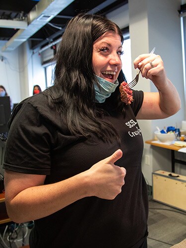
A research participant gives a thumbs up while holding a fork with a piece of steak with her affected arm.
The results provide “promising, albeit preliminary, evidence that spinal cord stimulation could be an assistive as well as a restorative approach for upper-limb recovery after stroke,” first author Marc P. Powell, PhD, Reach Neuro Inc., Pittsburgh, Pennsylvania, and colleagues wrote.
The findings were published online February 20 in Nature Medicine.
Top Cause of Paralysis
“Stroke is the largest cause of paralysis in the world,” with nearly three quarters of patients with stroke experiencing lasting deficits in motor control of their arm and hand, co-senior study author Marco Capogrosso, PhD, assistant professor of neurological surgery at the University of Pittsburgh, Pittsburgh, Pennsylvania, said during a press briefing.
Stroke can disrupt communication between the brain and the spinal cord, leading to motor deficits in the arm and hand. However, below the lesion, the spinal circuits that control movement remain intact and could be targeted to restore function, Capogrosso noted.
Spinal cord stimulation has shown promise in promoting long-lasting recovery of leg motor function in patients with spinal cord injury; but until now, it’s been largely unexplored for upper-limb recovery.
In this “first-in-human” study, the investigators percutaneously implanted two linear leads in the dorsolateral epidural space targeting neural circuits that control arm and hand muscles in two patients.
One of the patients was a woman (age, 31 years) who had experienced a right thalamic hemorrhagic stroke secondary to a cavernous malformation 9 years before enrolling in the pilot study.
The other patient was a woman (age, 47 years) who experienced a right ischemic middle cerebral artery (MCA) stroke secondary to a right carotid dissection, resulting in a large MCA territory infarct 3 years before entering the study.
In both patients, continuous stimulation of the targeted neural circuits led to significant and immediate improvement in arm and hand strength and dexterity. This enabled the patients to perform movements that they couldn’t perform without spinal cord stimulation.
The process also enabled fine motor skills, such as opening a lock and using utensils to eat independently — tasks that the younger woman had not been able to do for 9 years.
“Perhaps even more interesting, we found that after a few weeks of use, some of these improvements endure when the stimulation is switched off, indicating exciting avenues for the future of stroke therapies,” Capogrosso said in a news release.
No serious adverse events were reported.
‘Easily Translated’
Capogrosso said that, thanks to years of preclinical research, the investigators have developed a practical, easy-to-use stimulation protocol adapting existing clinical technologies that “could be easily translated to the hospital and quickly moved from the lab to the clinic.”
The researchers noted, however, that further studies in larger cohorts will be required to validate the safety and efficacy of this approach.
They are currently working with more patients with stroke to fine-tune placement of the leads and stimulation protocol, as well as determine which patients are best suited for the approach.
“Creating effective neurorehabilitation solutions for people affected by movement impairment after stroke is becoming ever more urgent,” co-senior author Elvira Pirondini, PhD, assistant professor of physical medicine and rehabilitation at the University of Pittsburgh, said in the release.
“Even mild deficits resulting from a stroke can isolate people from social and professional lives and become very debilitating, with motor impairments in the arm and hand being especially taxing and impeding simple daily activities, such as writing, eating, and getting dressed,” she added.
This research was funded by the National Institutes of Health BRAIN Initiative, with additional research support provided by the Department of Neurological Surgery and the Department of Physical Medicine and Rehabilitation at Pitt, and the Department of Mechanical Engineering and the Neuroscience Institute at Carnegie Mellon University. Three investigators have financial interests in Reach Neuro, Inc., which has an interest in the technology being evaluated in this study.
Nature Med. Published online February 20, 2023. Abstract.
[WEB] Helping Stroke Patients Regain Movement in Their Hands – The New York Times
Posted by Kostas Pantremenos in Paretic Hand, REHABILITATION on February 21, 2023
The results of an innovative study suggest electrical stimulation of the spinal cord could eventually help some of the many people disabled by strokes.

By Pam Belluck
- Feb. 20, 2023
Heather Rendulic was 23 when she suffered a stroke that disabled her left side. Ten years later, her left arm and hand remain so impaired that she cannot tie her shoes, type with two hands or cut her own food.
But for an extraordinary month, while participating in an innovative study, she suddenly was able to open a padlock with a key, draw a map of Italy, dip a chicken nugget in sauce and eat it with a fork — all with that left hand.
“It was like I actually had two arms, oh my gosh!” Ms. Rendulic said recently.
Researchers from the University of Pittsburgh and Carnegie Mellon University implanted electrodes along her spinal cord , delivering electrical stimulation while she tried different activities. With stimulation, her left arm had greater mobility, her fingers had more dexterity, and she could make intentional movements more quickly and fluidly.
The study, published Monday in the journal Nature Medicine, represents the first successful demonstration of spinal cord stimulation to address weakness and paralysis in the arms and hands of stroke patients.
The study was small and preliminary, involving only Ms. Rendulic and another patient. Several scientists said many questions remain about the technique’s effectiveness and applicability, but that the research suggested spinal cord stimulation could eventually help some of the many people who experience strokes.
“I think there’s enormous implications for improving quality of life,” said Dr. Lumy Sawaki-Adams, the program director in the clinical research division of the National Institute of Neurological Disorders and Stroke, who was not involved in the research. Still, she said, “we have to be cautious that we’re not offering hope to many people when I think we’re not there yet.”
Spinal cord stimulation has been used for decades to treat chronic pain. More recently, experiments delivering stimulation — either through surgically implanted electrodes or noninvasively through electrodes placed on the skin — have shown promise in helping patients with spinal cord injuries regain mobility in their legs and, in some cases, their arms and hands.
But the approach has been mostly unexplored for stroke, partly because of differences in the location and type of damage, neurological experts said.
Because strokes occur in the brain, it had been assumed that applying stimulation outside the brain would not provide “the same bang for the buck,” said Arun Jayaraman, the executive director of the technology and innovation hub at Shirley Ryan AbilityLab, a rehabilitation center in Chicago. He said the study, which he was not involved in, countered that assumption, instead suggesting that stimulating the spine, the pathway from the brain to hand and arm muscles, may help impaired limbs.
Each year, more than 12 million people worldwide and nearly 800,000 in the United States experience strokes, said Dr. Karen Furie, the vice chair of the American Stroke Association’s stroke brain health science subcommittee.
Initially, patients typically receive about six months of physical, occupational and other therapies, she said, but then progress often plateaus.
“We have virtually nothing to offer people who are years out and have longstanding disabilities,” said Dr. Furie, who is also the chair of neurology at Brown University’s Warren Alpert Medical School and was not involved in the study.
About three-quarters of stroke patients experience impairment, weakness or paralysis in their arms and hands, said Dr. Elliot Roth, an attending physician at Shirley Ryan AbilityLab’s Brain Innovation Center, who was not involved in the study. “For many people, it’s the toughest part of the stroke recovery process and tends to recover the slowest,” he said.
The patients who participated in the study had experienced different types of strokes and had varying degrees of impairment. Ms. Rendulic’s stroke was hemorrhagic, caused by bursting blood vessels. The other, more severely impaired patient, a 47-year-old woman whom researchers did not identify, experienced an ischemic stroke, which is more common and involves blocked blood vessels.
Researchers implanted strands of eight electrodes in two locations, corresponding to where neurosensory fibers from the arm and the hand enter the spinal cord.
Marco Capogrosso, an assistant professor of neurological surgery at the University of Pittsburgh, said that the approach derived from the fact that with strokes, some neural areas remain undamaged.
“So, if we can build this technology to amplify neural signals, maybe we have a chance to restore arm and hand movement,” said Dr. Capogrosso, who led the research with Elvira Pirondini, an assistant professor of physical medicine and rehabilitation at the University of Pittsburgh, and Douglas Weber, a professor of mechanical engineering at Carnegie Mellon’s Neuroscience Institute.
Five days a week for four hours each day, researchers activated the stimulation, calibrated it to determine optimal parameters for each patient and asked them to attempt various movements and tasks. Right away, the effect was noticeable.
“The very first day in the lab and the first time they turned it on, I was sitting in a chair, and they asked me to open and close my hand, and that’s something that’s really difficult for me,” Ms. Rendulic said. As her husband and mother watched, “I immediately was opening and closing my hand,” she said. “We all broke down in tears.”
Over four weeks, she was given increasingly challenging tasks, like gripping and moving a soup can. With stimulation, her left hand moved 14 small blocks over a barrier in a box, compared with six blocks without stimulation.
Typically, when Ms. Rendulic, 33, who works at home for a company’s human resources department, tries to make her left hand do something like grasp a pen, her arm feels like “it’s made of rock,” almost disconnected from her brain, she said. With stimulation “it was like my brain was able to find my left arm so much easier.”
The other patient, who was given simpler tasks because her left hand was almost completely paralyzed, improved in skills like reaching.
Researchers also tested a “sham” stimulation, activating electrodes randomly to see if patients responded to a kind of placebo effect rather than stimulation targeted specifically to their arms and hands. Both performed better with targeted stimulation.
The patients sensed the stimulation, but it didn’t cause pain, rigidity or safety problems, researchers reported.
The approved study protocol required removing the electrodes after 29 days. But one month later, the patients retained some improved abilities, surprising researchers. “We thought it was not possible” after only four weeks of stimulation, Dr. Pirondini said.
It is unclear exactly why the benefit can persist, Dr. Capogrosso said, but he hypothesized that “the same neural processes that allow these people to use this stimulation method also lead to a recovery of movement when the stimulation is off.” He added, “we’re not creating new fibers, but we’re definitely restrengthening what there is.”
Several experts noted that this pilot study was not designed to answer the most relevant question for patients: Can the improvements in laboratory tasks translate into skills that matter in daily life?
“It’s a first step among hundreds,” said Dr. Daniel Lu, a professor and vice chairman of neurosurgery at the University of California Los Angeles, who co-authored a 2016 study that showed that spinal stimulation from implanted electrodes improved hand strength and control in two spinal cord injury patients.
Dr. Lu said he believes stimulation is promising, but that its impact in the new study was difficult to evaluate because there was no comparison group and patients were not given the same regimen of intensive activities before stimulation — activities that might themselves have therapeutic benefit.
“Is it possible that you’re just exercising the patient, and the patient without the stimulation would have gotten the same effect?” he asked.
Another question neuroscientists raise is whether — or in what circumstances — it is better to surgically implant electrodes or place them on the skin, a less expensive method called transcutaneous stimulation. The new study’s authors consider surgical implantation superior because it is “much more specific,” said Dr. Weber, allowing it to “target the muscles that control the wrist and the hand.”
Others, like Chet Moritz, a professor of neurotechnology at the University of Washington, have reported improvements in spinal cord injury patients using electrodes on the skin, including benefits lasting months after stimulation ends. “It’s true we can’t tune the shoulder to this degree and the elbow to this degree and the wrist to that degree, but the nervous system seems to take care of that for us,” he said.
Several neurological experts predicted that both methods could eventually be helpful and appropriate for different patients, depending on their health and other factors. All the experts, including the study authors, said stimulation would be more effective if accompanied by rehabilitation therapies.
The study’s authors said their continuing research is evaluating patients of varying stroke severity, age and other characteristics to determine who would benefit from their approach. They have formed a company and said they envision that, as with similar technology for chronic pain, patients could adjust their stimulation via app or remote control.
If stimulation becomes regularly available to stroke patients, Ms. Rendulic would welcome it. “I did threaten to not show up to the surgery to get it removed,” she said. “I just wanted it all the time.”
While she has devised one-handed ways to do activities like driving and typing, everyday frustrations rankle, like needing her husband Mark, whom she calls “my left-hand man,” to slice steak for her.
“In the trial, I did get to cut up a steak, which was awesome,” she said. Then, fork in her left hand, she speared a piece and lifted it to her mouth — one previously impossible movement at a time.
Pam Belluck is a health and science writer whose honors include sharing a Pulitzer Prize and winning the Victor Cohn Prize for Excellence in Medical Science Reporting. She is the author of “Island Practice,” a book about an unusual doctor. @PamBelluck
[Abstract] Hybrid FES & Mechatronic Hand Control Method for Upper Limb Rehabilitation Systems
Posted by Kostas Pantremenos in Functional Electrical Stimulation (FES), Paretic Hand, Rehabilitation robotics on January 17, 2023
Abstract
This paper aims to describe a control concept algorithm developed as a new software for controlling both functional electrical stimulation (FES) and mechatronic components, inherited in an upper limb rehabilitation system for stroke people, but not limited to this specific affection. The main principle of the algorithm is to achieve a balanced control between FES and mechatronic exoskeleton, based on the input of the healthy hand via a sensory hand glove in order to help affected stroke patients to recover their affected hand mobility during the therapy.
Published in: 2022 E-Health and Bioengineering Conference (EHB)
[Abstract + References] Effects of Brain-Computer Interface Controlled Functional Electrical Stimulation on Motor Recovery in Stroke Survivors: a Systematic Review
Posted by Kostas Pantremenos in Functional Electrical Stimulation (FES) on September 20, 2022
Abstract
Purpose of Review
This systematic review aimed to investigate the effects of BCI-FES on motor recovery in patients with stroke.
Recent Findings
Nine studies met the eligibility criteria. Six studies were randomized controlled trials, and three were pilot studies. To date, the effectiveness of BCI-FES in patients with stroke has not been systematically reviewed.
Summary
The BCI-FES intervention may improve upper extremity function post-stroke. There is moderate evidence for positive effects of BCI-FES on gait and weak evidence for positive effects of BCI-FES on balance post-stroke. Further randomized controlled trials with a larger sample size are strongly warranted to confirm our findings.
References
Papers of particular interest, published recently, have been highlighted as: • Of importance •• Of major importance
- Thrift AG, Howard G, Cadilhac DA, et al. Global stroke statistics: an update of mortality data from countries using a broad code of “cerebrovascular diseases.” Int J Stroke. 2017;12(8):796–801. https://doi.org/10.1177/1747493017730782.Article PubMed Google Scholar
- Langhorne P, Coupar F, Pollock A. Motor recovery after stroke: a systematic review. Lancet Neurol. 2009;8(8):741–54. https://doi.org/10.1016/S1474-4422(09)70150-4.Article PubMed Google Scholar
- Kiper P, Szczudlik A, Agostini M, et al. Virtual reality for upper limb rehabilitation in subacute and chronic stroke: a randomized controlled trial. Arch Phys Med Rehabil. 2018;99(5):834-842.e4. https://doi.org/10.1016/j.apmr.2018.01.023.Article PubMed Google Scholar
- Dorsch S, Ada L, Canning CG, Al-Zharani M, Dean C. The strength of the ankle dorsiflexors has a significant contribution to walking speed in people who can walk independently after stroke: an observational study. Arch Phys Med Rehabil. 2012;93(6):1072–6. https://doi.org/10.1016/j.apmr.2012.01.005.Article PubMed Google Scholar
- Robertson JA, Eng JJ, Hung C. The effect of functional electrical stimulation on balance function and balance confidence in community-dwelling individuals with stroke. Physiother Can. 2010;62(2):114–9. https://doi.org/10.3138/physio.62.2.114.Article PubMed PubMed Central Google Scholar
- Daly JJ. Response of gait deficits to neuromuscular electrical stimulation for stroke survivors. Expert Rev Neurother. 2006;6(10):1511–22. https://doi.org/10.1586/14737175.6.10.1511.Article PubMed Google Scholar
- Hendricks HT, van Limbeek J, Geurts AC, Zwarts MJ. Motor recovery after stroke: a systematic review of the literature. Arch Phys Med Rehabil. 2002;83(11):1629–37. https://doi.org/10.1053/apmr.2002.35473.Article PubMed Google Scholar
- Kim T, Kim S, Lee B. Effects of action observational training plus brain-computer interface-based functional electrical stimulation on paretic arm motor recovery in patient with stroke: a randomized controlled trial. Occup Ther Int. 2016;23(1):39–47. https://doi.org/10.1002/oti.1403.Article PubMed Google Scholar
- • Rathee D, Chowdhury A, Meena YK, Dutta A, McDonough S, Prasad G. Brain-machine interface-driven post-stroke upper-limb functional recovery correlates with beta-band mediated cortical networks. IEEE Trans Neural Syst Rehabil Eng. 2019;27(5):1020–31. https://doi.org/10.1109/TNSRE.2019.2908125. This study demonstrated that BCI is used to induce plasticity according to activity by paying attention to tasks requiring individuals to activate or deactivate specific brain regions.Article PubMed Google Scholar
- Collinger JL, Vinjamuri R, Degenhart AD, et al. Motor-related brain activity during action observation: a neural substrate for electrocorticographic brain-computer interfaces after spinal cord injury. Front Integr Neurosci. 2014;8:17. Published 2014 Feb 19. https://doi.org/10.3389/fnint.2014.00017
- McFarland DJ, Wolpaw JR. Brain-computer interfaces for communication and control. Commun ACM. 2011;54(5):60–6. https://doi.org/10.1145/1941487.1941506.Article PubMed PubMed Central Google Scholar
- Etoh S, Noma T, Takiyoshi Y, et al. Effects of repetitive facilitative exercise with neuromuscular electrical stimulation, vibratory stimulation and repetitive transcranial magnetic stimulation of the hemiplegic hand in chronic stroke patients. Int J Neurosci. 2016;126(11):1007–12. https://doi.org/10.3109/00207454.2015.1094473.Article PubMed Google Scholar
- Peckham PH, Knutson JS. Functional electrical stimulation for neuromuscular applications. Annu Rev Biomed Eng. 2005;7:327–60. https://doi.org/10.1146/annurev.bioeng.6.040803.140103.CAS Article PubMed Google Scholar
- Dobkin BH, Dorsch A. New evidence for therapies in stroke rehabilitation. Curr Atheroscler Rep. 2013;15(6):331. https://doi.org/10.1007/s11883-013-0331-y.Article PubMed PubMed Central Google Scholar
- Urrútia G, Bonfill X. Declaración PRISMA: una propuesta para mejorar la publicación de revisiones sistemáticas y metaanálisis. Med Clin. 2010;135(11):507–11. https://doi.org/10.1016/j.medcli.2010.01.015.Article Google Scholar
- Liberati A, Altman DG, Tetzlaff J, et al. The PRISMA statement for reporting systematic reviews and meta-analyses of studies that evaluate health care interventions: explanation and elaboration. PLoS Med. 2009;6(7):e1000100. https://doi.org/10.1371/journal.pmed.1000100.Article PubMed PubMed Central Google Scholar
- Maher CG, Sherrington C, Herbert RD, Moseley AM, Elkins M. Reliability of the PEDro scale for rating quality of randomized controlled trials. Phys Ther. 2003;83(8):713–21.Article Google Scholar
- Moher D, Cook DJ, Jadad AR, et al. Assessing the quality of reports of randomised trials: implications for the conduct of meta-analyses. Health Technol Assess. 1999;3(12):i–98.CAS Article Google Scholar
- PEDro score – Strokengine. Strokengine.ca. https://strokengine.ca/en/glossary/pedro-score/. Published 2021. Accessed August 24, 2021.
- Lee SH, Kim SS, Lee BH. Action observation training and brain-computer interface controlled functional electrical stimulation enhance upper extremity performance and cortical activation in patients with stroke: a randomized controlled trial. Physiother Theory Pract. 2020;1–9. https://doi.org/10.1080/09593985.2020.1831114
- Chung E, Lee BH, Hwang S. Therapeutic effects of brain-computer interface-controlled functional electrical stimulation training on balance and gait performance for stroke: a pilot randomized controlled trial. Med (Baltimore). 2020;99(51):e22612. https://doi.org/10.1097/MD.0000000000022612.CAS Article Google Scholar
- Chung E, Park SI, Jang YY, Lee BH. Effects of brain-computer interface-based functional electrical stimulation on balance and gait function in patients with stroke: preliminary results. J Phys Ther Sci. 2015;27(2):513–6. https://doi.org/10.1589/jpts.27.513.Article PubMed PubMed Central Google Scholar
- Biasiucci A, Leeb R, Iturrate I, et al. Brain-actuated functional electrical stimulation elicits lasting arm motor recovery after stroke. Nat Commun. 2018;9(1):2421. https://doi.org/10.1038/s41467-018-04673-z.CAS Article PubMed PubMed Central Google Scholar
- Remsik AB, Dodd K, Williams L Jr, et al. Behavioral outcomes following brain-computer interface intervention for upper extremity rehabilitation in stroke: a randomized controlled trial. Front Neurosci. 2018;12:752. https://doi.org/10.3389/fnins.2018.00752.Article PubMed PubMed Central Google Scholar
- Jang YY, Kim TH, Lee BH. Effects of Brain-computer interface-controlled functional electrical stimulation training on shoulder subluxation for patients with stroke: a randomized controlled trial. Occup Ther Int. 2016;23(2):175–85. https://doi.org/10.1002/oti.1422.Article PubMed Google Scholar
- Young BM, Nigogosyan Z, Walton LM, et al. 2015 Dose-response relationships using brain-computer interface technology impact stroke rehabilitation. Front Hum Neurosci. 2015;9:361. https://doi.org/10.3389/fnhum.2015.00361.Article PubMed PubMed Central Google Scholar
- McCrimmon CM, King CE, Wang PT, Cramer SC, Nenadic Z, Do AH. Brain-controlled functional electrical stimulation therapy for gait rehabilitation after stroke: a safety study. J Neuroeng Rehabil. 2015;12:57. https://doi.org/10.1186/s12984-015-0050-4.Article PubMed PubMed Central Google Scholar
- •• Kruse A, Suica Z, Taeymans J, Schuster-Amft C. Effect of brain-computer interface training based on non-invasive electroencephalography using motor imagery on functional recovery after stroke – a systematic review and meta-analysis. BMC Neurol. 2020;20(1):385. https://doi.org/10.1186/s12883-020-01960-5. This review demonstrated an improvement in functional recovery post-stroke after BCI combined with conventional therapy for a duration of four weeks or longer, with a preference for high-intensity training of five times per week.Article PubMed PubMed Central Google Scholar
- Grefkes C, Fink GR. Connectivity-based approaches in stroke and recovery of function. Lancet Neurol. 2014;13(2):206–16. https://doi.org/10.1016/S1474-4422(13)70264-3.Article PubMed Google Scholar
- Wu J, Quinlan EB, Dodakian L, et al. Connectivity measures are robust biomarkers of cortical function and plasticity after stroke. Brain. 2015;138(Pt 8):2359–69. https://doi.org/10.1093/brain/awv156.Article PubMed PubMed Central Google Scholar
- Nicolo P, Rizk S, Magnin C, Pietro MD, Schnider A, Guggisberg AG. Coherent neural oscillations predict future motor and language improvement after stroke. Brain. 2015;138(Pt 10):3048–60. https://doi.org/10.1093/brain/awv200.Article PubMed Google Scholar
- Pundik S, McCabe JP, Hrovat K, et al. Recovery of post stroke proximal arm function, driven by complex neuroplastic bilateral brain activation patterns and predicted by baseline motor dysfunction severity. Front Hum Neurosci. 2015;9:394. https://doi.org/10.3389/fnhum.2015.00394.Article PubMed PubMed Central Google Scholar
- Schaechter JD, Moore CI, Connell BD, Rosen BR, Dijkhuizen RM. Structural and functional plasticity in the somatosensory cortex of chronic stroke patients. Brain. 2006;129(Pt 10):2722–33. https://doi.org/10.1093/brain/awl214.Article PubMed Google Scholar
- • Bouton CE. Merging brain-computer interface and functional electrical stimulation technologies for movement restoration. Handb Clin Neurol. 2020;168:303–9. https://doi.org/10.1016/B978-0-444-63934-9.00022-6. This review showed that BCI-FES treatment involves repeated attempts at functional activities to actively modulate brain activity during imagined movement, resulting in reward-based and use-dependent reinforcements and induction of neuroplastic change in the disrupted motor system.Article PubMed Google Scholar
- Alashram AR, Annino G, Padua E. Robot-assisted gait training in individuals with spinal cord injury: A systematic review for the clinical effectiveness of Lokomat. J Clin Neurosci. 2021;91:260–9. https://doi.org/10.1016/j.jocn.2021.07.019.CAS Article PubMed Google Scholar
- Alashram AR, Padua E, Hammash AK, Lombardo M, Annino G. Effectiveness of virtual reality on balance ability in individuals with incomplete spinal cord injury: A systematic review. J Clin Neurosci. 2020;72:322–7. https://doi.org/10.1016/j.jocn.2020.01.037.Article PubMed Google Scholar
- Alashram AR, Padua E, Annino G. Effects of whole-body vibration on motor impairments in patients with neurological disorders: a systematic review. Am J Phys Med Rehabil. 2019;98(12):1084–98. https://doi.org/10.1097/PHM.0000000000001252.Article PubMed Google Scholar
- Annino G, Alashram AR, Alghwiri AA, et al. Effect of segmental muscle vibration on upper extremity functional ability poststroke: A randomized controlled trial. Med (Baltimore). 2019;98(7):e14444. https://doi.org/10.1097/MD.0000000000014444.Article Google Scholar
- Alashram AR, Padua E, Romagnoli C, Annino G. Effectiveness of focal muscle vibration on hemiplegic upper extremity spasticity in individuals with stroke: A systematic review. NeuroRehabilitation. 2019;45(4):471–81. https://doi.org/10.3233/NRE-192863.Article PubMed Google Scholar
- Alashram A, Annino G, Al-qtaishat M, Padua E. Mental practice combined with physical practice to enhance upper extremity functional ability poststroke: a systematic review. J Stroke Med. 2020;3(2):51–61. https://doi.org/10.1177/2516608520943793.Article Google Scholar
- Alashram A. Optimizing gait ability after task-oriented circuit class training in posttraumatic brain injury: a case report. Indian J Phys Med Rehabil. 2019;30(4):112–6. https://doi.org/10.5005/jp-journals-10066-0053.Article Google Scholar
- Alashram A, Annino G, Mercuri N. Task-oriented motor learning in upper extremity rehabilitation post stroke. J Stroke Med. 2019;2(2):95–104. https://doi.org/10.1177/2516608519864760.Article Google Scholar
- Alashram AR, Annino G, Mercuri NB. Changes in spasticity following functional electrical stimulation cycling in patients with spinal cord injury: a systematic review [published online ahead of print, 2020 May 14]. J Spinal Cord Med. 2020;1–14. https://doi.org/10.1080/10790268.2020.1763713
- Alashram A, Alghwiri A, Padua E, Annino G. Efficacy of proprioceptive neuromuscular facilitation on spasticity in patients with stroke: a systematic review. Phys Ther Rev. 2021;26(3):168–76. https://doi.org/10.1080/10833196.2021.1892281.Article Google Scholar
- Alashram AR, Annino G, Mercuri NB. Rhythmic auditory stimulation in gait rehabilitation for traumatic brain and spinal cord injury. J Clin Neurosci. 2019;69:287–8. https://doi.org/10.1016/j.jocn.2019.08.080.Article PubMed Google Scholar
- Egger M, Smith G. meta-analysis bias in location and selection of studies. BMJ. 1998;316(7124):61–6. https://doi.org/10.1136/bmj.316.7124.61.CAS Article PubMed PubMed Central Google Scholar
- Higgins J, Green S. Cochrane handbook of systematic reviews of interventions. Chichester: Wiley; 2008. p. 187–241.
[Abstract + References] Safety of Transcranial Direct Current Stimulation: Evidence Based Update 2016
Posted by Kostas Pantremenos in REHABILITATION on August 8, 2022
Highlights
- •Report on tDCS safety using published Serious Adverse Effects in human trials, irreversible brain damage in animal models.
- •tDCS sessions defined and categorized by the electrode montage, stimulation intensity and duration.
- •Use of conventional tDCS in human trials has not yet produced any reports of a Serious Adverse Effect or irreversible injury.
Abstract
This review updates and consolidates evidence on the safety of transcranial Direct Current Stimulation (tDCS). Safety is here operationally defined by, and limited to, the absence of evidence for a Serious Adverse Effect, the criteria for which are rigorously defined. This review adopts an evidence-based approach, based on an aggregation of experience from human trials, taking care not to confuse speculation on potential hazards or lack of data to refute such speculation with evidence for risk. Safety data from animal tests for tissue damage are reviewed with systematic consideration of translation to humans. Arbitrary safety considerations are avoided. Computational models are used to relate dose to brain exposure in humans and animals. We review relevant dose–response curves and dose metrics (e.g. current, duration, current density, charge, charge density) for meaningful safety standards. Special consideration is given to theoretically vulnerable populations including children and the elderly, subjects with mood disorders, epilepsy, stroke, implants, and home users. Evidence from relevant animal models indicates that brain injury by Direct Current Stimulation (DCS) occurs at predicted brain current densities (6.3–13 A/m2) that are over an order of magnitude above those produced by conventional tDCS. To date, the use of conventional tDCS protocols in human trials (≤40 min, ≤4 milliamperes, ≤7.2 Coulombs) has not produced any reports of a Serious Adverse Effect or irreversible injury across over 33,200 sessions and 1000 subjects with repeated sessions. This includes a wide variety of subjects, including persons from potentially vulnerable populations.
References
- Sawyer D.W.
- Donowitz G.R.
- Mandell G.L.
- Nitsche M.A.
- Niehaus L.
- Hoffmann K.T.
- Hengst S.
- Liebetanz D.
- Paulus W.
- et al.
- Brunoni A.R.
- Nitsche M.A.
- Bolognini N.
- Bikson M.
- Wagner T.
- Merabet L.
- et al.
- Medeiros L.F.
- de Souza I.C.C.
- Vidor L.P.
- de Souza A.
- Deitos A.
- Volz M.S.
- et al.
- Kim S.J.
- Kim B.K.
- Ko Y.J.
- Bang M.S.
- Kim M.H.
- Han T.R.
- Fregni F.
- Nitsche M.A.
- Loo C.K.
- Brunoni A.R.
- Marangolo P.
- Leite J.
- et al.
- Peterchev A.V.
- Wagner T.A.
- Miranda P.C.
- Nitsche M.A.
- Paulus W.
- Lisanby S.H.
- et al.
- Fresnoza S.
- Paulus W.
- Nitsche M.A.
- Kuo M.-F.
- Liebetanz D.
- Klinker F.
- Hering D.
- Koch R.
- Nitsche M.A.
- Potschka H.
- et al.
- Batsikadze G.
- Moliadze V.
- Paulus W.
- Kuo M.-F.
- Nitsche M.A.
- Bikson M.
- Datta A.
- Rahman A.
- Scaturro J.
- Datta A.
- Bansal V.
- Diaz J.
- Patel J.
- Reato D.
- Bikson M.
- Domingo J.L.
- Llobet J.M.
- Corbella J.
- Kronberg G.
- Bikson M.
- Miranda P.C.
- Faria P.
- Hallett M.
- Nitsche M.A.
- Bikson M.
- Bestmann S.
- Liebetanz D.
- Koch R.
- Mayenfels S.
- König F.
- Paulus W.
- Nitsche M.A.
- Sundaram A.
- Stock V.
- Cruciani R.A.
- Knotkova H.
- Nitsche M.A.
- Paulus W.
- Shiozawa P.
- da Silva M.E.
- Raza R.
- Uchida R.R.
- Cordeiro Q.
- Fregni F.
- et al.
- DaSilva A.F.
- Volz M.S.
- Bikson M.
- Fregni F.
- Nitsche M.A.
- Doemkes S.
- Karaköse T.
- Antal A.
- Liebetanz D.
- Lang N.
- et al.
- Minhas P.
- Bansal V.
- Patel J.
- Ho J.S.
- Diaz J.
- Datta A.
- et al.
- Dmochowski J.P.
- Datta A.
- Huang Y.
- Richardson J.D.
- Bikson M.
- Fridriksson J.
- et al.
- Dmochowski J.P.
- Bikson M.
- Datta A.
- Richardson J.
- Fridriksson J.
- Parra L.C.
- Dmochowski J.P.
- Datta A.
- Bikson M.
- Su Y.
- Parra L.C.
- Pirulli C.
- Fertonani A.
- Miniussi C.
- Smit M.
- Schutter D.J.L.G.
- Nijboer T.C.W.
- Visser-Meily J.M.A.
- Kappelle L.J.
- Kant N.
- et al.
- Calabrese E.J.
- Calabrese E.J.
- Dhawan G.
- Kapoor R.
- Iavicoli I.
- Calabrese V.
- Bikson M.
- Datta A.
- Elwassif M.
- Baba T.
- Kameda M.
- Yasuhara T.
- Morimoto T.
- Kondo A.
- Shingo T.
- et al.
- Peruzzotti-Jametti L.
- Cambiaghi M.
- Bacigaluppi M.
- Gallizioli M.
- Gaude E.
- Mari S.
- et al.
- Kim Y.J.
- Ku J.
- Cho S.
- Kim H.J.
- Cho Y.K.
- Lim T.
- et al.
- Cho H.-S.
- Cha H.-G.
- Sattler V.
- Acket B.
- Raposo N.
- Albucher J.-F.
- Thalamas C.
- Loubinoux I.
- et al.
- Tahtis V.
- Kaski D.
- Seemungal B.M.
- Fusco A.
- Iosa M.
- Venturiero V.
- De Angelis D.
- Morone G.
- Maglione L.
- et al.
- Brunoni A.R.
- Amadera J.
- Berbel B.
- Volz M.S.
- Rizzerio B.G.
- Fregni F.
- Guarienti F.
- Caumo W.
- Shiozawa P.
- Cordeiro Q.
- Boggio P.S.
- Benseñor I.M.
- et al.
- Mielke D.
- Wrede A.
- Schulz-Schaeffer W.
- Taghizadeh-Waghefi A.
- Nitsche M.A.
- Rohde V.
- et al.
- Wachter D.
- Wrede A.
- Schulz-Schaeffer W.
- Taghizadeh-Waghefi A.
- Nitsche M.A.
- Kutschenko A.
- et al.
- Minhas P.
- Datta A.
- Bikson M.
- Rodríguez N.
- Opisso E.
- Pascual-Leone A.
- Soler M.D.
- Palm U.
- Feichtner K.B.
- Hasan A.
- Gauglitz G.
- Langguth B.
- Nitsche M.A.
- et al.
- Wang J.
- Wei Y.
- Wen J.
- Li X.
- Frank E.
- Wilfurth S.
- Landgrebe M.
- Eichhammer P.
- Hajak G.
- Langguth B.
- Woods A.J.
- Antal A.
- Bikson M.
- Boggio P.S.
- Brunoni A.R.
- Celnik P.
- et al.
- Edwards D.
- Cortes M.
- Datta A.
- Minhas P.
- Wassermann E.M.
- Bikson M.
- Datta A.
- Baker J.M.
- Bikson M.
- Fridriksson J.
- Datta A.
- Bikson M.
- Fregni F.
- Gillick B.T.
- Kirton A.
- Carmel J.B.
- Minhas P.
- Bikson M.
- Kessler S.K.
- Minhas P.
- Woods A.J.
- Rosen A.
- Gorman C.
- Bikson M.
- Mekonnen A.
- Salvador R.
- Ruffini G.
- Miranda P.C.
- Laakso I.
- Hirata A.
- Sadleir R.J.
- Vannorsdall T.D.
- Schretlen D.J.
- Gordon B.
- Rampersad S.M.
- Janssen A.M.
- Lucka F.
- Aydin Ü.
- Lanfer B.
- Lew S.
- et al.
- Mattai A.
- Miller R.
- Weisinger B.
- Greenstein D.
- Bakalar J.
- Tossell J.
- et al.
- Grecco L.A.C.
- Duarte N.A.C.
- Zanon N.
- Galli M.
- Fregni F.
- Oliveira C.S.
- Grecco L.A.C.
- de Almeida Carvalho Duarte N.
- Mendonça M.E.
- Cimolin V.
- Galli M.
- Fregni F.
- et al.
- Vestito L.
- Rosellini S.
- Mantero M.
- Bandini F.
- Kessler S.K.
- Turkeltaub P.E.
- Benson J.G.
- Hamilton R.H.
- Morales-Quezada L.
- Cosmo C.
- Carvalho S.
- Leite J.
- Castillo-Saavedra L.
- Rozisky J.R.
- et al.
- Poreisz C.
- Boros K.
- Antal A.
- Paulus W.
- Raimundo R.J.S.
- Uribe C.E.
- Brasil-Neto J.P.
- Nitsche M.A.
- Liebetanz D.
- Lang N.
- Antal A.
- Tergau F.
- Paulus W.
- McIntire L.K.
- McKinley R.A.
- Goodyear C.
- Nelson J.
- Tadini L.
- El-Nazer R.
- Brunoni A.R.
- Williams J.
- Carvas M.
- Boggio P.
- et al.
- Russo R.
- Wallace D.
- Fitzgerald P.B.
- Cooper N.R.
- Andrade C.
- Paneri B.
- Khadka N.
- Patel V.
- Thomas C.
- Tyler W.
- Parra L.C.
- et al.
- San-Juan D.
- Calcáneo Jde D.D.C.
- González-Aragón M.F.
- Bermúdez Maldonado L.
- Avellán A.M.
- Argumosa E.V.G.
- et al.
- Cosmo C.
- Baptista A.F.
- de Sena E.P.
- Auvichayapat N.
- Rotenberg A.
- Gersner R.
- Ngodklang S.
- Tiamkao S.
- Tassaneeyakul W.
- et al.
- Gillick B.T.
- Feyma T.
- Menk J.
- Usset M.
- Vaith A.
- Wood T.J.
- et al.
- Young S.J.
- Bertucco M.
- Sheehan-Stross R.
- Sanger T.D.
- Duarte Nde A.C.
- Grecco L.A.C.
- Galli M.
- Fregni F.
- Oliveira C.S.
- Grecco L.A.C.
- E Mendonça M.
- Duarte N.A.C.
- Zanon N.
- Fregni F.
- Oliveira C.S.
- Aree-uea B.
- Auvichayapat N.
- Janyacharoen T.
- Siritaratiwat W.
- Amatachaya A.
- Prasertnoo J.
- et al.
- Walbert T.
- Chasteen K.
- Young S.J.
- Bertucco M.
- Sanger T.D.
- Bhanpuri N.H.
- Bertucco M.
- Young S.J.
- Lee A.A.
- Sanger T.D.
- Anton S.D.
- Woods A.J.
- Ashizawa T.
- Barb D.
- Buford T.W.
- Carter C.S.
- et al.
- Salthouse T.A.
- Stephen L.J.
- Brodie M.J.
- Hsu W.-Y.
- Ku Y.
- Zanto T.P.
- Gazzaley A.
- Berryhill M.E.
- Jones K.T.
- Boggio P.S.
- Campanhã C.
- Valasek C.A.
- Fecteau S.
- Pascual-Leone A.
- Fregni F.
- Fertonani A.
- Brambilla M.
- Cotelli M.
- Miniussi C.
- Flöel A.
- Suttorp W.
- Kohl O.
- Kürten J.
- Lohmann H.
- Breitenstein C.
- et al.
- Goodwill A.M.
- Reynolds J.
- Daly R.M.
- Kidgell D.J.
- Hardwick R.M.
- Celnik P.A.
- Harty S.
- Robertson I.H.
- Miniussi C.
- Sheehy O.C.
- Devine C.A.
- McCreery S.
- et al.
- Holland R.
- Leff A.P.
- Josephs O.
- Galea J.M.
- Desikan M.
- Price C.J.
- et al.
- Manenti R.
- Brambilla M.
- Petesi M.
- Ferrari C.
- Cotelli M.
- Meinzer M.
- Lindenberg R.
- Antonenko D.
- Flaisch T.
- Flöel A.
- Meinzer M.
- Lindenberg R.
- Sieg M.M.
- Nachtigall L.
- Ulm L.
- Flöel A.
- Parikh P.J.
- Cole K.J.
- Park S.-H.
- Seo J.-H.
- Kim Y.-H.
- Ko M.-H.
- Puri R.
- Hinder M.R.
- Fujiyama H.
- Gomez R.
- Carson R.G.
- Summers J.J.
- Ross L.A.
- McCoy D.
- Coslett H.B.
- Olson I.R.
- Wolk D.A.
- Sandrini M.
- Brambilla M.
- Manenti R.
- Rosini S.
- Cohen L.G.
- Cotelli M.
- Zhou D.
- Zhou J.
- Chen H.
- Manor B.
- Lin J.
- Zhang J.
- Jones K.T.
- Stephens J.A.
- Alam M.
- Bikson M.
- Berryhill M.E.
- Manenti R.
- Brambilla M.
- Rosini S.
- Orizio I.
- Ferrari C.
- Borroni B.
- et al.
- Elder G.J.
- Taylor J.-P.
- Khedr E.M.
- Gamal N.F.E.
- El-Fetoh N.A.
- Khalifa H.
- Ahmed E.M.
- Ali A.M.
- et al.
- Boggio P.S.
- Ferrucci R.
- Mameli F.
- Martins D.
- Martins O.
- Vergari M.
- et al.
- Boggio P.S.
- Khoury L.P.
- Martins D.C.S.
- Martins O.E.M.S.
- de Macedo E.C.
- Fregni F.
- Cotelli M.
- Manenti R.
- Brambilla M.
- Petesi M.
- Rosini S.
- Ferrari C.
- et al.
- Ferrucci R.
- Mameli F.
- Guidi I.
- Mrakic-Sposta S.
- Vergari M.
- Marceglia S.
- et al.
- Suemoto C.K.
- Apolinario D.
- Nakamura-Palacios E.M.
- Lopes L.
- Leite R.E.P.
- Sales M.C.
- et al.
- Benninger D.H.
- Lomarev M.
- Lopez G.
- Wassermann E.M.
- Li X.
- Considine E.
- et al.
- Boggio P.S.
- Ferrucci R.
- Rigonatti S.P.
- Covre P.
- Nitsche M.
- Pascual-Leone A.
- et al.
- Doruk D.
- Gray Z.
- Bravo G.L.
- Pascual-Leone A.
- Fregni F.
- Fregni F.
- Boggio P.S.
- Santos M.C.
- Lima M.
- Vieira A.L.
- Rigonatti S.P.
- et al.
- Kaski D.
- Dominguez R.O.
- Allum J.H.
- Islam A.F.
- Bronstein A.M.
- Elder G.J.
- Firbank M.J.
- Kumar H.
- Chatterjee P.
- Chakraborty T.
- Dutt A.
- et al.
- Manenti R.
- Bianchi M.
- Cosseddu M.
- Brambilla M.
- Rizzetti C.
- Padovani A.
- et al.
- Cotelli M.
- Manenti R.
- Petesi M.
- Brambilla M.
- Cosseddu M.
- Zanetti O.
- et al.
- Gálvez V.
- Ho K.-A.
- Alonzo A.
- Martin D.
- George D.
- Loo C.K.
- Yamamoto J.
- Ikeda A.
- Kinoshita M.
- Matsumoto R.
- Satow T.
- Takeshita K.
- et al.
- Swartzwelder H.S.
- Lewis D.V.
- Anderson W.W.
- Wilson W.A.
- Bikson M.
- Ghai R.S.
- Baraban S.C.
- Durand D.M.
- Bikson M.
- Inoue M.
- Akiyama H.
- Deans J.K.
- Fox J.E.
- Miyakawa H.
- et al.
- Kabakov A.Y.
- Muller P.A.
- Pascual-Leone A.
- Jensen F.E.
- Rotenberg A.
- Sunderam S.
- Gluckman B.
- Reato D.
- Bikson M.
- Durand D.M.
- Bikson M.
- Reato D.
- Gasca F.
- Datta A.
- Bikson M.
- Marshall L.
- Parra L.C.
- Kamida T.
- Kong S.
- Eshima N.
- Fujiki M.
- Kamida T.
- Kong S.
- Eshima N.
- Abe T.
- Fujiki M.
- Kobayashi H.
- Dhamne S.C.
- Ekstein D.
- Zhuo Z.
- Gersner R.
- Zurakowski D.
- Loddenkemper T.
- et al.
- San-Juan D.
- Morales-Quezada L.
- Orozco Garduño A.J.
- Alonso-Vanegas M.
- González-Aragón M.F.
- Espinoza López D.A.
- et al.
- Fregni F.
- Thome-Souza S.
- Nitsche M.A.
- Freedman S.D.
- Valente K.D.
- Pascual-Leone A.
- Yook S.-W.
- Park S.-H.
- Seo J.-H.
- Kim S.-J.
- Ko M.-H.
- San-Juan D.
- Mayorga A.P.M.
- Anschel D.J.
- Avellán A.M.
- González-Aragón M.F.
- Cole A.J.
- Faria P.
- Fregni F.
- Sebastião F.
- Dias A.I.
- Leal A.
- Liu A.
- Bryant A.
- Jefferson A.
- Friedman D.
- Minhas P.
- Barnard S.
- et al.
- Ekici B.
- Moliadze V.
- Schmanke T.
- Andreas S.
- Lyzhko E.
- Freitag C.M.
- Siniatchkin M.
- Kuo H.-I.
- Paulus W.
- Batsikadze G.
- Jamil A.
- Kuo M.-F.
- Nitsche M.A.
- Rossi S.
- Hallett M.
- Rossini P.M.
- Pascual-Leone A.
- Safety of TMS Consensus Group
- Jennum P.
- Winkel H.
- Fuglsang-Frederiksen A.
- Dam M.
- Deng Z.-D.
- Lisanby S.H.
- Peterchev A.V.
- Tor P.-C.
- Bautovich A.
- Wang M.-J.
- Martin D.
- Harvey S.B.
- Loo C.
- Di Lazzaro V.
- Rothwell J.C.
- Kandratavicius L.
- Balista P.A.
- Lopes-Aguiar C.
- Ruggiero R.N.
- Umeoka E.H.
- Garcia-Cairasco N.
- et al.
- McIntyre D.C.
- Nathanson D.
- Edson N.
- Wu D.
- Wang J.
- Yuan Y.
- Fusco A.
- Assenza F.
- Iosa M.
- Izzo S.
- Altavilla R.
- Paolucci S.
- et al.
- Lee S.J.
- Chun M.H.
- Ayerbe L.
- Ayis S.
- Crichton S.
- Wolfe C.D.A.
- Rudd A.G.
- King R.B.
- Aström M.
- Asplund K.
- Aström T.
- Silverman I.E.
- Restrepo L.
- Mathews G.C.
- Opitz A.
- Paulus W.
- Will S.
- Antunes A.
- Thielscher A.
- Maeda A.
- Yamada M.
- Itoh Y.
- Otomo E.
- Hayakawa M.
- Miyatake T.
- Vernieri F.
- Assenza G.
- Maggio P.
- Tibuzzi F.
- Zappasodi F.
- Altamura C.
- et al.
- Nitsche M.A.
- Paulus W.
- Benvenuti A.
- Rucci P.
- Miniati M.
- Papasogli A.
- Fagiolini A.
- Cassano G.B.
- et al.
- Arul-Anandam A.P.
- Loo C.
- Mitchell P.
- Baccaro A.
- Brunoni A.R.
- Bensenor I.M.
- Fregni F.
- Gálvez V.
- Alonzo A.
- Martin D.
- Mitchell P.B.
- Sachdev P.
- Loo C.K.
- Brunoni A.R.
- Valiengo L.
- Zanao T.
- de Oliveira J.F.
- Bensenor I.M.
- Fregni F.
- Loo C.K.
- Alonzo A.
- Martin D.
- Mitchell P.B.
- Galvez V.
- Sachdev P.
- Brunoni A.R.
- Valiengo L.
- Baccaro A.
- Zanão T.A.
- de Oliveira J.F.
- Goulart A.
- et al.
- Loo C.
- Katalinic N.
- Mitchell P.B.
- Greenberg B.
- Lugon M.D.M.V.
- Batsikadze G.
- Fresnoza S.
- Grundey J.
- Kuo M.-F.
- Paulus W.
- et al.
- Fresnoza S.
- Stiksrud E.
- Klinker F.
- Liebetanz D.
- Paulus W.
- Kuo M.-F.
- et al.
- Batsikadze G.
- Paulus W.
- Kuo M.-F.
- Nitsche M.A.
- Shiozawa P.
- Cordeiro Q.
- Fregni F.
- Brunoni A.R.
- Parazzini M.
- Rossi E.
- Rossi L.
- Priori A.
- Ravazzani P.
- Merrill D.R.
- Bikson M.
- Jefferys J.G.R.
- Monte-Silva K.
- Kuo M.-F.
- Hessenthaler S.
- Fresnoza S.
- Liebetanz D.
- Paulus W.
- et al.
- Chang W.-J.
- Bennell K.L.
- Hodges P.W.
- Hinman R.S.
- Liston M.B.
- Schabrun S.M.
- Mortensen J.
- Figlewski K.
- Andersen H.
- Tippett D.C.
- Hillis A.E.
- Tsapkini K.
- Ho K.-A.
- Bai S.
- Martin D.
- Alonzo A.
- Dokos S.
- Loo C.K.
- Cosman J.D.
- Atreya P.V.
- Woodman G.F.
- Fitz N.S.
- Reiner P.B.
- de Berker A.O.
- Bikson M.
- Bestmann S.
- Jacobson L.
- Koslowsky M.
- Lavidor M.
- Brunoni A.R.
- Ferrucci R.
- Bortolomasi M.
- Scelzo E.
- Boggio P.S.
- Fregni F.
- et al.
- Hagenacker T.
- Bude V.
- Naegel S.
- Holle D.
- Katsarava Z.
- Diener H.-C.
- et al.
- Charvet L.E.
- Kasschau M.
- Datta A.
- Knotkova H.
- Stevens M.C.
- Alonzo A.
- et al.
- Kasschau M.
- Sherman K.
- Haider L.
- Frontario A.
- Shaw M.
- Datta A.
- et al.
- Steenbergen L.
- Sellaro R.
- Hommel B.
- Lindenberger U.
- Kühn S.
- Colzato L.S.
- Wexler A.
[VIDEO] What is Drop Foot? Foot Drop Causes, Symptoms, and Treatment. – YouTube
Posted by Kostas Pantremenos in Gait Rehabilitation - Foot Drop, Video on March 5, 2022
Dr. Scott Thompson OTD discusses drop foot causes, symptoms, and treatments. Foot drop, also known as dropped foot or drop foot, is the inability to raise the front part of the foot due to weakness or paralysis of the muscles that lift the foot.
0:00 Introduction
0:10 What is Foot Drop/Drop Foot?
0:40 Causes of Foot Drop/Drop Foot
2:07 Treating with surgery
2:34 Treating with electrical stimulation
3:33 Treating with braces or ankle foot orthoses (AFOs)
4:14 Treating with rehabilitation experts
4:45 Conclusion
Learn more about foot drop:
https://www.saebo.com/blog/foot-drop-…
https://www.saebo.com/blog/how-to-fin…
https://www.saebo.com/blog/get-back-f…
For more information on the SaeboStep:
https://www.saebo.com/shop/saebostep/https://www.saebo.com
Download your FREE Saebo Exercise Guide here:
https://www.saebo.com/stroke-exercise…
SUBSCRIBE AND FOLLOW US!
https://www.facebook.com/saeboinc
https://www.instagram.com/saeboinc/
https://www.linkedin.com/company/saeb…
https://www.pinterest.com/saeboforstr…
Saebo, Inc. is a medical device company primarily engaged in the discovery, development, and commercialization of affordable and novel clinical solutions designed to improve mobility and function in individuals suffering from neurological and orthopedic conditions. With a vast network of Saebo-trained clinicians spanning six continents, Saebo has helped over 500,000 clients around the globe achieve a new level of independence.
[ARTICLE] Lower Extremity Stroke Rehabilitation with Brain Computer Interfaces – Preliminary Results. Full Text
Posted by Kostas Pantremenos in Functional Electrical Stimulation (FES), Gait Rehabilitation - Foot Drop, REHABILITATION on February 22, 2022
INTRODUCTION
Stroke is the second largest cause of death worldwide. Stroke survivors usually suffers severe motor and sensory impairment that hinder the activities of daily living. The functional deficit of lower limb is the most common paresis after stroke. It is not common for the stroke patients to fully recovery after months and years of the treatments and therapies, which leaves them in some degrees of impairment. Many of them do not reach a walking level to perform all their daily activities [1, 2]. Gait recovery is one of the major therapy goals in the rehabilitation program for stroke patients and methods for gait analysis and rehabilitation has been developed [2]. The weakened muscle tone also becomes an obstacle in motor rehabilitation. Therapies such as active foot drop exercise, electromechanical assisted therapy and treadmill therapy are usually limited to the patients with mild or moderate impairment [2]. Functional electrical stimulation (FES) has also been used in motor rehabilitation therapy over the last decades. It is known that passive FES therapy itself can reduce muscle spasm and shorten the term of motor recovery [3]. Passive therapies such as continuous passive motion or cycling therapy have been employed for the patients and showed the functional improvements in the previous studies [4]. However, they do not include or cannot monitor the patients’ active engagement of the therapy.
Today, Brain-Computer Interface (BCI) technology can provide an objective tool for measuring Motor Imagery (MI), creating new possibilities for “closed-loop” feedback. The closed-loop feedback, that is linked with the desired mental activity, is important for every feedback system and made possible through MI-based BCI. It could significantly improve rehabilitation therapy outcomes of stroke patients [5].
Currently there are numerous articles that talk about rehabilitation of the upper limb with BCI technology, but the rehabilitation of the lower limb is completely new. The objective of this study was to analyze the effectiveness of BCI therapy in the functional rehabilitation of lower extremity in stroke survivors.
Methods
Participants
For this preliminary study 2 males and 1 female were included. The mean age of the participants was 59 (±4.3) years and they all were in a chronic stage with time since the stroke 13 (±17.3) years. Participant 1 (P1) was a female, 62 years old and time after stroke 33 years. Participant 2 (P2) and Participant 3 (P3) were males, 61 and 54 years old with time after stroke 4 and 2 years. No previous BCI training was performed with the subjects, the three participants were BCI naive.
The following inclusion criteria was applied: 1) residual hemiparesis in his/her lower limb, 2) the stroke occurred at least four days before the first assessment, 3) functional restriction of the lower extremities, which prevent the persons from activities of everyday life, 4) ability to perform 10 Meter Walk Test. Additionally, 6) able to understand written and spoken instructions, 7) stable neurological status, 8) willing to participate in the study and to understand and sign the informed consent, 9) able to attend meetings. Patients with 1) active or passive implanted medical devices such as pacemakers which do not allow the use of FES, 2) implanted metallic fragments in the extremities which can limit the use of FES, 3) cerebellar lesions, 4) multiple stroke history, 5) elevated intracranial pressure, 6) pronounced hemi-neglect, 7) history of disordered aneurism, 8) history of epilepsy or seizures that is uncontrolled by proper treatment, 9) under the influence of anesthesia or similar medication, 10) significant circulatory disturbances of the lower limbs, 11) severe lung diseases, infections, renal insufficiency, liver damage, heart diseases, 12) sensory disorders which can significantly affect the patient’s ability to feel pain and to react to unsuitable proprioceptive stimuli, 13) diseases of the peripheral nervous system affecting the upper limbs or 14) Botulinum toxin treatment of his/her paretic lower limb during this study were excluded from the study.
Study design
Each participant received 2 months of BCI training based on MI with FES and visual feedback. The BCI sessions were distributed in 3 times per week, 25 sessions in total. Each session lasted about 1 hour. Two functional assessments were performed before the therapy started (Pre1, one month before the therapy and Pre2, a few days before the therapy) and one more functional assessment was performed immediately after these 25 BCI sessions (Post1). The functional scales used in the assessment sessions are detailed in the next section.
Functional and Behavioral Assessments
The primary outcome measure was the 10 Meter Walk Test (10MWT) that assesses walking speed in meters per second over a short duration. Subject is instructed to walk in self-selected velocity (10MWTSS) and/or in fast velocity (10MWTFV) over the length of 10 meters. The time is measured between meter 2 and 8 to exclude the acceleration and deceleration speed. Secondary scores included Timed Up and Go Test (TUG) that is used to determine fall risk and measure the progress of balance, sit to stand and walking. Subject is sitting on a chair and is instructed to stand up, walk 3 meters, turn around, walk back to the chair and sit down again. The time is measured from the beginning until the end of the task [6]. These two scores are measured in time (minutes and seconds), where lower values represent lower level of impairment. Range of Motion (ROM) for active ankle dorsiflexion and flexion was also used to measure the movement ability of the affected ankle. The subject is in long sitting position with a pillow placed under his/her knee so that the hill is lifted off the surface. Goniometer is placed on the lateral malleolus with stationary arm is leaning up to the fibula head and movable arm is parallel to the longitudinal axis of the 5th metatarsal when the subject is asked to perform ankle dorsiflexion movement or/and ankle flexion movement [7]. The score is measured in degrees, where higher values are also linked to lower impairment.

[…]

