Archive for October, 2019

[BLOG POST] TBI Grief Is A Thief…and Post-TBI Grief Is Rarely Brief

Having something personal stolen from you isn’t just upsetting…it’s offensive, and well…it’s just not fair. That’s how TBI feels to many. Leaving a TBI survivor to start over, the thief (TBI) often leaves no trace. Still, other times there is more than enough evidence.

Talking about grief, versus experiencing grief…or living with grief daily are totally different things. Grief can be an overwhelming sense of loss, a heavy mental weight pressing down on your very soul. After a traumatic brain injury, grief is an understatement. But it’s a place to start a discussion of what grief is, and how it’s different to people that may have been through similar situations.

Finding your way through the grieving process is like navigating without a map (or a GPS) – because there’s no set arrival time, and no itinerary – you just go along at your own pace, feel what you feel, and hope for the best. Nobody wants to hear that! With that being said, here’s an excerpt from a “tip card” by Lash & Associates Publishing titled “Loss, Grief, and Mourning.”

Tips for persons with brain injury to grieve and mourn…

✓ Be gentle with yourself – grieving can be physically, spiritually, and emotionally draining.

✓ Do not diminish how you feel about what has happened and don’t allow others to underrate your loss either. Your loss is real.

✓ Take time to work through your feelings about what has happened and how it affects you.

✓ Recognize that you may have secondary losses (e.g., loss of income, loss of friends, and loss of lifestyle).


✓ Recognize that your family is also experiencing grief. They need time to work through their emotions and may do it differently than you do.

✓ Find appropriate and safe ways to express your grief. It is essential to your well-being.

✓ Take time to reflect on who you were before your injury, who you are now, and who you want to be in the future.

✓ Ask for help – you do not need to do this alone.

✓ Keep life in perspective so that grieving and mourning do not totally overwhelm you.

Bereavement, Grieving, and Mourning

They are not the same. These words are used inter­changeably; however, they have different meanings. Dr. Alan Wolfelt, of the Center for Loss and Life Transition in Fort Collins, CO, defines bereavement, grieving, and mourning as follows.

Bereavement is the “call”.

It is the event that causes a loss (death, injury, ending of a relationship, etc.).

Grieving is the “internal response” to loss.

It is how one feels on the inside (sad, angry, confused, afraid, alone, etc.).

Mourning is the “external response” to the loss.

It is how one expresses feelings about the loss (funerals, ceremonies, rituals, talking, writing, etc.).

Primary and secondary losses are also a part of the process, in a “domino effect” of sorts. The initial injury of the TBI survivor is considered the primary loss…the other losses that follow affect the survivor, their family, friends, co-workers, and more. Everyone’s lives are changed.

Also, a whole range of emotions come with these losses, and mourning due to the situation can range from complicated, to extraordinary.

The journey of grief is complex, and acceptance is a big part of getting to the point with your life that you can go forward and find some happiness and reward. Embracing the new isn’t replacing the old…it’s acknowledging the old but moving ahead without it! It would be too easy to say “don’t let it get you down” …and survivors hear that more than they’d like. Although it’s meant as encouragement, many folks just don’t know how to put it into words in a more empathetic way. The point is that they didn’t experience what the TBI survivor did, but they deal with a lot of the aftermath on a daily basis – and they are just trying to build up and encourage the survivor.

In closing, grief is different in every single instance because every injury is different, every survivor is different…and every family is different. The difference is inevitable, but embracing each other’s differences after TBI is the best way to help each other feel included, and a part of the survivor community. Work to accept your differences as well, and you’ll be better prepared to have empathy for what other survivors have overcome too.

If you’d like to purchase the Lash tip card “Loss, Grief and Mourning After Brain Injury, by Janelle Breese Biagioni, you can click this link (price is $1.00 each, and is great to share with others).  https://www.lapublishing.com/loss-grief-mourning-tbi/
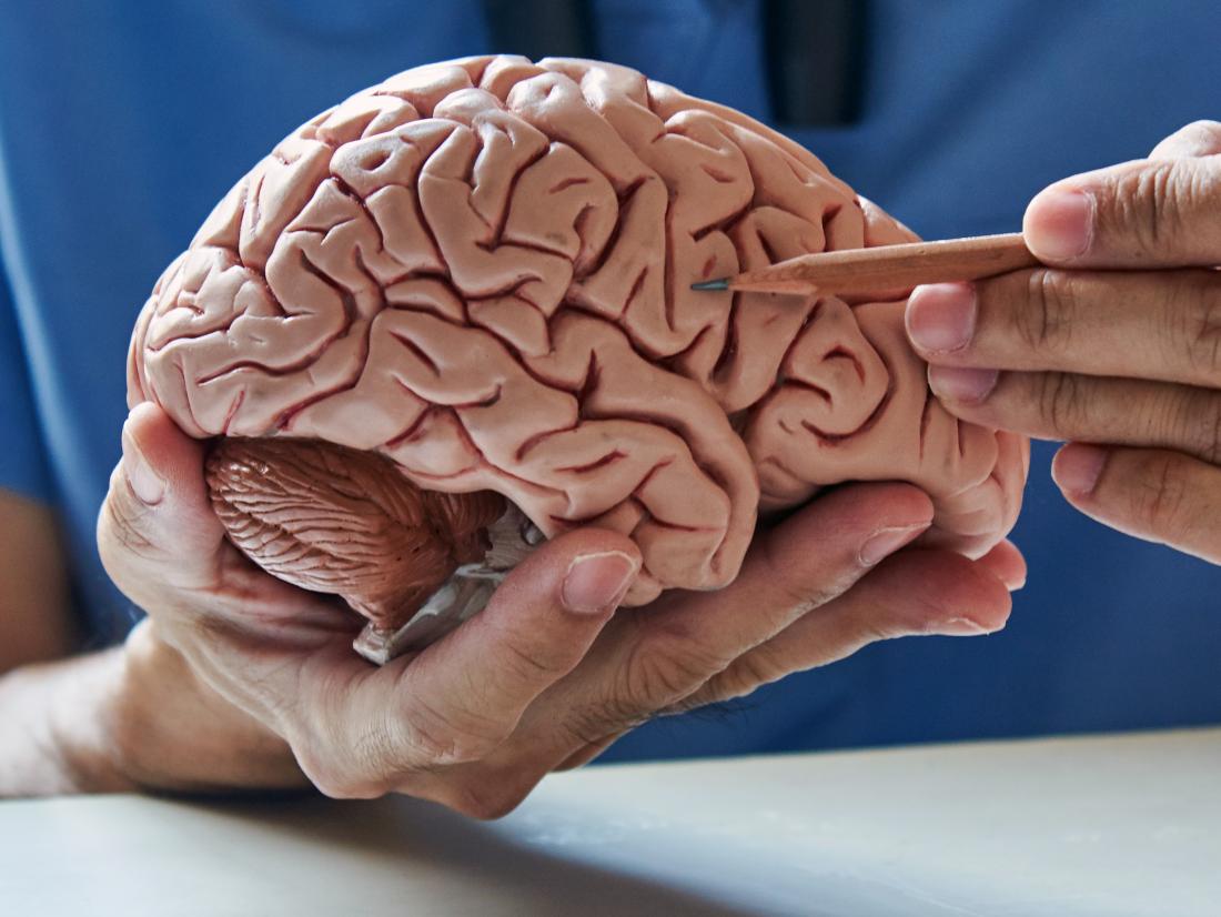
 

via TBI Grief Is A Thief…and Post-TBI Grief Is Rarely Brief

, , , , , ,

Leave a comment

[WEB PAGE] Neurotransmitters: What they are, functions, and psychology

via Neurotransmitters: What they are, functions, and psychology

, , , , , , , , , , , , , ,

Leave a comment

[WEB PAGE] What are the benefits of increased GABA levels in the brain?

Gamma-aminobutyric acid (GABA) is a neurotransmitter, or chemical messenger, in the brain. It blocks specific signals in the central nervous system, slowing down the brain. This provides a protective and calming effect on the brain and body.

The body produces GABA, and it may also be present in some fermented foods, such as kimchi, miso, and tempeh. These are not foods that most people include in their daily diets, so some people take GABA supplements to achieve the benefits.

In this article, we examine how increased levels of GABA may impact the brain and body, and whether taking GABA supplements could have the same benefits.

What is GABA?

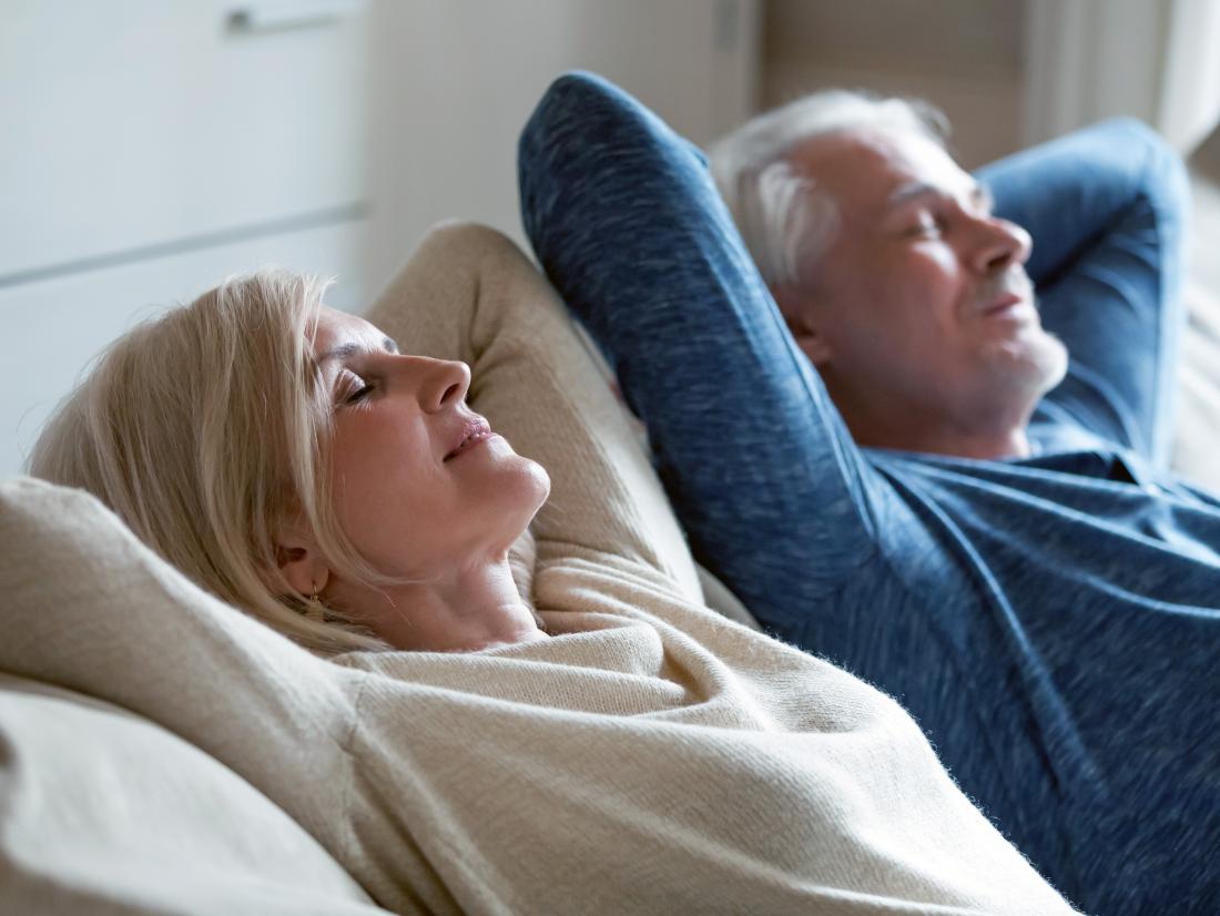
a couple looking relaxed because of gaba activity.

GABA activity can relieve stress, reduce stress, and improve sleep.

GABA is a neurotransmitter that inhibits or slows the brain’s functions. This activity produces effects such as:

  • relieving anxiety
  • reducing stress
  • improving sleep
  • preventing brain damage

The brain naturally releases GABA at the end of a day to promote sleepiness and allow a person to rest. Some of the medications doctors prescribe to induce sleep and reduce anxiety may also increase the action of GABA.

Medical benefits of increased GABA

Some experts have suggested that increased levels of GABA may have benefits, but the evidence is not clear. According to a 2019 review, GABA has anti-microbial, anti-seizure, and antioxidant properties and may help treat and prevent conditions such as:

Medications to increase GABA

Doctors may prescribe medicines that increase the amount of GABA or stimulate the same neurotransmitters in the brain to treat some medical conditions, such as epilepsy.

For example, benzodiazepines (Valium, Xanax) act on many of the same neurotransmitter receptors as GABA. According to one study, people who have depression may have reduced GABA levels in the brain. The use of benzodiazepines may be beneficial in those instances.

Doctors also prescribe the medication gabapentin (Neurontin), which is chemically similar to GABA to reduce seizures and muscle pain.

However, doctors are not clear whether the therapeutic effects of these medications are related to their effect on GABA receptors or whether they work in other ways.

GABA as a supplement

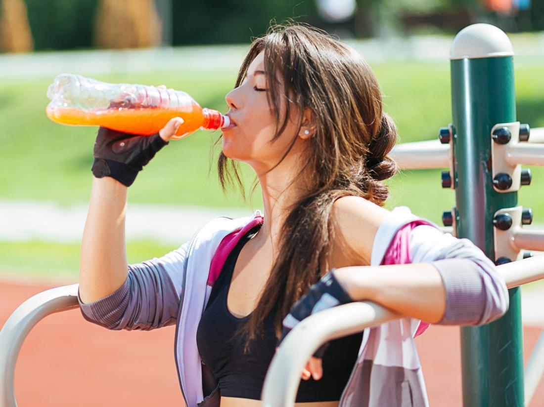
a woman enjoying the benefits of taurine in an energy drink she is drinking

Many sports drinks contain GABA.

Some people take supplements of GABA for their supposed stress- and anxiety-relieving benefits.

The Food and Drug Administration (FDA) has approved GABA for use as a supplement and as a food additive. Manufacturers may add GABA to:

  • sports drinks
  • snack bars
  • chewing gum
  • candies, and more

Manufacturers produce GABA supplements by fermenting a form of lactic acid bacteria.

However, the FDA do not regulate dietary supplements in the same way as medications. Therefore, consumers should exercise caution as to where they purchase the product from and only buy from reputable vendors and companies.

How to use GABA supplements

Some people may take a supplement in pill form, while others may add it to foods, such as protein drinks.

Researchers have not established a daily recommended intake or a suggested upper limit for GABA. Anyone wanting to take GABA as a supplement should consider talking to their doctor first.

At present, there is not enough research to evaluate the possible side effects of taking GABA supplements. However, if a person does experience side effects that might be GABA-related, they should discontinue the use of the supplement and contact their doctor.

Benefits of taking GABA supplements

Some researchers have voiced concerns about the supposed positive benefits of taking GABA supplements. An article in the journal Frontiers in Psychology notes that experts remain unclear whether GABA offers real benefits or whether the effects that people report experiencing are a placebo response.

Other researchers do not believe that GABA supplements cross the blood-brain barrier, which they would have to do to have any effect on the body.

However, some studies report positive effects from taking GABA supplements. These include:

Enhanced thinking and task performance abilities

study from 2015 found that taking 800 milligrams (mg) of GABA supplementation per day enhanced a person’s ability to prioritize and plan actions. Although the study was small, involving just 30 healthy volunteers, it showed how GABA supplementation might promote enhanced thinking.

Stress reduction

An older study from 2012 found that taking 100 mg of GABA daily helped reduce stress due to mental tasks. Like many other studies related to GABA, the study was small and involved just 63 participants.

Workout recovery and muscle building

a man and a woman working out together outside.

GABA supplements may improve workout recovery and muscle building.

The participants performed the same resistance training exercises twice a week, and the researchers measured the results. The researchers found that the combination of whey protein and GABA increased levels of growth hormone compared to whey protein alone.

Although this was another small study, the researchers concluded that GABA supplements might help to build muscle and assist in workout recovery. They recommended that researchers conduct more studies.

Summary

GABA naturally plays an essential role in promoting sleep, relieving anxiety, and protecting the brain.

Scientists have not been able to prove the positive effects of GABA supplementation on a large scale, and their use may have limited effectiveness.

If a person has received a diagnosis for conditions such as depression, anxiety, or attention deficit hyperactivity disorder, they may wish to talk to their doctor about medically-proven treatment before taking GABA supplements.

via What are the benefits of increased GABA levels in the brain?

, , , , , , , , ,

Leave a comment

[WEB PAGE] MEDRhythms Launches Trial of Post-Stroke Walking Device

Published on 

http://www.dreamstime.com/stock-image-clinical-trial-concept-image29482071

MEDRhythms Inc has launched a randomized controlled trial (RCT) at five top rehab hospitals and research centers across the country to examine the impact of a digital therapeutic device on stroke survivors who have post-stroke walking impairments, in support of the company’s eventual FDA submission.

“This clinical trial marks an important milestone toward MEDRhythms’ mission to make this high-quality intervention available to those who need and deserve to have it,” says Brian Harris, Co-Founder and CEO of Portland, Me-based MEDRhythms, in a media release. “As this new industry grows, it is important for digital therapeutics to demonstrate efficacy with the support of rigorous clinical trials, and this RCT is an integral step in MEDRhythms’ evidence generation strategy to do so.”

MEDRhythms’ clinical trial will be conducted at the Shirley Ryan AbilityLab in Chicago, the Kessler Foundation in New Jersey, Mt. Sinai Hospital in New York, Spaulding Rehabilitation Hospital in Boston, and the Boston University Neuromotor Recovery Laboratory in Boston. This trial was launched following completion of a successful feasibility study in the target population, which was conducted at the Boston University Neuromotor Recovery Lab. The results of this feasibility study will be announced at the American Physical Therapy Association’s annual Combined Sections Meeting in February 2020 in Denver, Colorado.

“Right now, the MEDRhythms digital therapeutic technology is a novel treatment for a subset of individuals that have few, if any, effective treatment options,” states David Putrino, the Director of Abilities Research Center (ARC) for the Department of Rehabilitation and Human Performance at the Mount Sinai Health System and the Principal Investigator at MEDRhythms’ Mount Sinai clinical trial site.

“The mission of the ARC is to identify and validate novel technologies that have the potential to significantly enhance the rehabilitation of people who are recovering from brain injuries and neurological conditions, including chronic stroke. The digital therapeutics industry has the potential to transform rehabilitation and disrupt healthcare, and it is imperative for companies in this space to run full-scale, multisite RCTs like MEDRhythms is doing.”

The digital therapeutic for post-stroke walking rehabilitation is one of a full pipeline of products that include therapeutics for indications such as Parkinson’s disease, multiple sclerosis, aging, and fall prevention, for which the company is actively exploring partnerships, per the release.

[Source(s): MEDRhythms, Business Wire]


via MEDRhythms Launches Trial of Post-Stroke Walking Device – Rehab Managment

, , , ,

Leave a comment

[ARTICLE] Robot-Assisted Stair Climbing Training on Postural Control and Sensory Integration Processes in Chronic Post-stroke Patients: A Randomized Controlled Clinical Trial – Full Text

Background: Postural control disturbances are one of the important causes of disability in stroke patients affecting balance and mobility. The impairment of sensory input integration from visual, somatosensory and vestibular systems contributes to postural control disorders in post-stroke patients. Robot-assisted gait training may be considered a valuable tool in improving gait and postural control abnormalities.

Objective: The primary aim of the study was to compare the effects of robot-assisted stair climbing training against sensory integration balance training on static and dynamic balance in chronic stroke patients. The secondary aims were to compare the training effects on sensory integration processes and mobility.

Methods: This single-blind, randomized, controlled trial involved 32 chronic stroke outpatients with postural instability. The experimental group (EG, n = 16) received robot-assisted stair climbing training. The control group (n = 16) received sensory integration balance training. Training protocols lasted for 5 weeks (50 min/session, two sessions/week). Before, after, and at 1-month follow-up, a blinded rater evaluated patients using a comprehensive test battery. Primary outcome: Berg Balance Scale (BBS). Secondary outcomes:10-meter walking test, 6-min walking test, Dynamic gait index (DGI), stair climbing test (SCT) up and down, the Time Up and Go, and length of sway and sway area of the Center of Pressure (CoP) assessed using the stabilometric assessment.

Results: There was a non-significant main effect of group on primary and secondary outcomes. A significant Time × Group interaction was measured on 6-min walking test (p = 0.013) and on posturographic outcomes (p = 0.005). Post hoc within-group analysis showed only in the EG a significant reduction of sway area and the CoP length on compliant surface in the eyes-closed and dome conditions.

Conclusion: Postural control disorders in patients with chronic stroke may be ameliorated by robot-assisted stair climbing training and sensory integration balance training. The robot-assisted stair climbing training contributed to improving sensorimotor integration processes on compliant surfaces. Clinical trial registration (NCT03566901).

Introduction

Postural control disturbances are one of the leading causes of disability in stroke patients, leading to problems with transferring, maintaining body position, mobility, and walking (Bruni et al., 2018). Therefore, the recovery of postural control is one of the main goals of post-stroke patients. Various and mixed components (i.e., weakness, joint limitation, alteration of tone, loss of movement coordination and sensory organization components) can affect postural control. Indeed, the challenge is to determine the relative weight placed on each of these factors and their interaction to plan specific rehabilitation programs (Bonan et al., 2004).

The two functional goals of postural control are postural orientation and equilibrium. The former involves the active alignment of the trunk and head to gravity, the base of support, visual surround and an internal reference. The latter involves the coordination of movement strategies to stabilize the center of body mass during self-initiated and externally triggered stability perturbations. Postural control during static and dynamic conditions requires a complex interaction between musculoskeletal and neural systems (Horak, 2006). Musculoskeletal components include biomechanical constraints such as the joint range of motion, muscle properties and limits of stability (Horak, 2006). Neural components include sensory and perceptual processes, motor processes involved in organizing muscles into neuromuscular synergies, and higher-level processes essential to plan and execute actions requiring postural control (Shumway-Cook and Woollacott, 2012). A disorder in any of these systems may affect postural control during static (in quite stance) and dynamic (gait) tasks and increase the risk of falling (Horak, 2006).

Literature emphasized the role of impairments of sensory input integration from visual, somatosensory and vestibular systems in leading to postural control disorders in post-stroke patients (Bonan et al., 2004Smania et al., 2008). Healthy persons rely on somatosensory (70%), vision (10%) and vestibular (20%) information when standing on a firm base of support in a well-lit environment (Peterka, 2002). Conversely, in quite stance on an unstable surface, they increase sensory weighting to vestibular and vision information as they decrease their dependence on surface somatosensory inputs for postural orientation (Peterka, 2002). Bonan et al. (2004) investigate whether post-stroke postural control disturbances may be caused by the inability to select the pertinent somatosensory, vestibular or visual information. Forty patients with hemiplegia after a single hemisphere chronic stroke (at least 12 months) performed computerized dynamic posturography to assess the patient’s ability to use sensory inputs separately and to suppress inaccurate inputs in case of sensory conflict. Six sensory conditions were assessed by an equilibrium score, as a measure of body stability. Results show that patients with hemiplegia seem to rely mostly on visual input. In conditions of altered somatosensory information, with visual deprivation or visuo-vestibular conflict, the patient’s performance was significantly lower than healthy subjects. The mechanism of this excessive visual reliance remains unclear. However, higher-level inability to select the appropriate sensory input rather than to elementary sensory impairment has been advocated as a potential mechanism of action (Bonan et al., 2004).

Sensory strategies and sensory reweighting processes are essential to generate effective movement strategies (ankle, hip, and stepping strategies) which can be resolved through feed-back or feed-forward postural adjustments. The cerebral cortex shapes these postural responses both directly via corticospinal loops and indirectly via the brainstem centers (Jacobs and Horak, 2007). Moreover, the cerebellar- and basal ganglia-cortical loop is responsible for adapting postural responses according to prior experience and for optimizing postural responses, respectively (Jacobs and Horak, 2007).

Rehabilitation is the cornerstone in the management of postural control disorders in post-stroke patients (Pollock et al., 2014). To date, no one physical rehabilitation approach can be considered more effective than any other approach (Pollock et al., 2014). Specific treatments should be chosen according to the individual requirements and the evidence available for that specific treatment. Moreover, it appears to be most beneficial a mixture of different treatment for an individual patient (Pollock et al., 2014). Considering that, rehabilitation involving repetitive, high intensity, task-specific exercises is the pathway for restoring motor function after stroke (Mehrholz et al., 2013Lo et al., 2017) robotic assistive devices for gait training have been progressively being used in neurorehabilitation to Sung et al. (2017). In the current literature, three primary evidence have been reported.

Firstly, a recent literature review highlights that robot-assisted gait training is advantageous as add-on therapy in stroke rehabilitation, as it adds special therapeutic effects that could not be afforded by conventional therapy alone (Morone et al., 2017Sung et al., 2017). Specifically, robot-assisted gait training was beneficial for improving motor recovery, gait function, and postural control in post-stroke patients (Morone et al., 2017Sung et al., 2017). Stroke patients who received physiotherapy treatment in combination with robotic devices were more likely to reach better outcomes compared to patients who received conventional training alone (Bruni et al., 2018).

Second, the systematic review by Swinnen et al. (2014) supported the use of robot-assisted gait therapy to improve postural control in subacute and chronic stroke patients. A wide variability among studies was reported about the robotic-device system and the therapy doses (3–5 times per week, 3–10 weeks, 12–25 sessions). However, significant improvements (Cohen’s d = 0.01 to 3.01) in postural control scores measured with the Berg Balance Scale (BBS), the Tinetti test, postural sway tests, and the Timed Up and Go (TUG) test were found after robot-assisted gait training. Interestingly, in five studies an end-effector device (gait trainer) was used (Peurala et al., 2005Tong et al., 2006Dias et al., 2007Ng et al., 2008Conesa et al., 2012). In two study, the exoskeleton was used (Hidler et al., 2009Westlake and Patten, 2009). In one study, a single joint wearable knee orthosis was used (Wong et al., 2012). Because the limited number of studies available and methodological differences among them, more specific randomized controlled trial in specific populations are necessary to draw stronger conclusions (Swinnen et al., 2014).

Finally, technological and scientific development has led to the implementation of robotic devices specifically designed to overcome the motor limitation in different tasks. With this perspective, the robot-assisted end-effector-based stair climbing (RASC) is a promising approach to facilitate task-specific activity and cardiovascular stress (Hesse et al., 20102012Tomelleri et al., 2011Stoller et al., 20142016Mazzoleni et al., 2017).

To date, no studies have been performed on the effects of RASC training in improving postural control and sensory integration processes in chronic post-stroke patients.

The primary aim of the study was to compare the effects of robot-assisted stair climbing training against sensory integration balance training on static and dynamic balance in chronic stroke patients. The secondary aims were to compare the training effects on sensory integration processes and mobility. The hypothesis was that the task-specific and repetitive robot-assisted stairs climbing training might act as sensory integration balance training, improving postural control because sensorimotor integration processes are essential for balance and walking.[…]


Continue —->  Frontiers | Robot-Assisted Stair Climbing Training on Postural Control and Sensory Integration Processes in Chronic Post-stroke Patients: A Randomized Controlled Clinical Trial | Neuroscience

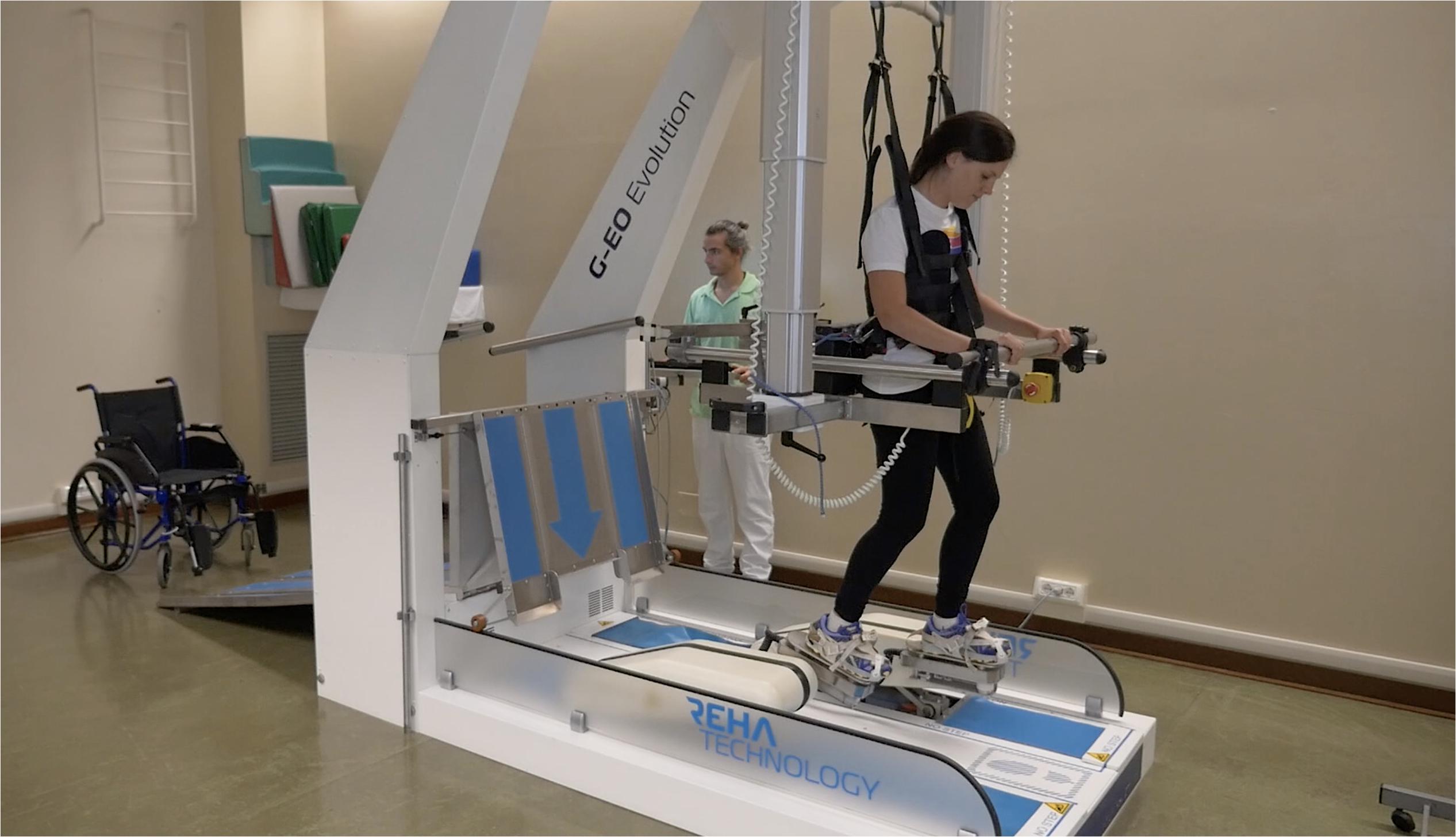
Figure 1. The G-EO system used in the Robot-Assisted Stair-Climbing Training (Written informed consent was obtained from the individual pictured, for the publication of this image).


, , , , , , , , , ,

Leave a comment

[Editorial] Virtual reality in stroke rehabilitation: virtual results or real values?

1Laboratory for the Study of Mind and Action in Rehabilitation Technologies, IRCCS Fondazione Santa Lucia, Rome, Italy.

Seven Capital Devices for the Future of Stroke Rehabilitation was the title of a review published seven years ago by our group, in which we analyzed the most promising technologies for neurorehabilitation1. They were: robots, virtual reality, brain computer interfaces, wearable devices for human movement analysis, noninvasive brain stimulators (such as transcranial direct current stimulation and transcranial magnetic stimulation), neuroprostheses, and computers/tablets for electronic clinical records and planning1.

Seven years later, we can now take stock of the situation. We must be honest: on one hand, we can surely affirm that the above-proposed technologies have really been the most developed and applied in these last years, but on the other hand, we should say that questions about their efficacy are still open, as reported by Cochrane reviews highlighting the need of further studies2,3.

However, every month, new studies claiming the efficacy of technological rehabilitation are published, and this continuously-growing amount of literature reveals the lack of definitive proof; otherwise all these studies would have been unnecessary. This “efficacy paradox” could potentially give us many more years of research without any conclusive results, especially because the more technology is adaptable to the needs of the patients (as clinicians want), the less the protocol to test the efficacy of that technology is standardizable (as researchers want)4.

Furthermore, the pressure on researchers to publish, the optimism about the use of technologies of some clinicians, the hopes of patients and their caregivers about new miraculous approaches, and the commercial interests of technology companies, may lead to some misleading claims in the mass media. For example, in many scientific and journalistic papers, some electromechanical devices without any intelligence on board are improperly called “robots”, nonimmersive video games are called “virtual reality”, the expressions “mind power” or “force of thought” are associated with brain computer interfaces1. Market analysts expect that the greatest developing field for robots in the next five years will be rehabilitation, compared with other fields5. Conversely, computers, the Internet and smartphones have changed our lives and were not directly developed for rehabilitation, but this clinical field may benefit from all the developed know-how. Virtual reality should be differentiated by video games, referring to a high-end user-computer interface involving real-time stimulation based on the three “I’s”: immersive experience, interaction, and imagination6.

In this scenario, the recent study by Ogun and colleagues clearly shows all the potentials of using a Leap Motion controller interfaced with 3D immersive virtual reality to improve the upper extremity functions in patients with ischemic stroke7. The Leap Motion controller is an optical tracking system including three infrared light emitters and two infrared cameras for tracking hand and finger kinematics, interfacing them with a virtual environment developed as a human-computer interface. In 2014, our group published the first feasibility pilot study proposing the use of Leap Motion in neurorehabilitation, noting its advantageous features: it is precise, markerless, low-cost, small, and easy to use8.

Ogun and colleagues have confirmed our intuition: they found that virtual reality rehabilitation guided by a Leap Motion controller appeared to be effective in improving upper extremity function and self-care skills (but not functional independence), more than conventional therapy, in a wide sample of patients7.

Many studies have reported that the sense of presence, of body ownership and agency elicited by virtual reality are similar to those in the real environment, and daily life activities have been replicated in virtual environments for training patients. But what is the real value of virtual reality in rehabilitation if it is just a replication of a real environment? Virtual reality can also elicit amusement, arousal and valence, even more than in the real environment, as happens in virtual reality-based video games. Amusement can improve participation, arousal can improve brain activities, valence can improve learning9. It seems to be time for a generation of amusing and immersive virtual reality for improving real outcomes in neurorehabilitation.

REFERENCES

1. Iosa M, Morone G, Fusco A, Bragoni M, Coiro P, Multari M, et al. Seven capital devices for the future of stroke rehabilitation. Stroke Res Treat. 2012;2012:187965. https://doi.org/10.1155/2012/187965 [ Links ]

2. Mehrholz J, Pohl M, Platz T, Kugler J, Elsner B. Electromechanical and robot-assisted arm training for improving activities of daily living, arm function, and arm muscle strength after stroke. Cochrane Database Syst Rev. 2018 Sep;9:CD006876. https://doi.org/10.1002/14651858.CD006876.pub5 [ Links ]

3. Laver KE, Lange B, George S, Deutsch JE, Saposnik G, Crotty M. Virtual reality for stroke rehabilitation. Cochrane Database Syst Rev. 2017 Nov;11:CD008349. https://doi.org/10.1002/14651858.CD008349.pub4 [ Links ]

4. Iosa M, Morone G, Cherubini A, Paolucci S. The Three laws of neurorobotics: a review on what neurorehabilitation robots should do for patients and clinicians. J Med Biol Eng. 2016;36(1):1–11. https://doi.org/10.1007/s40846-016-0115-2 [ Links ]

5. Ugalmugale S, Mupid S. Healthcare assistive robot market size by product. City: Global Market Insights, 2017. [ Links ]

6. Burdea GC, Coiffet P. Virtual reality technology. 2nd ed. Hoboken, NJ: John Wiley & Sons; 2003. [ Links ]

7. Ögün1 MN, Kurul R, Yaşar MF, Turkoglu SA, Avcı S, Yildiz N. Effect of leap motion-based 3D immersive virtual reality usage on upper extremity function in ischemic stroke patients. Arq Neuropsiquiatr 2019;77(10):681-88. https://doi.org/10.1590/0004-282X20190129 [ Links ]

8. Iosa M, Morone G, Fusco A, Castagnoli M, Fusco FR, Pratesi L, et al. Leap motion controlled videogame-based therapy for rehabilitation of elderly patients with subacute stroke: a feasibility pilot study. Top Stroke Rehabil. 2015 Aug;22(4):306–16. https://doi.org/10.1179/1074935714Z.0000000036 [ Links ]

9. Tieri G, Morone G, Paolucci S, Iosa M. Virtual reality in cognitive and motor rehabilitation: facts, fiction and fallacies. Expert Rev Med Devices. 2018 Feb;15(2):107–17. https://doi.org/10.1080/17434440.2018.1425613 [ Links ]


via Virtual reality in stroke rehabilitation: virtual results or real values?

, , , ,

Leave a comment

[Abstract] Variable impedance control of finger exoskeleton for hand rehabilitation following stroke

Abstract

Purpose

The purpose of this paper is to propose a variable impedance control method of finger exoskeleton for hand rehabilitation using the contact forces between the finger and the exoskeleton, making the output trajectory of finger exoskeleton comply with the natural flexion-extension (NFE) trajectory accurately and adaptively.

Design/methodology/approach

This paper presents a variable impedance control method based on fuzzy neural network (FNN). The impedance control system sets the contact forces and joint angles collected by sensors as input. Then it uses the offline-trained FNN system to acquire the impedance parameters in real time, thus realizing tracking the NFE trajectory. K-means clustering method is applied to construct FNN, which can obtain the number of fuzzy rules automatically.

Findings

The results of simulations and experiments both show that the finger exoskeleton has an accurate output trajectory and an adaptive performance on three subjects with different physiological parameters. The variable impedance control system can drive the finger exoskeleton to comply with the NFE trajectory accurately and adaptively using the continuously changing contact forces.

Originality/value

The finger is regarded as a part of the control system to get the contact forces between finger and exoskeleton, and the impedance parameters can be updated in real time to make the output trajectory comply with the NFE trajectory accurately and adaptively during the rehabilitation.


via Variable impedance control of finger exoskeleton for hand rehabilitation following stroke | Emerald Insight

, , , , , , , , , ,

Leave a comment

[ARTICLE] An acceptance and commitment therapy-based intervention for PTSD following traumatic brain injury: a case study

Introduction: A case study is presented to illustrate the management of Post-Traumatic Stress Disorder (PTSD) in the context of Traumatic Brain Injury (TBI), using an Acceptance and Commitment Therapy (ACT) based approach. A 48-year-old female presented to Neuropsychology with cognitive difficulties, significant distress and trauma symptoms following a car accident. ACT is a third wave cognitive-behavioral approach aimed at increasing psychological flexibility as a means of reducing distress: it is a trans-diagnostic model that may be suited to the complex and multi-factorial difficulties experienced by this client group.

Methods: A guided self-help approach based on ACT was implemented by the client working with a Clinical Psychologist within a Community Neuropsychology service, over 12 appointments.

Results: Outcome measures were administered pre and post-intervention as well as at three and then 12-month follow-ups. Improvements were seen across ACT outcome measures, psychological measures and quality of life ratings and were consistent with subjective reporting.

Discussion: Outcomes were positive in all domains post-intervention and at follow-up, indicating that this may be a feasible intervention for PTSD following TBI.

via An acceptance and commitment therapy-based intervention for PTSD following traumatic brain injury: a case study: Brain Injury: Vol 0, No 0

, , , , , ,

Leave a comment

[ARTICLE] Follow-up after 5.5 years of treatment with methylphenidate for mental fatigue and cognitive function after a mild traumatic brain injury – Full Text

Objective: Prolonged mental fatigue and cognitive impairments are common after a mild traumatic brain injury (TBI). This sets limits for rehabilitation and for regaining the capacity for work and participation in social life.

Method: This follow-up study, over a period of approximately 5.5 years was designed to evaluate the effect and safety of methylphenidate treatment for mental fatigue after a mild TBI. A comparison was made between those who had continued, and those who had discontinued the treatment. The effect was also evaluated after a four-week treatment break.

Results: Significant improvement in mental fatigue, depression, and anxiety for the group treated with methylphenidate (p < .001) was found, while no significant change was found for the group without methylphenidate. The methylphenidate treatment group also improved their processing speed (p = .008). Withdrawal produced a pronounced and significant deterioration in mental fatigue, depression, and anxiety and a slower processing speed. This indicates that the methylphenidate effect is reversible if discontinued and that continued methylphenidate treatment can be a prerequisite for long-term improvement. The effect was found to be stable and safe over the years.

Conclusion: We suggest methylphenidate to be a possible treatment option for patients with post-TBI symptoms including mental fatigue and cognitive symptoms.

Introduction

Long-term mental fatigue and cognitive impairment are common after a mild, moderate or severe traumatic brain injury (TBI) and these can have a significant impact on work, well-being and quality of life (1). Fatigue and concentration deficits are acknowledged as being one of the most distressing and long-lasting symptoms following mild TBI (1). There is currently no approved treatment (2), although the most widely used research drug for cognitive impairments after TBI is methylphenidate (3). A few studies have used methylphenidate for mental fatigue after TBI with promising results including our own (4,5). Other clinical trials of drugs have reported improvements in mental fatigue ((−)-osu6162 (6)) or none ((−)-osu616, modafinil (79)).

In our feasibility study of methylphenidate (not placebo controlled) we reported decreased mental fatigue, improved processing speed and enhanced well-being with a “normal” dose of methylphenidate compared to no methylphenidate for people suffering from post-traumatic brain injury symptoms (4). We tested methylphenidate in two different dosages and found that the higher dose (20 mg three times/day) had the better effect compared to the lower dose. We also found methylphenidate to be well tolerated by 80% of the participants. Adverse events were reported as mild and the most commonly reported side-effects included restlessness, anxiety, headache, and increased heart rate; no dependence or misuse were detected (10). However, a careful monitoring for adverse effects is needed, as many patients with TBI are sensitive to psychotropic medications (11).

Participants who experienced a positive effect with methylphenidate were allowed to continue the treatment. We have reported the long-term positive effects on mental fatigue and processing speed after 6 months (12) and 2 years (13). No serious adverse events were reported (13)(Figure 1). In a 30-week double-blind-randomized placebo-controlled trial, Zhang et al. reported that methylphenidate decreased mental fatigue and improved cognitive function in the participants who had suffered a TBI. Moreover, social and rehabilitation capacity and well-being were improved (5). Other studies evaluating methylphenidate treatment after TBI have focused only on cognitive function reporting improved cognitive function with faster information processing speed and enhanced working memory and attention span (1421). A single dose of methylphenidate improved cognitive function and brain functionality compared to placebo in participants suffering from post-TBI symptoms (22,23). Most of these have been short-term studies covering a period between 1 day and 6 weeks and included participants suffering from mild or more severe brain injuries.

This clinical follow-up study was designed to evaluate the long-term effect and safety of methylphenidate treatment. We also evaluated the effect after a four-week treatment break and compared the subjective and objective effects with and without methylphenidate. Patients who had discontinued methylphenidate during this long-term study were also included in this follow-up, as it was our intention to compare the long-term effects on mental fatigue in patients with and without methylphenidate treatment.

[…]


Continue —->  Follow-up after 5.5 years of treatment with methylphenidate for mental fatigue and cognitive function after a mild traumatic brain injury: Brain Injury: Vol 0, No 0

, , , , ,

Leave a comment

[NEWS] Botox is Now Approved for Lower-Limb Spasticity in Children

Published on 

FDAapprovedAAA1

The US Food and Drug Administration (FDA) has approved onabotulinumtoxinA (Botox, Allergan) to ease lower-limb spasticity in children and adolescents aged 2 years to 17 years, excluding spasticity caused by cerebral palsy (CP), Allergan announces.

“Lower limb spasticity can impact many aspects of a child’s life and have a drastic influence on their overall development and quality of life,” David Nicholson, Allergan’s chief research and development officer, says in a news release.

The FDA approved Botox for lower-limb spasticity on the basis of safety and efficacy data from a phase 3 study involving more than 300 children aged 2 years or older with lower-limb spasticity.

Participants in the trial had CP, but the approved indication excludes lower-limb spasticity caused by CP, owing to marketing exclusivity by another company, according to Allergan.

The approved recommended dose per treatment session is 4 to 8 units/kg divided among affected muscles of the lower limb. The total dose for pediatric patients should not exceed 8 units/kg body weight, or 300 units, whichever is lower.

When treating both lower limbs or upper and lower limbs in combination, the total dose for pediatric patients should not exceed 10 units/kg, or 340 units, whichever is lower, in a 3-month interval, the company states.

“Pediatric lower limb spasticity inhibits normal muscular movement and function and can result in delayed or impaired motor development, as well as difficulty with posture and positioning,” Mark Gormley, Jr, MD, of Gillette Children’s Specialty Healthcare–St. Paul, comments, in the release.

“Botox has a well-established safety and efficacy profile, and supports children and adolescents successfully manage both their upper and lower limb spasticity,” said Gormley.

Botox was approved for pediatric upper-limb spasticity in June.

[Source: Medscape]


via Botox is Now Approved for Lower-Limb Spasticity in Children – Rehab Managment

, , , , , ,

Leave a comment