With the continuous development of artificial intelligence technology, “brain-computer interfaces” are gradually entering the field of medical rehabilitation. As a result, brain-computer interfaces (BCIs) have been included in many countries’ strategic plans for innovating this field, and subsequently, major funding and talent have been invested in this technology. In neurological rehabilitation for stroke patients, the use of BCIs opens up a new chapter in “top-down” rehabilitation. In our study, we first reviewed the latest BCI technologies, then presented recent research advances and landmark findings in BCI-based neurorehabilitation for stroke patients. Neurorehabilitation was focused on the areas of motor, sensory, speech, cognitive, and environmental interactions. Finally, we summarized the shortcomings of BCI use in the field of stroke neurorehabilitation and the prospects for BCI technology development for rehabilitation.
1. Introduction
According to WHO clinical criteria, stroke is defined as “a rapidly developing sign of focal (or global) brain dysfunction lasting more than 24 hours (unless interrupted by death), with no apparent nonvascular cause.” Stroke is the world’s second leading cause of death and third leading cause of injury and can cause severe cognitive, emotional, and sensorimotor impairment in patients [1]. Most stroke victims survive the initial event, and the greatest impact of stroke disease is usually the long-term effect it has on the patient and their family [2, 3]. Unfortunately, there are significant gaps between countries in the quality of stroke research and the effectiveness of medical interventions [4]. Over the last decade, advances in the medical treatment of stroke patients have resulted in a substantial reduction in mortality rates. However, one-third of the 16 million patients worldwide remain disabled each year [5].
In traditional rehabilitation, the gold standard in care for poststroke recovery is a combination of specialized training and general aerobic exercise. Bimanual arm training (BAT) and constraint-induced movement therapy (CIMT) are two of the most established methods for treating stroke-related sports injuries [6]. These rehabilitation techniques are bottom-up interventions that focus on distal limb modulation to cause subsequent improvements in the neural circuits involved in motor recovery. However, even with intensive task-specific training and physical activity, 15-30% of people who have had a stroke are permanently disabled. As a result, many bottom-up interventions are ineffective in stroke patients who have very limited upper limb mobility (Fugl-Meyer score 20) [7]. We need to explore and develop more effective stroke rehabilitation strategies that supplement or replace traditional rehabilitation training.
The remodeling of neurological function after stroke may facilitate the development of new interventions for poststroke rehabilitation, and recent therapeutic options have shifted to facilitating neural circuit reorganization in order to restore motor function. These top-down approaches to rehabilitation are largely due to the mechanisms of brain plasticity [8]. The advancement of artificial intelligence methods and a better understanding of brain plasticity are also critical for functional movement recovery. The human brain’s ability to adapt to change and environmental stimuli (brain injury, treatment, and experience) by reorganizing its structure, function, and connections is known as brain plasticity [9]. The basic structural reserve and anatomical plasticity of the brain are important parameters for significant motor recovery [10]. Therefore, the key challenge is to figure out how to optimize neuroplasticity during treatment while also reinforcing connections across the infarcted region and promote creation of new connections, thus facilitating long-term functional recovery.
With the advancement of science and technology, artificial intelligence technologies, such as brain-computer interface (BCI), virtual reality (VR), and augmented reality (AR), are rapidly developing and are gradually being applied in the field of medicine. Due to its direct action on the brain, BCI induces brain plasticity and promotes functional reorganization of the brain, proving to be a superior approach in poststroke rehabilitation, especially for improving motor function in stroke patients. The limited neurorehabilitation modalities are no longer adequate to meet increasing rehabilitation needs of patients with central injuries, and BCI has been shown to be effective in improving motor function and enhancing the lives of stroke patients. In this review, we first examined the latest BCI technologies, including how BCIs are acquired, how signals are processed, and how other artificial intelligence technologies are combined with BCIs, such as functional electrical stimulation (FES) technology, virtual reality, exoskeletons, orthotics, and intelligent wheelchairs. We then presented the specific applications, mechanisms of action, and efficacy of BCI in the treatment of poststroke neural remodeling, such as in BCI-based neurorehabilitation of stroke patients in motor, sensory, verbal, cognitive, and environmental interactions. Finally, we summarized our recent research findings and shortcomings, as well as an outlook on the development of BCI technology in the field of rehabilitation.
2. BCI Technology
The word “brain-computer interface” was first formally identified as “a communication device that does not depend on the usual output pathways of the peripheral nerves and muscles of the brain” at the First International Conference on Brain-Computer Interface Technology in June 1999 [11]. The brain-computer interface (BCI) is a new technology that enables interaction with one’s environment through brain signals. This technology takes physiological measurements of mental states directly from the brain and converts them into control signals that can be used to control external devices or computers [12]. The BCI recognizes a set of patterns in brain signals by going through four successive stages: signal acquisition, feature extraction, feature transformation, and device output [13] (Figure 1).[…]
Figure 1Brain-computer interface for the acquisition, extraction, and conversion of signals from the brain for the ultimate application of controlling external devices: virtual reality, functional electrical stimulation, exoskeleton robots, and intelligent wheelchairs.
BACKGROUND:Traumatic brain injuries often result in impaired social functioning that may cause uncertainty, isolation, and precipitation of significant stress in social situations. Involvement in directed group treatment helps participants to develop new capabilities for feeling relationally connected, improving verbal and nonverbal communication skills, as well as building the capacity for empathic intersubjectivity. We suggest that group intervention to improve communication and social skills may provide an efficient and effective way for a patient to return to successful social participation.
OBJECTIVE:The purpose of this paper is to describe and discuss the principles of a clinical group intervention for increasing the social participation of persons with traumatic brain injury.
METHODS:Since 2000, several intervention periods, each with 10–20 group meetings, including some individual sessions for guidance, have been carried out under our direction in Finland. The intervention periods include education, reflection, and practical experiential exercises and can be multidisciplinary with both speech-language pathologists and neuropsychologists providing oversight and direction. The main goal of the described group interventions is to support community participation and social reintegration. In this article, we describe guiding principles and provide examples of the clinical group interventions drawn from our experience.
CONCLUSION:As determined by clinical observations and patient reports, the group intervention for social participation has proven to be beneficial. The participants report gaining more understanding of and insight into social situations, nonverbal and verbal communication, as well as affective interactive experience. Practicing social skills in a group situation is inherently self-motivating and encourages a constructive, positive impetus toward greater social participation. Based on our experience with this approach, it appears that this experiential form of group intervention is an effective bridge between structured cognitive-communication rehabilitation and successful real-life social participation.
“I actually don’t see it as two different things. I think my job as a physician is to help people, but also to better understand diseases so treatments can improve,” said the SmartState endowed chair for brain imaging.
A neurologist, Bonilha splits his research interests between two important problems – epilepsy and aphasia. Epilepsy is one of the world’s oldest recognized conditions, going back to 4000 B.C., notes the World Health Organization. Doctors and researchers are still trying to fully understand the disease. Aphasia is a condition in which a person can’t produce or can’t understand language. It’s caused by damage to the brain, usually because of stroke.
Since joining the MUSC faculty in 2012, Bonilha has built a multidisciplinary lab with a varied group of researchers whose backgrounds include language pathology, neurology, biomedical imaging, biomedical engineering and neuropsychology.
“One of the main themes of the lab is to better understand the complexity of brain networks and how our brain functions emerge from the organization and the dynamics within the networks of neurons,” he said.
Training new researchers is an important part of his job. It’s not just a matter of more hands making lighter work; through the research, they see how these diseases affect people and begin thinking in a cross-disciplinary fashion.
“They form their careers and their thinking in terms of how to be researchers in the field,” he said. “Really, how we make an impact is by having multiple people thinking about these issues – talented and passionate investigators collaborating across disciplines.”
Teamwork across MUSC, and even across the state, is key as well. He works with the Stroke Recovery Research Center, housed in the College of Health Professions, as well as the Center for the Study of Aphasia Recovery (C-STAR), housed at the University of South Carolina (UofSC) led by Julius Fridriksson, Ph.D. Before completing his neurology residency at MUSC, Bonilha was an assistant research professor in the Department of Communication Sciences and Disorders in the Arnold School of Public Health at UofSC, and those professional ties helped to launch the Bonilha lab and have fostered continued collaboration.
“We are a strong group because of our collaborations. In fact, South Carolina is probably one of the best places in the country for aphasia research because of the pioneering work at UofSC and the synergism between our groups,” Bonilha said.
Aphasia
“There are no clear guidelines right now regarding which speech treatments are better for each patient,” said Janina Wilmskoetter, Ph.D., a postdoctoral scholar in Bonilha’s lab.
About 2 million people in the U.S. live with aphasia, according to the National Aphasia Association. Although aphasia can result from brain trauma or brain tumors, most often it is the result of a stroke. There are several subtypes of aphasia, but even within the same subtype, there is enough variability that the most effective treatment isn’t immediately apparent.
Language therapy is a well-established treatment for aphasia, but there are different types and it remains unclear how to best match a person with aphasia with the form of language therapy that would be most beneficial. “The choice of language therapy remains trial and error,” Wilmskoetter explained.
Bonilha’s team is working to provide clarity. He’s currently principal investigator on two aphasia studies funded by the National Institute on Deafness and Other Communication Disorders (NIDCD). The first study uses brain scans to understand the structural brain features common in patients who show improvement and how their brains change as a result of therapy, with the eventual goal of developing guideposts to match the right type of therapy to the right patient.
The second study is testing the results of a little-used type of language therapy called speech entrainment. It’s a therapy that Bonilha, along with collaborators at the University of South Carolina, has studied for several years. Bonilha and his team are expanding their understanding of how well this technique works with a five-year, multi-site study dubbed SpARc, or Speech Entrainment for Aphasia Recovery. Stroke survivors with non-fluent aphasia – meaning they can understand language and know what they want to communicate but have trouble getting out more than a few words at a time – who enroll in the study at MUSC, UofSC or the University of Utah will be divided into four groups: a control group that receives no treatment and groups that receive the therapy for three, four-and-a-half or six weeks.
Anna Doyle, manager of aphasia clinical trials in the Bonilha lab, said participants watch a video of a closeup of a person’s mouth speaking, then attempt to speak the words along with the video while wearing headphones to block out the sound of their own voices. Speech entrainment therapy takes advantage of a natural communication pattern in which people’s speech becomes more similar to each other’s over the course of a conversation, but in the therapy, all stimuli are blocked out except for the video and sound of the speaker’s mouth and voice.
Doyle, who worked as a speech language pathologist before turning to research, said she particularly likes this approach. Speaking can be frustrating for people with non-fluent aphasia because they’re aware of their errors yet can’t fix them. In this method, they simply keep going, mimicking the video speaker as best as they can without focusing on errors.
“The goal is for them to get out as many correct words as they can,” she said.
Participants will be assessed three months after treatment on the number of verbs per minute they can speak while engaging in both procedural and narrative storytelling. In other words, they’ll be asked to explain an everyday task, like how to make scrambled eggs, and to retell a common fairy tale, like the story of Little Red Riding Hood.
Verbs per minute was chosen as a measurement because verbs tend to be harder to use, Doyle said. It’s easier to point out an object and give it a name than to say what it does, especially once verb conjugation enters the picture. Verbs add significant meaning to a sentence and are part of what makes speech fluent, so it is a good measure to judge improvements in fluent speech for those with non-fluent aphasia.
This five-year study has just gotten underway with its first subject. Doyle said the COVID-19 pandemic somewhat delayed the study’s start and forced the organizers to get creative in delivering the therapy. Originally designed as an in-person therapy, the study will now be conducted virtually.
“I think it’s really great that we’re able to do it online, because our population is more vulnerable,” she said. “They’re really happy to be able to do it in the comfort of their own homes.”
While the SpARc study gets started, Wilmskoetter is using structural MRI scans to analyze the results of two aphasia studies led by C-STAR with the goal of understanding who is likely to respond to treatment and, ultimately, how to promote recovery in more patients.
White matter tractography is performed based on an individual’s structural MRI scans. This shows two types of MRIs delineating a chronic stroke lesion. The larger image is a visualization of white matter fiber reconstruction based on deterministic tractography. Image provided
One study, now completed, used transcranial direct current stimulation (tDCS), or electric stimulation of the brain, during language therapy to see whether it improved results. The second ongoing study, called Prediction of Outcome of Language Rehabilitation, or POLAR, compares semantic treatment to phonological treatment. In semantic treatment, patients learn to analyze the meaning of words. In phonological treatment, patients learn to analyze the sounds in the words they want to say.
Wilmskoetter isn’t directly analyzing the outcomes of each study to determine effectiveness of a particular treatment. Instead, she is comparing patients’ MRI scans with their results to determine which features in residual brain tissue can predict which patients are more likely to recover. Residual brain tissue is that which is not directly affected by the stroke.
From there, she’s looking at what happens in the brain when someone gets better. Researchers think that for people with lots of healthy white matter connecting language areas that are not affected by the stroke, their brains can recruit more nearby brain areas to take over additional language functions.
With all this information, the lab then wants to make the leap to figuring out how to elicit these positive brain structural changes after stroke to improve recovery.
Overall, the idea behind all of the lab’s work is to find how to match the best treatment to the right patient, she said, so that clinicians can look at a patient and determine that with this type of stroke and this type of brain health, one treatment or the other would be best.
“The idea is to find the best solution early on,” Wilmskoetter said.
Epilepsy
The second line of research in the Bonilha lab is epilepsy. Epilepsy is one of the most common neurological disorders, affecting about 50 million people worldwide, according to the World Health Organization. Epilepsy can be caused by head injury or stroke, but in about half of cases, the cause is unknown, the WHO states. According to the Centers for Disease Control and Prevention, more than 53,000 people in South Carolina live with epilepsy.
Medication is often enough to control epilepsy in two thirds of patients. For the other third, surgery may hold the hope for cure.
The problem, said Ezequiel Gleichgerrcht, M.D., Ph.D., an epilepsy fellow who also conducts research in the Bonilha lab, is that doctors still can’t know whether a particular patient will benefit from surgery. About 30% of patients who receive surgery do not become seizure free.
“We still don’t understand why some people are different regarding their treatment response. And so far, none of the studies that use just the standard of care imaging – looking visually at the MRI or looking at the EEG or predicting the pathology they found when they look under the microscope – none of those variables alone or in combination predict accurately who will be seizure-free after the surgery,” he said.
Thus, the Bonilha lab is leading a five-year National Institutes of Health study taking place across five sites to come up with better predictions of who will benefit from epilepsy surgery.
The study builds on previous research conducted in the Bonilha lab. Past studies have been retrospective – meaning they looked back at previous patients’ records – and they were confined to MUSC patients. A single site study is somewhat limited in its findings, Gleichgerrcht said. More recently, a multi-site study with data from six busy academic epilepsy centers, led by MUSC’s Bonilha lab, demonstrated that the single-site findings could be replicated in independent samples using artificial intelligence methods.
This new study will gather data from Hofstra/Northwell in New York, Emory in Atlanta, the University of Pittsburgh and the University of Pennsylvania, and it will be prospective, meaning it will look at the records of current and future patients, once recruited.
“This is one of many outcomes that can be measured – seizure freedom. It’s a very important outcome because it’s the one that defines a lot of the patient’s quality of life. However, there are other contributors to quality of life.”
Ezequiel Gleichgerrcht, M.D., Ph.D.
The study won’t determine the care the patients receive. Their doctors will continue to make their best assessments as to whether the patients are good candidates for surgery and whether they should undergo laser ablation or resection.
“I think that’s what makes it a very attractive study. You’re still doing standard of care. No one gets more or less than if they were not participating,” Gleichgerrcht said.
Epilepsy patients already get MRI scans. For those who agree to participate in the study, their scans will be fed into a machine learning system. The computer has already developed an algorithm for likely success based on scans during previous studies, but as more scans and surgery results are entered, the computer will continually learn and hopefully find common features among those for whom the surgery worked.
The hope is that one day, an automated algorithm could offer a prediction, on an individual patient basis, about whether surgery would likely be successful, Gleichgerrcht said. There’s a long way to go before that point, he said, but this study is an important step forward.
And although this study doesn’t cover other beneficial outcomes, it’s possible that eventually other benefits could be predicted as well.
“This is one of many outcomes that can be measured – seizure freedom. It’s a very important outcome because it’s the one that defines a lot of the patient’s quality of life. However, there are other contributors to quality of life,” he said, listing among them memory and the ability to hold down a job.
Lab
Bonilha said Wilmskoetter, Doyle and Gleichgerrcht are great examples of the next generation of researchers who will become leaders in the field and generate medical discoveries to help people who suffer from these diseases.
“Being a clinician scientist is part of the commitment to medical discovery, which ultimately means improving treatment and improving outcomes.”/Uni Release. The material in this public release comes from the originating organization and may be of a point-in-time nature, edited for clarity, style and length. View in full here.
BACKGROUND: People with intellectual disability and motor impairment have reduced opportunities to participate in recreational and communication activities.
OBJECTIVE: This study reports on the evaluation of a new tablet-based program to help seven participants with mild/moderate or moderate intellectual disability, motor impairment, and limited communication skills to access leisure events and video calls independently.
METHODOLOGY: The program relied on the use of a tablet fitted with the WhatsApp Messenger and MacroDroid applications. The leisure and communication options (i.e., music, films, and video calls) were presented in sequences of three. The participant could choose the first, second or third element of the sequence by touching/covering the tablet’s proximity sensor once, twice or three times. The program was evaluated according to a non-concurrent multiple baseline design across participants.
RESULTS: During the baseline (i.e., when the program was not in use), the participants failed to access leisure events or video calls independently. During the post-intervention (i.e., with the program), their mean percentages of session time spent with the two types of engagement were within the 80–90 range.
CONCLUSION: We conclude that the new tablet-based program can be a fairly efficient and beneficial tool to enable people with intellectual disability and motor impairments to access leisure events and video calls independently.
Guest Post: Top Tips For Dating After Receiving A Traumatic Brain Injury
Brandon Leuangpaseuth is a freelance copywriter from San Diego, CA. Brandon is an avid traveler, book enthusiast and loves animals. He loves exploring new places and going on long walks on the beach. You can connect with him on LinkedIn @bleuangpaseuth.
Today he wants to share with you his personal experience of dating after receiving a traumatic brain injury, including his top tips for how to successfully settle down with the right person.
In 2015, I was hit by a car and I received a severe traumatic brain injury. A brain injury that left me without the ability to remember what I had done the day prior, constant fatigue, and the need to sleep more than usual. I have openly written and shared my journey to recovery since the incident on Jumbled Brain’s blog… From my struggles to graduating from college while dealing with the detrimental effects of my brain injury to struggling to work a full-time job (but finding a great career that worked with my TBI!) to coping with the everyday changes, my brain injury has had on my life.
Now, the next strenuous chapter I face while living with a brain injury is…dating (honestly, dating without a brain injury was already hard enough!) Dating with a brain injury opens up a slew of other obstacles that make it even more arduous. In this article, I want to spread how I learned to cope with the new obstacles my brain injury has had on my dating life.
The Importance of Communication
My doctor once told me a brain injury is only invisible to the outside world if I did not tell people about my head injury. What he meant by that other people will not know I suffer some negative effects from an unseen disability, unless I inform them of it. It can definitely be a tough conversation to have. At first, I struggled with telling people about my brain injury because I was ashamed and I wanted nothing more than to be normal. As time passed, I’ve come to terms with my head injury.
I made it a point to tell everyone who I dated after the car accident about my brain injury. I want to let my partner know what they were getting into before they started to date me. That I do have a lingering invisible disability that would impact the relationship. I’ve had some partners that said it was not a big deal until they had to deal with some of the issues I faced when dealing with a brain injury.
I have to let them know that I can be quite forgetful and I get horrific TBI exhaustions that make me take an enormous amount of naps throughout a day. Sometimes I can’t drive for long hours or stay out too late because my body would get too fatigued and I would need to sleep.
After I communicated with my partner, they would understand that I needed a nap whenever I got brain fog.
Being honest with my partner about some of the hurdles I face and that the effects it would have on the relationship have been extremely helpful. Telling my partner ahead of time some situations where the effects of my brain injury would come into play has helped my partner understand me better.
Dealing With the Dreaded Fatigue and Brain Fog
Fatigue is the absolute biggest issue I face. Hands down it is the worst part of my brain injury. My partner would get mad at me when I would fall asleep watching a movie in every theater date we had or when I would say I could not stay out any longer because I was getting some serious brain fog. Of course, I want to spend time or being out late nights with my partner…but it is just harder with a brain injury. I would get pretty sad when I had to tell my partner that I can’t stay out late on some date nights because I was too tired.
The best solution for me was to plan ahead of time. I’m a freelance writer so I would write in the mornings and take my naps throughout the day if I had a big date that night or an outing. I would also pack some bottles of black teas to keep with me in case I needed to stay awake.
I would also make sure to use ride-sharing applications on some night outs because I know I’d be too exhausted to drive later.
I can’t stress it enough that preparation is key when you are dating with a brain injury.
Being Confident in Myself
Right after I received the brain injury, I had a lot of confidence issues. I used to pride myself on being a pretty academic and intelligent guy, but when I could not even remember what I did the day prior and I had difficulty forming cogent thoughts…I started to second guess that belief.
Thoughts of “who would date somebody with a TBI” started to pop into my head…
I felt like damaged goods.
–and my own thoughts and how I felt about myself flowed out into my dating life. People around me can sense my lack of confidence whenever I interacted with them.
If I didn’t even want to date myself, who would want to date me…
So, the first step was rebuilding some confidence in myself. I started to routinely hit the gym, cleaned up my diet and really worked on reframing how I thought about my brain injury. I realized that if someone didn’t completely accept me for who I was, a guy with a brain injury, why would I want to date that person? I had to learn how to be loved for who I was and accept all parts of me. If a girl was not interested in dating me because of my disability, then it was her loss! With this mindset, I started to be more confident with myself in my dating life.
Here’s to Dating With A Brain Injury
If you have a brain injury and you are struggling with dating, hang in there. Hopefully, my tips can make it a little bit easier to dating someone when you have a brain injury. Being honest with your partner, being prepared for dates and reframing how you think about your brain injury can go a long way.
I wish you the best of luck on your dating journey and I hope you stay safe!
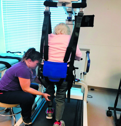
An 82-year-old female who suffered a left corona radiata CVA and presented with decreased left-sided neuromuscular control and coordination uses the body weight support device over the treadmill. The therapist assists with advancement and placement of her left lower extremity during functional ambulation.
By Devin Cooney, MOR, OTR/L; Brittany Merkh, PT, DPT; Kelly Tender, MS, CCC-SLP
According to the American Stroke Association, one in four people worldwide will have a stroke. It is the number five cause of death and a leading cause of long-term disability in the United States. More than 7 million stroke survivors are currently living in the United States.1
Many stroke survivors choose to go to an acute inpatient rehabilitation facility for their specific rehabilitation needs. In 2018, 1,494 individuals and their families selected Kessler Institute for Rehabilitation in Marlton, NJ, for their start of recovery. Thirty percent of those individuals were diagnosed with a stroke. Although their rehabilitation needs were different, they shared a common goal: to gain the skills, strengths, and strategies to rebuild their lives.
Recently, rehabilitation has shifted to include greater use of technology. The integration of technology-based rehabilitation within stroke recovery may empower an individual toward being more engaged in their own care in addition to forcing greater outcomes and providing objective data.2 Technology can be incorporated in stroke recovery from “head to toe,” including rehabilitation of cognitive-communication, recovery of swallow function, improvement of upper and lower extremity use, and restoration of the ability to walk.
Communication and Swallow Function
Depending on the area of the brain in which a stroke occurs, a survivor may experience problems related to speech, reading, writing, and/or understanding words (aphasia). Technology is often incorporated into speech-language pathology treatment for individuals affected by aphasia at Kessler-Marlton. Technology such as smartphones, tablets, text-to-speech or word prediction software, augmentative-alternative communication devices, video telecommunication, and online support communities provide additional avenues for survivors affected by aphasia to communicate.3
Use of technology is also widely incorporated in the management of dysphagia. There are many causes of dysphagia, but a very common cause is stroke. Swallowing problems can lead to poor nutrition, pneumonia, and poorer quality of life. A specialized swallowing test (videofluoroscopic swallow study and/or fiber optic endoscopic evaluation of swallowing) is recommended to better assess the problems and determine a treatment plan. Swallowing exercises may be needed to improve strength and coordination of swallowing muscles. One example of technology utilized at Kessler-Marlton is surface electromyography (sEMG) biofeedback to provide visual monitoring of the sEMG signal to guide performance in swallowing therapy, increase active participation, provide objective data, and track outcomes.4,5 Use of sEMG in conjunction with swallow exercise has been shown to improve functional swallowing outcomes.6 Additional supportive interventions include neuromuscular stimulation, patterned electrical stimulation, pressure biofeedback to measure and target tongue strength, and dysphagia applications to offer education and personalized exercise programs to patients and their families.
Upper Extremity and Vision Restoration
Stroke survivors may have difficulty performing activities of daily living for various reasons. However, for the purpose of this article the focus will be on the recovery of upper extremity use and visual/visuo-spatial impairments. Occupational therapy (OT) is vital in stroke recovery, and the goal is to increase, improve, or restore independence within activities of daily living. While manual therapies are typically used with the majority of patients, therapists at Kessler Marlton incorporate supportive technology and equipment including an integrated therapy system with a touchscreen display that can be used for oculomotor therapy, motor control training, and cognitive learning; a computerized, task-oriented upper extremity workstation; and a hand rehabilitation system that uses a wireless orthosis to deliver electrical stimulation.
The integrated therapy systems is a large technological board with a touch screen. It can be used to target a wide range of impairments including vision-related activities. These activities focus on visuomotor coordination, reaction time, visual processing, and visuospatial perception. This technology engages patients and can be personalized to individual needs.
In addition to vision, OT focuses on the rehabilitation of the upper extremity when indicated. To facilitate this, the computerized upper extremity workstation is often utilized. It is a computerized training system with a full workstation as well as a computer program. It utilizes games and objective data to motivate and engage patients. This system offers the ability to complete both gross and fine motor activities including different grip and pinch patterns. Different planes of movement or positions can also be completed to downgrade or upgrade patient challenge.
When appropriate, the hand rehabilitation system with wireless orthosis may be utilized to target the upper extremity. While neuromuscular electrical stimulation (NMES) can be used on different body parts and muscle groups, this device specifically targets the muscles of the hand. This system’s program settings such as pinching, grasping, and releasing can be utilized individually or while completing functional activities.
Restoration of the Lower Extremity
Many people who are affected by stroke lose function of their lower extremity to some capacity. The goal of the physical therapist (PT) is to facilitate improved functional independence and maximize safety with overall mobility while primarily focusing on lower extremity recovery. Recently, with the use of technology, rehabilitation and mobility can be initiated sooner, which translates into improved overall outcomes.
Within the inpatient rehabilitation setting at Kessler-Marlton, PTs have access to a multitude of technology options, including an adaptive cycling machine. The cycling device is multimodal and allows patients to participate passively, motor-supported, or actively. The passive mode allows for early mobilization of patients diagnosed with stroke, as it is able to assist with reducing muscle tone in those patients with hypertonicity. With patients who are not ready to perform ambulation itself, the passive mode also allows for repetitive motions that mimic the back-and-forth motion of walking. These repetitive, rhythmic movements help to stimulate the brain to reorganize and relearn motor tasks.
As patients improve and regain motor control and neuromuscular strength, the adaptive cycling machine can be used in the motor-supported mode, which allows the motor to assist to stimulate both strength and endurance until the patient is able to participate in active mode. In active mode, the patient is using her or his own strength to pedal.
Partial Weight-Bearing
As early mobilization is an important factor in recovery, another technological device that assists the user with partial weight-bearing is of great importance. This technology is a body weight-supported gait training device in which the patient is supported by an overhead suspension system and harness. This system allows the patient, who might not necessarily be able to stand on his or her own, the opportunity to force weight-bearing through the affected lower extremity to force motor recovery. In the upright position, it not only affords the patient the opportunity to weight-bear, but it also promotes proper upright posturing and provides the patient a safe, fall-free environment in which to practice initiating mobility. This system allows the therapist to provide hands-on assistance at the lower extremity or at the pelvis to achieve proper gait pattern. The device can be used for over-ground training or for training over the treadmill, which challenges a patient’s coordination and timing of the different phases of the gait cycle. As a patient improves, the overhead system can be adjusted to allow increased weight-bearing and increased degrees of freedom.
As the patient moves into a more ambulatory level, he or she still may demonstrate impairments in the amount or quality of movement in their lower extremity. The use of functional electrical stimulation (FES) may be used as a recovery tool. The FES device this facility uses is a wireless foot drop system that helps to stimulate the nerves and muscles of the affected lower extremity, most often at the ankle and in the thigh, to re-educate the brain and restore muscle function during walking. This system comes with a lower leg cuff to stimulate the ankle muscles in patients with difficulty clearing their toes or patients with foot drop. The system also comes with a thigh cuff to stimulate muscles in the upper leg to provide stability while in stance phase.
Although speech, occupational, and/or physical therapy itself has been shown to improve patient outcomes, the integration of technology in therapeutic intervention following stroke can maximize patient motivation and progress. For example, the inclusion of technology in physical therapy has been able to allow patients the opportunity to participate in therapy at earlier phases. This early mobilization is crucial to jump-start a patient’s recovery. The use of technology throughout the patient’s recovery process has been an integral part in demonstrating improved outcomes which allows physical therapists to facilitate recovery by working on the often stated patient-centered goal of returning to walking. RM
Devin Cooney, MOR, OTR/L, has worked at Kessler Institute for Rehabilitation—Marlton for the past 3 years. The majority of her career has been spent in the acute rehabilitation setting focusing on ADLs and IADLs to encourage safe return to home.
Brittany Merkh, PT, DPT, is Co-Chair of the Stroke Program at Kessler Institute for Rehabilitation in Marlton, New Jersey. Most of her career has been spent in acute rehabilitation, treating patients with a variety of diagnoses.
Kelly Tender, MS, CCC-SLP, is senior speech-language pathologist and leader within the SLP department. Her certifications and training include MDTP, VitalStim, PENS, and sEMG. She is a member of the hospital’s stroke committee. For more information, contact RehabEditor@medqor.com.
2. Des Roches C, Kiran S. Technology-based rehabilitation to improve communication after acquired brain injury. Front Neurosci. 2017;11. doi:10.3389/fnins.2017.00382. Accessed November 21, 2019.
5. Crary M, Carnaby (Mann) G, Groher M, Helseth E. Functional benefits of dysphagia therapy using adjunctive sEMG biofeedback. Dysphagia. 2004;19(3). doi: https://www.doi.org/10.1007/s00455-004-0003-8.
6. Bogaardt H, Grolman W, Fokkens W. The use of biofeedback in the treatment of chronic dysphagia in stroke patients. Folia Phoniatrica et Logopaedica. 2009;61(4):200-205. doi: https://www.doi.org/10.1159/000227997
Disabilities resulting from a TBI depend upon the severity of the injury, the location of the injury, and the age and general health of the patient. Some common disabilities include problems with cognition (thinking, memory, and reasoning), sensory processing (sight, hearing, touch, taste, and smell), communication (expression and understanding), and behavior or mental health (depression, anxiety, personality changes, aggression, acting out, and social inappropriateness).
Within days to weeks of the head injury approximately 40 percent of TBI patients develop a host of troubling symptoms collectively called postconcussion syndrome (PCS). A patient need not have suffered a concussion or loss of consciousness to develop the syndrome and many patients with mild TBI suffer from PCS. Symptoms include headache, dizziness, vertigo (a sensation of spinning around or of objects spinning around the patient), memory problems, trouble concentrating, sleeping problems, restlessness, irritability, apathy, depression, and anxiety. These symptoms may last for a few weeks after the head injury. The syndrome is more prevalent in patients who had psychiatric symptoms, such as depression or anxiety, before the injury. Treatment for PCS may include medicines for pain and psychiatric conditions, and psychotherapy and occupational therapy todevelop coping skills.
Cognition is a term used to describe the processes of thinking, reasoning, problem solving, information processing, and memory. Most patients with severe TBI, if they recover consciousness, suffer from cognitive disabilities, including the loss of many higher level mental skills. The most common cognitive impairment among severely head-injured patients is memory loss, characterized by some loss of specific memories and the partial inability to form or store new ones. Some of these patients may experience post-traumatic amnesia (PTA), either anterograde or retrograde. Anterograde PTA is impaired memory of events that happened after the TBI, while retrograde PTA is impaired memory of events that happened before the TBI.
Many patients with mild to moderate head injuries who experience cognitive deficits become easily confused or distracted and have problems with concentration and attention. They also have problems with higher level, so-called executive functions, such as planning, organizing, abstract reasoning, problem solving, and making judgments, which may make it difficult to resume pre-injury work-related activities. Recovery from cognitive deficits is greatest within the first 6 months after the injury and more gradual after that.
The most common cognitive impairment among severely head-injured patients is memory loss, characterized by some loss of specific memories and the partial inability to form or store new ones.
Patients with moderate to severe TBI have more problems with cognitive deficits than patients with mild TBI, but a history of several mild TBIs may have an additive effect, causing cognitive deficits equal to a moderate or severe injury.
Many TBI patients have sensory problems, especially problems with vision. Patients may not be able to register what they are seeing or may be slow to recognize objects. Also, TBI patients often have difficulty with hand-eye coordination. Because of this, TBI patients may be prone to bumping into or dropping objects, or may seem generally unsteady. TBI patients may have difficulty driving a car, working complex machinery, or playing sports. Other sensory deficits may include problems with hearing, smell, taste, or touch. Some TBI patients develop tinnitus, a ringing or roaring in the ears. A person with damage to the part of the brain that processes taste or smell may develop a persistent bitter taste in the mouth or perceive a persistent noxious smell. Damage to the part of the brain that controls the sense of touch may cause a TBI patient to develop persistent skin tingling, itching, or pain. Although rare, these conditions are hard to treat.
Language and communication problems are common disabilities in TBI patients. Some may experience aphasia, defined as difficulty with understanding and producing spoken and written language; others may have difficulty with the more subtle aspects of communication, such as body language and emotional, non-verbal signals.
In non-fluent aphasia, also called Broca’s aphasia or motor aphasia, TBI patients often have trouble recalling words and speaking in complete sentences. They may speak in broken phrases and pause frequently. Most patients are aware of these deficits and may become extremely frustrated. Patients with fluent aphasia, also called Wernicke’s aphasia or sensory aphasia, display little meaning in their speech, even though they speak in complete sentences and use correct grammar. Instead, they speak in flowing gibberish, drawing out their sentences with non-essential and invented words. Many patients with fluent aphasia are unaware that they make little sense and become angry with others for not understanding them. Patients with global aphasia have extensive damage to the portions of the brain responsible for language and often suffer severe communication disabilities.
TBI patients may have problems with spoken language if the part of the brain that controls speech muscles is damaged. In this disorder, called dysarthria, the patient can think of the appropriate language, but cannot easily speak the words because they are unable to use the muscles needed to form the words and produce the sounds. Speech is often slow, slurred, and garbled. Some may have problems with intonation or inflection, called prosodic dysfunction. An important aspect of speech, inflection conveys emotional meaning and is necessary for certain aspects of language, such as irony. These language deficits can lead to miscommunication, confusion, and frustration for the patient as well as those interacting with him or her.
Most TBI patients have emotional or behavioral problems that fit under the broad category of psychiatric health. Family members of TBI patients often find that personality changes and behavioral problems are the most difficult disabilities to handle. Psychiatric problems that may surface include depression, apathy, anxiety, irritability, anger, paranoia, confusion, frustration, agitation, insomnia or other sleep problems, and mood swings. Problem behaviors may include aggression and violence, impulsivity, disinhibition, acting out, noncompliance, social inappropriateness, emotional outbursts, childish behavior, impaired self-control, impaired self awareness, inability to take responsibility or accept criticism, egocentrism, inappropriate sexual activity, and alcohol or drug abuse/addiction. Some patients’ personality problems may be so severe that they are diagnosed with borderline personality disorder, a psychiatric condition characterized by many of the problems mentioned above. Sometimes TBI patients suffer from developmental stagnation, meaning that they fail to mature emotionally, socially, or psychologically after the trauma. This is a serious problem for children and young adults who suffer from a TBI. Attitudes and behaviors that are appropriate for a child or teenager become inappropriate in adulthood. Many TBI patients who show psychiatric or behavioral problems can be helped with medication and psychotherapy.
By Devin Cooney, MOR, OTR/L; Brittany Merkh, PT, DPT; Kelly Tender, MS, CCC-SLP
According to the American Stroke Association, one in four people worldwide will have a stroke. It is the number five cause of death and a leading cause of long-term disability in the United States. More than 7 million stroke survivors are currently living in the United States.1
Many stroke survivors choose to go to an acute inpatient rehabilitation facility for their specific rehabilitation needs. In 2018, 1,494 individuals and their families selected Kessler Institute for Rehabilitation in Marlton, NJ, for their start of recovery. Thirty percent of those individuals were diagnosed with a stroke. Although their rehabilitation needs were different, they shared a common goal: to gain the skills, strengths, and strategies to rebuild their lives.
Recently, rehabilitation has shifted to include greater use of technology. The integration of technology-based rehabilitation within stroke recovery may empower an individual toward being more engaged in their own care in addition to forcing greater outcomes and providing objective data.2 Technology can be incorporated in stroke recovery from “head to toe,” including rehabilitation of cognitive-communication, recovery of swallow function, improvement of upper and lower extremity use, and restoration of the ability to walk.
Communication and Swallow Function
Depending on the area of the brain in which a stroke occurs, a survivor may experience problems related to speech, reading, writing, and/or understanding words (aphasia). Technology is often incorporated into speech-language pathology treatment for individuals affected by aphasia at Kessler-Marlton. Technology such as smartphones, tablets, text-to-speech or word prediction software, augmentative-alternative communication devices, video telecommunication, and online support communities provide additional avenues for survivors affected by aphasia to communicate.3
Use of technology is also widely incorporated in the management of dysphagia. There are many causes of dysphagia, but a very common cause is stroke. Swallowing problems can lead to poor nutrition, pneumonia, and poorer quality of life. A specialized swallowing test (videofluoroscopic swallow study and/or fiber optic endoscopic evaluation of swallowing) is recommended to better assess the problems and determine a treatment plan. Swallowing exercises may be needed to improve strength and coordination of swallowing muscles. One example of technology utilized at Kessler-Marlton is surface electromyography (sEMG) biofeedback to provide visual monitoring of the sEMG signal to guide performance in swallowing therapy, increase active participation, provide objective data, and track outcomes.4,5 Use of sEMG in conjunction with swallow exercise has been shown to improve functional swallowing outcomes.6 Additional supportive interventions include neuromuscular stimulation, patterned electrical stimulation, pressure biofeedback to measure and target tongue strength, and dysphagia applications to offer education and personalized exercise programs to patients and their families.
Upper Extremity and Vision Restoration
Stroke survivors may have difficulty performing activities of daily living for various reasons. However, for the purpose of this article the focus will be on the recovery of upper extremity use and visual/visuo-spatial impairments. Occupational therapy (OT) is vital in stroke recovery, and the goal is to increase, improve, or restore independence within activities of daily living. While manual therapies are typically used with the majority of patients, therapists at Kessler Marlton incorporate supportive technology and equipment including an integrated therapy system with a touchscreen display that can be used for oculomotor therapy, motor control training, and cognitive learning; a computerized, task-oriented upper extremity workstation; and a hand rehabilitation system that uses a wireless orthosis to deliver electrical stimulation.
The integrated therapy systems is a large technological board with a touch screen. It can be used to target a wide range of impairments including vision-related activities. These activities focus on visuomotor coordination, reaction time, visual processing, and visuospatial perception. This technology engages patients and can be personalized to individual needs.
In addition to vision, OT focuses on the rehabilitation of the upper extremity when indicated. To facilitate this, the computerized upper extremity workstation is often utilized. It is a computerized training system with a full workstation as well as a computer program. It utilizes games and objective data to motivate and engage patients. This system offers the ability to complete both gross and fine motor activities including different grip and pinch patterns. Different planes of movement or positions can also be completed to downgrade or upgrade patient challenge.
When appropriate, the hand rehabilitation system with wireless orthosis may be utilized to target the upper extremity. While neuromuscular electrical stimulation (NMES) can be used on different body parts and muscle groups, this device specifically targets the muscles of the hand. This system’s program settings such as pinching, grasping, and releasing can be utilized individually or while completing functional activities.
Restoration of the Lower Extremity
Many people who are affected by stroke lose function of their lower extremity to some capacity. The goal of the physical therapist (PT) is to facilitate improved functional independence and maximize safety with overall mobility while primarily focusing on lower extremity recovery. Recently, with the use of technology, rehabilitation and mobility can be initiated sooner, which translates into improved overall outcomes.
Within the inpatient rehabilitation setting at Kessler-Marlton, PTs have access to a multitude of technology options, including an adaptive cycling machine. The cycling device is multimodal and allows patients to participate passively, motor-supported, or actively. The passive mode allows for early mobilization of patients diagnosed with stroke, as it is able to assist with reducing muscle tone in those patients with hypertonicity. With patients who are not ready to perform ambulation itself, the passive mode also allows for repetitive motions that mimic the back-and-forth motion of walking. These repetitive, rhythmic movements help to stimulate the brain to reorganize and relearn motor tasks.
As patients improve and regain motor control and neuromuscular strength, the adaptive cycling machine can be used in the motor-supported mode, which allows the motor to assist to stimulate both strength and endurance until the patient is able to participate in active mode. In active mode, the patient is using her or his own strength to pedal.
Partial Weight-Bearing
As early mobilization is an important factor in recovery, another technological device that assists the user with partial weight-bearing is of great importance. This technology is a body weight-supported gait training device in which the patient is supported by an overhead suspension system and harness. This system allows the patient, who might not necessarily be able to stand on his or her own, the opportunity to force weight-bearing through the affected lower extremity to force motor recovery. In the upright position, it not only affords the patient the opportunity to weight-bear, but it also promotes proper upright posturing and provides the patient a safe, fall-free environment in which to practice initiating mobility. This system allows the therapist to provide hands-on assistance at the lower extremity or at the pelvis to achieve proper gait pattern. The device can be used for over-ground training or for training over the treadmill, which challenges a patient’s coordination and timing of the different phases of the gait cycle. As a patient improves, the overhead system can be adjusted to allow increased weight-bearing and increased degrees of freedom.
As the patient moves into a more ambulatory level, he or she still may demonstrate impairments in the amount or quality of movement in their lower extremity. The use of functional electrical stimulation (FES) may be used as a recovery tool. The FES device this facility uses is a wireless foot drop system that helps to stimulate the nerves and muscles of the affected lower extremity, most often at the ankle and in the thigh, to re-educate the brain and restore muscle function during walking. This system comes with a lower leg cuff to stimulate the ankle muscles in patients with difficulty clearing their toes or patients with foot drop. The system also comes with a thigh cuff to stimulate muscles in the upper leg to provide stability while in stance phase.
Although speech, occupational, and/or physical therapy itself has been shown to improve patient outcomes, the integration of technology in therapeutic intervention following stroke can maximize patient motivation and progress. For example, the inclusion of technology in physical therapy has been able to allow patients the opportunity to participate in therapy at earlier phases. This early mobilization is crucial to jump-start a patient’s recovery. The use of technology throughout the patient’s recovery process has been an integral part in demonstrating improved outcomes which allows physical therapists to facilitate recovery by working on the often stated patient-centered goal of returning to walking. RM
Devin Cooney, MOR, OTR/L, has worked at Kessler Institute for Rehabilitation—Marlton for the past 3 years. The majority of her career has been spent in the acute rehabilitation setting focusing on ADLs and IADLs to encourage safe return to home.
Brittany Merkh, PT, DPT, is Co-Chair of the Stroke Program at Kessler Institute for Rehabilitation in Marlton, New Jersey. Most of her career has been spent in acute rehabilitation, treating patients with a variety of diagnoses.
Kelly Tender, MS, CCC-SLP, is senior speech-language pathologist and leader within the SLP department. Her certifications and training include MDTP, VitalStim, PENS, and sEMG. She is a member of the hospital’s stroke committee. For more information, contact RehabEditor@medqor.com.
2. Des Roches C, Kiran S. Technology-based rehabilitation to improve communication after acquired brain injury. Front Neurosci. 2017;11. doi:10.3389/fnins.2017.00382. Accessed November 21, 2019.
5. Crary M, Carnaby (Mann) G, Groher M, Helseth E. Functional benefits of dysphagia therapy using adjunctive sEMG biofeedback. Dysphagia. 2004;19(3). doi: https://www.doi.org/10.1007/s00455-004-0003-8.
6. Bogaardt H, Grolman W, Fokkens W. The use of biofeedback in the treatment of chronic dysphagia in stroke patients. Folia Phoniatrica et Logopaedica. 2009;61(4):200-205. doi: https://www.doi.org/10.1159/000227997
At times I’m just too tired to explain how I’m feeling. (You might have noticed I write less often than I used to and that’s one of the reasons for it.) Sometimes there is a storm of emotions inside me which I realise are irrational but I can’t quell them. So to avoid saying anything that I would later regret, I find myself clamming up. But my face rarely gets the memo and goes into full on sulky mode. I’m so bored of this brain injury, I wish I could direct my anger at it and evict it from my head. Sadly it doesn’t work like that though.
At the time clamming up feels like the lesser of two evils. But maybe it isn’t.
My silence carries more weight than I intend it to. All I’m doing it trying to contain my poisonous tongue. Some people go quiet for dramatic effect, waiting for someone to ask “What’s wrong, you’ve hardly said a word today?” I guarantee you, that when you are struggling with a brain injury there is no such thing as dramatic effect.
Even when I’m trying to be mindful, holding my tongue is the best I can do. I might be sulking about my partner James having to work so much, and having less time with him. As he is the only one bringing in a income, I know I can’t begrudge him for being so conscientious. In fact, I know he would like nothing more than being able to work less, or even take early retirement. But currently neither are an option. So I try to remind myself of this and empathise with his position. And it works, but not for my brain injury. It just keeps complaining and dragging me down.
How silence leaves too many unanswered questions.
My grumpiness is too obvious, and I know it makes James feel guilty. But he has nothing to feel guilty about. He has been truly amazing the entire time. Superman hasn’t got a patch on this guy. If the world had more people like him in it, people wouldn’t need heaven.
But he still doubts himself as he can read my face. So eventually I manage to spill, but I start with a disclaimer: “I know I’m being stupid, and here’s the reason why it’s stupid …..blah,blah,blah…. but I can’t help it. I’m upset about ….x,y,z….. because…..”
This really does help the situation, it’s just a shame it takes me so long to be in a position where I can do it. James is getting used to my behaviour, but he is only human (although he’s as close to an angel as you can get.) When I am clamming up, his mind is running a million miles an hour, thinking of all the things I might be annoyed about. I do feel bad for torturing him like this as he doesn’t deserve it. So here’s a shout out for all the carers who some how put up with the nonsense some of us survivors put them through.
I’m tired and I’ve run out of words again so I’m going to leave it there. I think you get the point, and I’m sure I’m not the only one who is going through this.
Parenting is a challenging life role for all people, yet one of the most valued roles within society. Brain Injury frequently occurs at a life stage where people are yet to complete their parenting responsibilities. For people with acquired brain injury (ABI), facing cognitive, physical, communication, behavioural and psychological challenges, parenting can present complex challenges. In addition, persons with ABI often face societal and environmental barriers. These fact sheets have been developed to assist parents with an ABI and their partners to improve their knowledge and skills to meet the ongoing challenges of parenting.


 Brandon Leuangpaseuth is a freelance copywriter from San Diego, CA. Brandon is an avid traveler, book enthusiast and loves animals. He loves exploring new places and going on long walks on the beach. You can connect with him on LinkedIn @ bleuangpaseuth.