Archive for August, 2018

[WEB SITE] ELEX – Exoskeleton – The Robotic Exoskeleton Kit

Project submitted by: Markus Scherzinger, Illya Gents and Gerardo Garcia.

Introduction

The goal of this project was to explore and learn about robotic exoskeleton technology by redesigning several parts of the EduExo to extend its functionality. The work was submitted to us by Markus ScherzingerIllya Gents and Gerardo Garcia. It was a student project called “ELEX  development of an elbow exoskeleton” they conducted as part of their Bachelor studies in robotics at the Hochschule Furtwangen University in Furtwangen, Germany. The project was supervised by Prof. Hans-Georg Enkler.
The project might be of special interest to you if you would like to redesign mechanical parts, or extend the electronics of your EduExo.

The team kindly provided us with their project report that contains many technical details, the student’s perspective and their step-by-step progress. You can find a download link below. Thank you very much for sharing your results!

Implementation

The first major alteration was a mechanical redesign of most of the EduExo parts, to allow size adjustments and integration of the electronics on-board. Mechanical design work was conducted using the software SolidWorks, and the parts were printed in an Alphacam Type Dimension bst 768 3D printer.

The redesign included a new upper arm segment with a length adjustment mechanism.

Another major mechanical extension was the addition of housing for the upper and lower arms that enable the integration of all electronic components into the exoskeleton arm.

Besides the new mechanical parts, the team also worked on implementing new electronic components. The main new feature was a display that provides information such as remaining battery power to the exoskeleton user.

Results

Overall, the work resulted in an extended elbow exoskeleton that self-contained all the electronics and a length adjustment mechanism.

If you want to learn more about the project, please find the original report below. In the report, the authors provide many details about their motivation and the technical implementation. If you are interested in expanding your EduExo as well, this document could be a good starting point!

Resources

Download

Student Report
The student’s project report. Provided with permission by the authors. Copyright is with the authors of the report.
ELEX_Handbook.pdf
Adobe Acrobat Document 2.1 MB

via ELEX – Exoskeleton – EduExo

, , , ,

Leave a comment

[Abstract] The use of antidepressant drugs in pregnant women with epilepsy: A study from the Australian Pregnancy Register

Summary

Objective

To study interactions between first‐trimester exposure to antidepressant drugs (ADDs) and antiepileptic drugs (AEDs), and a history of clinical depression and/or anxiety, on pregnancy outcomes and seizure control in pregnant women with epilepsy (WWE).

Methods

We examined data from the Australian Pregnancy Register of Antiepileptic Drugs in Pregnancy, collected from 1999 to 2016. The register is an observational, prospective database, from which this study retrospectively analyzed a cohort. Among the AED‐exposed outcomes, comparisons were made among 3 exposure groups: (1) pregnancy outcomes with first‐trimester exposure to ADDs; (2) outcomes with mothers diagnosed with depression and/or anxiety but who were not medicated with an ADD; and (3) those with mothers who were not diagnosed with depression and/or anxiety and were not medicating with ADD. Prevalence data was analyzed using Fisher’s exact test.

Results

A total of 2124 pregnancy outcomes were included in the analysis; 1954 outcomes were exposed to AEDs in utero, whereas 170 were unexposed. Within the group of WWE taking AEDs, there was no significant difference in the prevalence of malformations in infants who were additionally exposed to ADDs (10.2%, 95% confidence interval [CI] 3.9‐16.6), compared to individuals in the non–ADD‐medicated depression and/or anxiety group (7.7%, 95% CI 1.2‐14.2), or those without depression or anxiety (6.9%, 95% CI 5.7‐8.1; = 0.45). The malformation rates in pregnancy outcomes unexposed to AEDs were also similar in the above groups (= 0.27). In WWE medicated with AEDs and ADDs, the frequency of convulsive seizures (= 0.78), or nonconvulsive seizures (= 0.45) throughout pregnancy, did not differ across comparative groups.

Significance

Co‐medicating with ADDs in WWE taking AEDs does not appear to confer a significant added teratogenic risk, and it does not affect seizure control.


via The use of antidepressant drugs in pregnant women with epilepsy: A study from the Australian Pregnancy Register – Sivathamboo – – Epilepsia – Wiley Online Library

, , , , , , ,

Leave a comment

[WEB PAGE] What are the various treatment options for paralysis?

When there is a loss of muscular functioning in an area or sensory loss on area resulting usually from any damage to central nervous system, there is paralysis. Some of the probable causes of this dangerous condition are polio, stroke, excessive trauma or multiple sclerosis, etc. There may be complete paralysis or partial paralysis. It is mainly of two kinds, namely, paraplegia and quadriplegia. Paralysis is the consequence when the brain fails to send signals to various regions of the body. This may result from a variety of reasons. Stroke accounts for 30% of paralysis cases and is the major cause. However, one can choose paralysis treatment depending on the severity of the condition and the region which is paralyzed.

How is paralysis diagnosed?

On the event of any failure of muscular functioning or sensory loss on certain area, it is important to visit a medical practitioner immediately. To diagnose the condition, he prescribes a series of tests including CT Scan, MRI, X-Ray, Electromyography. If at all it is necessary, the patient may be suggested a neurologist. After paralysis is confirmed, the treatment begins. Certain types of paralysis may be cured and this mainly includes partial paralysis. You can ask the doctor whether the recovery is possible or not. No matter what the cause of the condition, the treatment procedure will be almost the same. Whatever treatment you choose for recovery, the treatment provider will try and restore brain and body connection. This is the only way to bring about recovery.

Some of the basic treatment options for paralysis

Wearable device running on electricity is the most basic treatment for paralysis. This wearable electronic device is also used for stroke treatment. It improves arm functioning and restores motion in the arms. When you wear this device, it delivers electrical current to activate the muscles of arms and legs. This technique of motion restoration is also termed as FES or Functional Electrical Stimulation. It can recover the feet or lower legs from paralysis. The use of FES along with specific exercises can bring about a relief.

Some of the best treatment options for paralysis 

If anyone of your loved one is suffering from paralysis, read the following section to learn how to reduce the symptoms:

  • Surgery can address physical barriers. It may be that there is an object fixed in the brain or spinal cord of the person. It needs to be got rid of. Through the surgery, certain portions of the spinal cord can also be fused together.
  • Some paralysis medication may be used to reduce swelling, inflammation and infection on the area. If there is chronic pain, it may be addressed with medicines.
  • Continuous monitoring of the person is mandatory to ensure that this condition does not get worse
  • Psychotherapy can help a lot. Support groups may teach you how to cope with this critical situation.
  • To restore muscular and nerve functioning, you may be asked to do certain exercises. Occupational therapy can also help a lot. Work on the injuries and practice them as much as possible. Physical therapy may reverse paralysis by rewiring the brain.
  • Some people got great results from alternative treatments like chiropractic care, massage therapy and acupuncture treatment.

If there are breathing difficulties, problem in the bowel movement, take immediate treatment for them. Again, surgery is an effective sleep apnea treatment. Whether it is sleep apnea or paralysis, immediate medical attention is required.

via What are the various treatment options for paralysis?- The New Indian Express

, , , ,

Leave a comment

[WEB SITE] ReWalk Exoskeleton – Rehabilitation Technology – PhysioFunction

Rewalk Exoskeleton

Rewalk

What is the ReWalk?

The ReWalk is a robotic Exoskeleton that can be worn for personal use at home and out in the community.

The robotic device provides hip and knee motion to enable individuals with spinal cord injury to stand upright, walk, turn, climb and descend stairs. The system can be customised to provide optimal fit to ensure safety, function and joint function.

ReWalk allows people to walk independently as the robotic device mimics the natural gait pattern of the legs.

What are the benefits?

The benefits of using the ReWalk include:

  • Ability to walk upright rather than sit in a wheelchair
  • Improve mobility and quality of life measures such as:

  • Improvements in bowel and bladder function

  • Maintenance of bone mass

  • Reduction of some medications for certain ailments

  • Emotional and psychosocial benefits

How can you trial and purchase a ReWalk for home use?

At our Midlands ‘Centre of Excellence Clinic’ in Northampton.

Firstly, we will book you into our clinic for an initial assessment where you will be able to trial the device*.

Providing you are suitable for the device, you will be given the option to purchase your own ReWalk and at the same time we will discuss the rehab package on offer that will help you achieve maximum use of your ReWalk.

If you live outside the Midlands and need accommodation, we can also help find you an accessible place to stay.

*Prior to the assessment we will need to establish your suitability for the device as ReWalk is intended for use by individuals with lower limb disabilities whose hands and shoulders can support crutches or a walker. Your height will need to be between 160cm – 190cm (5’3″ to 6’2″). Weight requirement is up to 100kg (220lbs). Other factors such as bone density and range of motion will be considered and evaluated.

via ReWalk Exoskeleton | Rehabilitation Technology | PhysioFunction

, , ,

2 Comments

[Abstract + References] A Tendon-driven Upper-limb Rehabilitation Robot – IEEE Conference Publication

Abstract

Rehabilitation robots are playing an increasingly important role in daily rehabilitation of patients. In recent years, exoskeleton rehabilitation robots have become a research hotspot. However, the existing exoskeleton rehabilitation robots are mainly rigid exoskeletons. During rehabilitation training using such exoskeletons, the patient’s joint rotation center is fixed, which cannot adapt to the actual joint movements, resulting in secondary damage to the patients. Therefore, in this paper, a tendon-driven flexible upper-limb rehabilitation robot is proposed; the structure and connectors of the rehabilitation robot are designed considering the physiological structure of human upper limbs; we also built the prototype and performed experiments to validate the designed robot. The experimental results show that the proposed upper-limb rehabilitation robot can assist the human subject to conduct upper-limb rehabilitation training.

I. Introduction

Central nervous system diseases, such as stroke, spinal cord injury and traumatic brain injury, tend to cause movement disorder [1]. Clinical studies have shown that intensive rehabilitation training after cerebral injury help patients recover motoric functions because of the brain plasticity [1], [2]. Traditional movement therapy is highly dependent on physiotherapists and the efficacy is limited by professional knowledge and skill levels of physiotherapists [3]. Upper-limbs recover more slowly than lower limbs because of the complex function of neurons. Meanwhile, the rehabilitation therapies are unaffordable for most patients. Robotic rehabilitation opened another way of rehabilitation training and its efficacy has been validated in clinical trials [3], [4]. Many upper-limb robot devices have been developed for rehabilitation or assistance in various forms. One of the famous devices was MIT-MANUS developed by MIT. This kind of devices are stationary external system where the patient inserts their hand or arm and is robotically assisted or resisted in completing predetermined tasks [3], [5]. Other examples of this type of devices include Lum et al.^{\prime}s MIME [6], Kahn et al.’s ARM Guide [7] and a 2-DOF upper-limb rehabilitation robot developed by Tsinghua


References

1. M. Hallett, “Plasticity of the human motor cortex and recovery from stroke”, Brain Research Reviews, vol. 36, pp. 169-174, 2001.

2. J. D. Schaechter, “Motor rehabilitation and brain plasticity after hemiparetic stroke”, Progress in Neurobiology, vol. 73, pp. 61-72, 2004.

3. Q. Yang, D. Cao, J. Zhao, “Analysis on State of the Art of upper-limb Rehabilitation Robots”, Jiqiren/robot, vol. 35, pp. 630, 2013.

4. P. Maciejasz, J. Eschweiler, K. Gerlach-Hahn, A. Jansen-Troy, S. Leonhardt, “A survey on robotic devices for upper-limb rehabilitation”, Journal of Neuroengineering & Rehabilitation, vol. 11, pp. 3, 2014.

5. C. J. Nycz, M. A. Delph, G. S. Fischer, “Modeling and design of a tendon actuated flexible robotic exoskeleton for hemiparetic upper-limb rehabilitation”, International Conference of the IEEE Engineering in Medicine and Biology Society, pp. 3889-3892, 2015.

6. P. S. Lum, C. G. Burgar, P. C. Shor, “Use of the MIME robotic system to retrain multijoint reaching in post-stroke hemiparesis: why some movement patterns work better than others”, Engineering in Medicine and Biology Society 2003. Proceedings of the International Conference of the IEEE, vol. 2, pp. 1475-1478, 2003.

7. D. J. Reinkensmeyer, L. E. Kahn, M. Averbuch, A. Mckenna-Cole, B. D. Schmit, W. Z. Rymer, “Understanding and treating arm movement impairment after chronic brain injury: progress with the ARM guide”, Journal of Rehabilitation Research & Development, vol. 37, pp. 653-662.

8. Y. Zhang, Z. Wang, L. Ji, S. Bi, “The clinical application of the upper extremity compound movements rehabilitation training robot”, International Conference on Rehabilitation Robotics, pp. 91-94, 2005.

9. H. Fukushima, “Health and wellbeing in the 21st century (No. 4): Early rehabilitation and conditions for which it is appropriate [J]” in Social-human environmentology, pp. 6, 2004.

10. T. G. Sugar, J. He, E. J. Koeneman, J. B. Koeneman, R. Herman, H. Huang et al., “Design and control of RUPERT: a device for robotic upper extremity repetitive therapy”, IEEE Transactions on Neural Systems & Rehabilitation Engineering a Publication of the IEEE Engineering in Medicine & Biology Society, vol. 15, no. 3, pp. 336-46, 2007.

11. J. C Perry, J. Rosen, S. Burns, “Upper-Limb Powered Exoskeleton Design”, Mechatronics IEEE/ASME Transactions on, vol. 12, pp. 408-417, 2007.

12. A. U. Pehlivan, O. Celik, M. K. O’Malley, “Mechanical design of a distal arm exoskeleton for stroke and spinal cord injury rehabilitation”, IEEE International Conference on Rehabilitation Robotics IEEE Int Conf Rehabil Robot, pp. 5975428, 2011.

13. S Koo, T. P. Andriacchi, “The Knee Joint Center of Rotation is Predominantly on the Lateral Side during Normal Walking[J]”, Journal of Biomechanics, vol. 41, no. 6, pp. 1269, 2008.

14. Y. Mao, S. K. Agrawal, “Transition from mechanical arm to human arm with CAREX: A cable driven ARm EXoskeleton (CAREX) for neural rehabilitation”, Proc. IEEE Int. Conf. Robot. Autom., pp. 2457-2462, 2012.

15. Y. Mao, X. Jin, G. G. Dutta, J. P. Scholz, S. K. Agrawal, “Human movement training with a cable driven ARm EXoskeleton (CAREX)”, IEEE Trans. Neural Syst. Rehabil. Eng., vol. 23, no. 1, pp. 84-92, Jan. 2015.

16. DJ Reinkensmeyer, JL Emken, SC. Cramer, “Robotics motor learning and neurologic recovery”, Annual Review of Biomedical Engineering, vol. 6, no. 1, pp. 497-525, 2004.

17. QZ Yang, CF Cao, JH. Zhao, “Analysis of the status of the research of the upper-limb rehabilitative robot”, Robot, vol. 35, no. 5, pp. 630-640, 2013.

18. XZ Jiang, XH Huang, CH Xiong et al., “Position Control of a Rehabilitation Robotic Joint Based on Neuron Proportion-Integral and Feedforward Control”, Journal of Computational & Nonlinear Dynamics, vol. 7, no. 2, pp. 024502, 2012.

19. ZC Chen, Z. Huang, “Motor relearning in the application of the rehabilitation therapy for stroke”, Chinese Journal of Rehabilitation Medicine, vol. 22, no. 11, pp. 1053-1056, 2007.

20. JC Perry, J Rosen, S. Burns, “Upper-Limb Powered Exoskeleton Design[J]”, IEEE/ASME Transactions on Mechatronics, vol. 12, no. 4, pp. 408-417, 2007.

21. C LV, Research on rehabilitation robot for upper-limb hemiplegia, Shanghai China:, 2011.

22. Y K Woo, G H Cho, E Y. Yoo, Effect of PNF Applied to the Unaffected Side on Muscle Tone of Affected Side in Patients with Hemiplegia[J], vol. 9, no. 2, 2002.

23. JH Liang, JP Tong, X. Li, “Observation of the curative effect of continuous passive movement of joints in the treatment of lower limb spasticity”, Theory and practice of rehabilitation in China, vol. 14, no. 11, pp. 1067-1067, 2008.


via A Tendon-driven Upper-limb Rehabilitation Robot – IEEE Conference Publication

, , , , , , , , ,

Leave a comment

[Abstract + References] Design of Isometric and Isotonic Soft Hand for Rehabilitation Combining with Noninvasive Brain Machine Interface

Abstract

Comparing with the traditional way for hand rehabilitation, such as simple trainers and artificial rigid auxiliary, this paper presents an isometric and isotonic soft hand for rehabilitation supported by the soft robots theory which aims to satisfy the more comprehensive rehabilitation requirements. Salient features of the device are the ability to achieve higher and controllable stiffness for both isometric and isotonic contraction. Then we analyze the active control for isometric and isotonic movement through electroencephalograph (EEG) signal. This paper focuses on three issues. The first is using silicon rubber to build a soft finger which can continuously stretch and bend to fit the basic action of the fingers. The second is changing stiffness of the finger through the coordination between variable stiffness cavity and actuating cavity. The last is to classify different EEG states based on isometric and isotonic contraction using common spatial pattern feature extraction (CSP) methods and support vector machine classification methods (SVM). On this basis, an EEG-based manipulator control system was set up.


I. Introduction

In recent years, stroke has became one of the major health problems which significantly affect the daily life of the elderly, and hand rehabilitation is introduced as an auxiliary treatment. Though various kinds of mechanical devices for hand rehabilitation have been developed, some deficiencies still exist in the current rigid rehabilitation hand, such as the degrees of freedom is not enough, complexity, unsafe status, overweight, being uncomfortable, unfitness and so on. Therefore, with the growth of aging population, it is highly needed to develop some new devices to satisfy the comprehensive rehabilitation requirements. Meanwhile, inspired by the mollusks in nature, soft robot is made of soft materials that can withstand large strains. It is a new type of continuum robot with high flexibility and environmental adaptability. The soft robot has a broad application prospects in military detection techniques, such as instance search, rescue, medical application and other fields.

References

1. J Zhang, H Wang, J Tang et al., “Modeling and design of a soft pneumatic finger for hand rehabilitation [C]”, IEEE International Conference on Information and Automation, pp. 2460-2465, 2015.

2. H Godaba, J Li, Y Wang et al., “A Soft Jellyfish Robot Driven by a Dielectric Elastomer Actuator [J]”, IEEE Robotics & Automation Letters, vol. 1, no. 2, pp. 624-631, 2016.

3. Y Yang, Y. Chen, “Novel design and 3D printing of variable stiffness robotic fingers based on shape memory polymer [C]”, IEEE International Conference on Biomedical Robotics and Biomechatronics, pp. 195-200, 2016.

4. M Wehner, R L Truby, D J Fitzgerald et al., “An integrated design and fabrication strategy for entirely soft autonomous robots [J]”, Nature, vol. 536, no. 7617, pp. 451, 2016.

5. P Polygerinos, Z Wang, K C Galloway et al., “Soft robotic glove for combined assistance and at-home rehabilitation [J]”, Robotics & Autonomous Systems, vol. 73, no. C, pp. 135-143, 2014.

6. M Tian, Y Xiao, X Wang et al., “Design and Experimental Research of Pneumatic Soft Humanoid Robot Hand [M]/ /” in Robot Intelligence Technology and Applications 4. Springer International Publishing, 2017.

7. K Y Hong, J H Lim, F Nasrallah et al., “A soft exoskeleton for hand assistive and rehabilitation application using pneumatic actuators with variable stiffness [C]”, IEEE International Conference on Robotics and Automation, pp. 4967-4972, 2015.

8. J.R Wolpaw, N Birbaumer, WJ Heetderks, DJ Mcfarland, PH Peckham, G Schalk et al., “Brain-computer interface technology: a review of thefirst international meeting”, IEEE Transactions on Rehabilitation Engineering A Publication of the IEEE Engineering in Medicine & Biology Society, vol. 8, no. 2, pp. 164, 2000.

9. C Ethier, ER Oby, MJ Bauman, LE. Miller, “Restoration of grasp following paralysis through brain-controlled stimulation of muscles”, Nature, vol. 485, no. 7398, pp. 368, 2012.

10. C JL, B W, D JE, W W, T EC, W DJ et al., “High-performance neuroprosthetic control by an individual with tetraplegia”, Lancet, vol. 381, no. 9866, pp. 557-564, 2013.

11. UA Qidwai, M. Shakir, Fuzzy Classification-Based Control of Wheelchair Using EEG Data to Assist People with Disabilities, vol. 7666, pp. 458-467, 2012.

12. UA Qidwai, M. Shakir, Fuzzy Classification-Based Control of Wheelchair Using EEG Data to Assist People with Disabilities, vol. 7666, pp. 458-467, 2012.

13. D Broetz, C Braun, C Weber, S.R Soekadar, A Caria, N. Birbaumer, “Combination of brain-computer interface training and goal-directed physical therapy in chronic stroke: a case report”, Neurorehabilitation & Neural Repair, vol. 24, no. 7, pp. 674, 2010.

14. BH. Dobkin, “Brain-computer interface technology as a tool to augment plasticity and outcomes for neurological rehabilitation”, Journal of Physiology, vol. 579, no. Pt 3, pp. 637, 2007.

15. S.R Soekadar, N Birbaumer, LG. Cohen, Brain-Computer Interfaces in the Rehabilitation of Stroke and Neurotrauma, Japan:Springer, 2011.

16. LR Hochberg, B Daniel, J Beata, NY Masse, JD Simeral, V Joern et al., “Reach and grasp by people with tetraplegia using a neurally controlled robotic arm”, Nature, vol. 485, no. 7398, pp. 372-375, 2013.

17. S R Soekadar, M Witkowski, C Gómez et al., Hybrid EEG/EOG-based brain/neural hand exoskeleton restores fully independent daily living activities after quadriplegia [J], vol. 1, no. 1, pp. eaag3296, 2016.

18. L B. Rosenberg, Force feedback interface having isotonic and isometric functionality: CA US 5825308 A [P], 1998.

19. L B. Rosenberg, Isotonic-isometric haptic feedback interface: US US71 02541 [P], 2006.

20. J T Gwin, D P. Ferris, “An EEG-based study of discrete isometric and isotonic human lower limb muscle contractions [J]”, Journal of NeuroEngineering and Rehabilitation, vol. 9, no. 1, pp. 35, 9 2012-06-09.

21. S Bouisset, F Goubel, B. Maton, “[Isometric isotonic contraction and anisotonic isometric contraction: an electromyographic comparison] [J]”, Electromyography & Clinical Neurophysiology, vol. 13, no. 5, pp. 525, 1973.


via Design of Isometric and Isotonic Soft Hand for Rehabilitation Combining with Noninvasive Brain Machine Interface – IEEE Conference Publication

, , , , , , , , ,

Leave a comment

[ARTICLE] Assessment of the Efficacy of ReoGo-J Robotic Training Against Other Rehabilitation Therapies for Upper-Limb Hemiplegia After Stroke: Protocol for a Randomized Controlled Trial – Full Text

Background: Stroke patients experience chronic hemiparesis in their upper extremities leaving negative effects on quality of life. Robotic therapy is one method to recover arm function, but its research is still in its infancy. Research questions of this study is to investigate how to maximize the benefit of robotic therapy using ReoGo-J for arm hemiplegia in chronic stroke patients.

Methods: Design of this study is a multi-center parallel group trial following the prospective, randomized, open-label, blinded endpoint (PROBE) study model. Participants and setting will be 120 chronic stroke patients (over 6 months post-stroke) will be randomly allocated to three different rehabilitation protocols. In this study, the control group will receive 20 min of standard rehabilitation (conventional occupational therapy) and 40 min of self-training (i.e., sanding, placing and stretching). The robotic therapy group will receive 20 min of standard rehabilitation and 40 min of robotic therapy using ReoGo®-J device. The combined therapy group will receive 40 min of robotic therapy and 20 min of constraint-induced movement therapy (protocol to improve upper-limb use in ADL suggests). This study employs the Fugl-Meyer Assessment upper-limb score (primary outcome), other arm function measures and the Stroke Impact Scale score will be measured at baseline, 5 and 10 weeks of the treatment phase. In analysis of this study, we use the mixed effects model for repeated measures to compare changes in outcomes between groups at 5 and 10 Weeks. The registration number of this study is UMIN000022509.

Conclusions: This study is a feasible, multi-site randomized controlled trial to examine our hypothesis that combined training protocol could maximize the benefit of robotic therapy and best effective therapeutic strategy for patients with upper-limb hemiparesis.

Introduction

Severe, persistent paresis occurs in over 40% of stroke patients (1) and is reported to significantly decrease their quality of life (2). Thus, much research has been conducted to develop interventions, with many specifically targeting upper extremity hemiplegia. Among the many examples of neuroscience-based rehabilitation (neuro-rehabilitation) strategies, there is strong evidence supporting robotic therapy, constraint-induced movement therapy (CIMT), and task-oriented training (34).

Robotic therapy is considered an effective intervention for mild to severe hemiplegic arm (56), and is cost-effective for chronic stroke patients in terms of both manpower and medical costs (78). However, its effects may be limited for some patients. Some researchers have found that robotic therapy effectively improves arm function as measured by the Fugl-Meyer Assessment (FMA) (9) and Action research arm test (ARAT) (10), but does not improve the use of the affected arm in activities of daily living (ADL) as measured by the Motor activity log (MAL)-14 (11) and by analysis of data from an accelerometer attached to the affected arm (61214).

On the contrary, CIMT is the most well-established intervention for improving the use of the affected arm in ADL (15). CIMT consists of three components: (1) a repeated task-oriented approach, (2) a behavioral approach to transfer the function gained during training to actual life (also called the “transfer package”), and (3) constraining use of the affected arm. Some researchers consider the transfer package the most important component of CIMT. In fact, research has shown that usage of the affected arm in daily life is significantly different between patients treated with and without the transfer package component (1617). However, many therapists question whether CIMT could benefit their patients because of the shortage of sites possessing the clinical resources to provide the intervention for the long duration required for effectiveness (18).

Therefore, there is an urgent need to establish an effective therapeutic approach, especially for upper-limb hemiplegia during the chronic stage of stroke recovery for which there are few clinical resources (In Japan, the insurance system only allows 260 min per month). Therefore, we will compare the efficacy of several therapy methods. As a control, we will monitor changes in arm function in patients undergoing a short, standard rehabilitation by a therapist and standard self-training (control group). This will be compared to similar self-training including robotic therapy with the ReoGo-J device as an adjuvant therapy (RT group). Finally, the robotic therapy will be compared to combined therapy including robotic therapy and CIMT (CT group). Through these comparisons, we will investigate the effect of robotic therapy, both alone and in combination with CIMT, which we hypothesize will complement each other in chronic stroke rehabilitation. Here, we report the structure and protocol of a multi-center, randomized controlled trial.[…]


Continue —> Frontiers | Assessment of the Efficacy of ReoGo-J Robotic Training Against Other Rehabilitation Therapies for Upper-Limb Hemiplegia After Stroke: Protocol for a Randomized Controlled Trial | Neurology

, , , , , , , , ,

Leave a comment

[VIDEO] Toyota’s newest robotic brace helps people walk again

Toyota has introduced a motorized leg that can help people with limited mobility walk again. https://yhoo.it/2L9CzR7

via Toyota’s newest robotic brace helps people walk again

, , , ,

Leave a comment

[WEB SITE] Breaking Down the Barriers of Stroke – Physical Therapy Products

A therapist and patient discuss features of a lower extremity functional electrical stimulation device. The patient’s understanding of the benefits advanced technology can provide is crucial to enhance the individual’s engagement in usage of these interventions.

by Justine Mamone, PT, DPT, and Michael Scarneo, PT, DPT, NCS

The World Health Organization (WHO) estimates that more than 1 billion people—about 15% of the world population—are living with some form of disability. The WHO has identified this as a global public health issue as it relates to barriers with accessing health services, education, and employment, and overall poorer health outcomes.1 Stroke has been identified as the leading cause of long-term disability in the United States, costing an estimated $34 billion each year per the Center for Disease Control and Prevention (CDC), with the prevalence only expected to increase as the aging population grows.

It has been shown that those who seek immediate medical care within 3 hours of identifying their first signs or symptoms of a stroke often demonstrate decreased disability 3 months post-stroke.2 Similarly, early rehabilitation for stroke survivors has been shown to improve functional outcomes and minimize the likelihood of long-term disability.2,3


Technology Snapshot:
Dynamic Stair Trainer and Body Weight Support
Today’s technology for locomotor training has advanced beyond what tools such as parallel bars and gait belts traditionally have provided. Part of this advance can be seen in devices such as the DST8000 Triple Pro Dynamic Stair Trainer from Clarke Health Care Products, Oakdale, Pa. DST8000 features include one side with electronically adjustable steps that can be controlled by push button in 1-centimeter increments, rising from flat plane up to 6½ inches. The other side has a walking surface that rises to 26º angled incline.Locomotor training has also benefitted from advances in partial body weight support systems, such as the LiteGait from Mobility Research, Tempe, Ariz,. The LiteGait supports the user with a harness suspended over a wheeled base to assist with over-ground walking. Therapists can use a handheld control with the LiteGait to adjust weight-bearing incrementally. The Andago from Hocoma, Norwell, Mass, is another advanced harness system over a wheeled base, which uses robotic technology to sense patient movement during over-ground gait therapy. For bigger budgets, ceiling-mounted systems that use an overhead trolley are part of the technology mix, including the SafeGait 360 from Gorbel Medical, Victor, NY, which can distinguish between a fall and movement that is initiated by a patient.


Technology’s Role in Recovery

Early rehabilitation is crucial in not only minimizing long-term disability, but also in persevering independence and optimizing quality of life. The optimal time frame to begin rehabilitation is unclear. However, research supports that there is a window after a stroke occurs when there is enhanced neuroplasticity and the brain is the most susceptible to change.4 During that time, intensive and dynamic therapeutic interventions are implemented in an acute rehabilitation setting to maximize an individual’s rehabilitation potential.

The WHO developed a model of care, The International Classification of Function (ICF), to streamline the terminology and provide a comprehensive framework in providing care for individuals with various diagnoses, including stroke.5,6 The ICF model is used to identify the needs of everyone seen in an acute rehabilitation facility, such as Kessler Institute for Rehabilitation in New Jersey. Within that plan of care a vast number of interventions are functional and goal-oriented to address the specific needs of each individual, including the use of advanced technology. Technology can be included in a plan of care not only to drive recovery through task-specific training, but to prevent secondary complications that arise and to preserve the mobility of those affected by a neurological insult.

Neuromuscular Electrical Stimulation

Many of the stroke survivors seen in acute rehabilitation have impaired walking ability resulting in the need for physical assistance, the use of bracing and assistive devices, and concerns regarding joint integrity and overall safety. As a result, much of the technology used in rehabilitation has been studied to improve quality of movement, overall function, and independence. Additionally, the use of technology allows for the opportunity for functional, high-intensity mass practice under safe, controlled conditions where compensatory strategies can be minimized. One of the most commonly used pieces of technology is neuromuscular electrical stimulation (NMES) specifically used to stimulate the ankle dorsiflexors to assist in foot clearance during the swing phase of gait.

The option of using NMES is to obtain a neuroprosthetic effect which provides the therapist the opportunity to facilitate and train patients in exhibiting a more normal gait pattern as compared to gait training with the use of an ankle-foot orthosis. In addition, the use of NMES may also yield a neuro-therapeutic effect in which there is carryover in gait quality and mechanics when no longer in use.7 The literature has identified a neuro-therapeutic effect as noted in lower extremity motor function, improved gait mechanics when use acutely as compared to several weeks post-CVA, significant improvements on the Berg Balance Scale, Timed Up and Go (TUG), and decreased spasticity and subsequent improvements in range of motion.7,8


Product Resources

The following companies offer technologies that can be used for the rehabilitation of gait and balance disorders:

Accelerated Care Plus
www.acplus.com

APDM
www.apdm.com

Allard USA Inc
www.allardusa.com

Biodex
www.biodex.com

Clarke Health Care Products
www.clarkehealthcare.com

GAITRite/CIR Systems Inc
www.gaitrite.com

Gorbel Inc-Medical Division/SafeGait
www.safegait.com

Hocoma
www.hocoma.com

ICARE (SportsArt)
www.gosportsart.com

Micromedical
www.micromedical.com

Mobility Research
www.litegait.com

OPTP
www.optp.com

Perry Dynamics
www.perrydynamics.com

ProtoKinetics
www.protokinetics.com

Tekscan
www.tekscan.com

Vista Medical
www.boditrak.com


Robotic Technologies

Robotic-assisted gait training (RAGT) was researched and designed for utilization among the spinal cord injury population. In recent years, more and more studies have addressed the utilization of robotic exoskeletons with individuals post-stroke. This type of technology allows for increased training intensity and reduced demands on the therapists during locomotor training, allowing the therapist to address more specific impairments that would otherwise be difficult to complete in a safe and functional way.11 Additionally, RAGT provides the opportunity to minimize ineffective gait patterns, normalize gait speeds, and reduce the need for bracing in early gait training that would be difficult to control for with conventional interventions.

As expected, recent studies have shown that these devices have positive effects on gait recovery as compared to conventional gait training alone.12 Also, notably when utilized in a more acute phase of recovery, such as in an inpatient rehabilitation setting, there were more meaningful improvements.11 Improvements inclusive of increases in the individual’s self-selected walking speed and improvements in outcomes measures, such as the TUG and Functional Gait Assessment (FGA) when measured post-RAGT use.13There has also been evidence to improvements in spasticity management, bowel and bladder function, and bone density with varying populations in addition to improvements in balance, gait quality, and lower extremity strength.14

Locomotor Training

Functional Electrical Stimulation (FES) bicycle ergometers are utilized to improve aerobic capacity, neuromuscular recovery, and upper and/or lower extremity strength by increasing the intensity to a level not otherwise attainable and by stimulating plegic muscles, respectively. Safe and functional locomotor training requires the motor control, strength, and cardiovascular endurance to withstand the natural demands of walking, all of which can be addressed with the use of the aforementioned technology. Studies have identified that with the use of FES there is earlier onset of walking by 2 to 3 days, greater discharges to home as compared to conventional therapist, improvements in gait speed and walking distance tolerated, greater force production and limb symmetry.16-18

Breaking Down Barriers to Technology Acceptance

Advanced technologies have provided a greater number of options to physical therapists and increased possibilities of alternative interventions to provide to stroke survivors in an inpatient rehabilitation setting. The new and exciting interventions that the advent of technology has brought to the world of stroke rehabilitation continues to have increasing evidence to support utilization. An evidenced-based approach to identifying appropriate therapeutic interventions is the approach used when developing a physical therapy plan of care. Despite the amount of literature to support the use of advanced technology, there may be barriers and limitations to implementing some of these forms of technology.

For example, in a recent study, Auchstaetter et al found that barriers impacting the use of FES specifically included therapist preference for specific interventions, lack of knowledge/training/expertise, perception of intervention not being appropriate for specific patients, and lack of resources inclusive of time, equipment, and assistance.19 It can be inferred that, although these findings are specific to FES usage, that these may be barriers to utilization among many of the assistive technologies highlighted here. There are also patient-specific barriers that play a role in the utilization, including impaired cognition and communication limiting the individual’s ability to report pain and distress during use, behavioral issues, pre-morbid orthopedic issues, and hemodynamic instability which would result in poor tolerance.20

The limitations in knowledge and training may be directly related to the therapist’s ability to remain current with clinical advances and research which may prevent seamless integration of technology into daily practice. In a clinical setting, there may also be challenges with regard to access to these new technologies due to cost-effectiveness or benefits for populations served or with regard to clinical setting. Hughes et al stated that therapists take a pragmatic view when it comes to using assistive technologies and find these aspects of technology to be barriers to use. The Technology Acceptance Model developed by Hughes suggests that with any new technological advances several factors influence integration, including perceived usefulness and perceived ease of use.21 Taking these factors into consideration, stronger clinical evidence in various treatment environments, educational opportunities, and having a collaborative partnership with technology vendors will enhance knowledge transfer and increase usage of assistive technologies across all clinical settings.

Among the advanced technologies at Kessler Institute for Rehabilitation is the EKSO GT, a robotic assisted gait trainer used to promote upright posture while modifying parameters to facilitate safe gait training.

Understanding the Path to Improved Outcomes

Greater understanding yielding greater clinician acceptance is critical to usage of assistive technologies. Additionally, the patient’s understanding of the benefit that technology can provide is crucial to enhance the individual’s engagement in usage of these interventions. As clinicians, we are limited in identifying the individual’s perception of their improvement or their adherence to interventions and with few outcome measures that address these areas.22 Psychology of the individual receiving the intervention, engagement and participation are key factors in rehabilitation performance, neuroplasticity, and ultimate recovery.23 Integration of technology has become standard in every daily life, and as the cultural gap between our treatment population and assistive technology usage closes, patients will be more eager in seeking out these interventions.23

It is anticipated that with greater understanding of the enhanced benefits that technology can provide by the entire therapy team inclusive of the patient, there will be improved patient outcomes. Despite the few barriers to use, technology has shown a great deal of promise with regard to functional outcomes and psycho-social benefits. Continued research and advances in technology will aid in providing our patients with a greater number of options and interventions to maximize function and minimize long-term disability. PTP

Justine Mamone, PT, DPT, is a board-certified clinical specialist in Neurologic Physical Therapy, and an inpatient clinical specialist physical therapist, at Kessler Institute for Rehabilitation.

Michael Scarneo, PT, DPT, NCS, is a senior physical therapist at Kessler Institute for Rehabilitation. For more information, contact PTPEditor@medqor.com.

References

  1. World Health Organization. Summary: World report on disability, 2011. Geneva, Switzerland: World Health Organization; 2011.
  2. Benjamin EJ, Blaha MJ, Chiuve SE, et al, on behalf of the American Heart Association Statistics Committee and Stroke Statistics Subcommittee. Heart disease and stroke statistics—2017 update: a report from the American Heart Association. Circulation. 2017;135(10):e146-e603.
  3. Stroke Facts | cdc.gov. https://www.cdc.gov/stroke/facts.htm. Published 2018. Accessed May 24, 2018.
  4. Lynch E, Hillier S, Cadilhac D. When should physical rehabilitation commence after stroke: a systematic review. Int J Stroke. 2014;9(4):468-478.

  5. Silva S, Corrêa F, Faria C, Buchalla C, Silva P, Corrêa J. Evaluation of post-stroke functionality based on the International Classification of Functioning, Disability, and Health: a proposal for use of assessment tools. J Phys Ther Sci. 2015;27(6):1665-1670.

  6. Brewer L, Horgan F, Hickey A, Williams D. Stroke rehabilitation: recent advances and future therapies. QJM. 2012;106(1):11-25.

  7. Knutson JS, Fu MJ, Sheffler LR, Chae J. Neuromuscular electrical stimulation for motor restoration in hemiplegia. Phys Med Rehabil Clin N Am. 2015;26(4):729-745.

  8. Howlett OA, Lannin NA, Ada L, McKinstry C. Functional electrical stimulation improves activity after stroke: a systematic review with meta-analysis. Arch Phys Med Rehabil. 2015:96(5):934-943.

  9. Louie DR, Eng JJ. Powered robotic exoskeletons in post-stroke rehabilitation of gait: a scoping review. J Neuroeng Rehabil. 2016;13(1):53.

  10. Nilsson A, Vreede KS, Häglund V, Kawamoto H, Sankai Y, Borg J. Gait training early after stroke with a new exoskeleton – the hybrid assistive limb: a study of safety and feasibility. J Neuroeng Rehabil. 2014;11:92.

  11. Srivastava S, Kao PC, Reisman DS, Scholz JP, Agrawal SK, Higginson JS. Robotic assist-as-needed as an alternative to therapist-assisted gait rehabilitation. Int J Phys Med Rehabil. 2016;4(5):370.

  12. Bruni MF, Melegari C, De Cola MC, Bramanti A, Bramanti P, Calabrò RS. What does best evidence tell us about robotic gait rehabilitation in stroke patients: A systematic review and meta-analysis. J Clin Neurosci. 2018;48:11-17.

  13. Chang WH, Kim Y-H. Robot-assisted therapy in stroke rehabilitation. J Stroke. 2013;15(3):174-181.

  14. Ambrosini E, Ferrante S, Ferrigno G, Molteni F, Pedrocchi A. Cycling induced by electrical stimulation improves muscle activation and symmetry during pedaling in hemiparetic patients. IEEE Trans Neural Syst Rehabil Eng. 2012;20(3):320-330.

  15. Aaron SE, Vanderwerker CJ, Embry AE, Newton JH, Lee SCK, Gregory CM. FES-assisted cycling improves aerobic xapacity and locomotor function postcerebrovascular accident. Med Sci Sports Exerc. 2018;50(3):400-406.

  16. Yan T, Hui-Chan CW, Li LS. Functional electrical stimulation improves motor recovery of the lower extremity and walking ability of subjects with first acute stroke: a randomized placebo-controlled trial. Stroke. 2005;36(1):80-85.

  17. Auchstaetter N, Luc J, Lukye S, Lynd K, Schemenauer S, Whittaker M, Musselman KE. Physical therapists’ use of functional electrical stimulation for clients with stroke: frequency, barriers, and facilitators. Phys Ther. 2016:96(7):995-1005.

  18. Chua KSG, Kuah CWK. Innovating with rehabilitation technology in the real world. Am J Phys Med Rehabil. 2017;96(10 Suppl 1):S150-S156.

  19. Hughes AM, Burridge JH, Demain SH, et al. Translation of evidence-based assistive technologies into stroke rehabilitation: users’ perceptions of the barriers and opportunities. BMC Health Serv Res. 2014;14:124.

  20. Meadmore KL, Hughes AM, Freeman CT, Benson V, Burridge JH. Participant feedback in the evaluation of novel stroke rehabilitation technologies. J Rehab Robotics. 2013;1:82-92.

  21. Morone G, Paolucci S, Cherubini A, et al. Robot-assisted gait training for stroke patients: current state of the art and perspectives of robotics. Neuropsychiatr Dis Treat. 2017;13:1303-1311.


Objective Data for Evaluating Gait & Balance

An expanding category of tech-enabled devices is helping gait evaluation stay on the straight and narrow.

Compiled by Physical Therapy Products staff

Once restricted to observational analysis, today’s clinicians now have access to technologies that provide objective data for developing an accurate picture of a patient’s recovery. To spotlight the latest features and benefits of systems that collect objective gait measurements, Physical Therapy Products profiles four solutions that provide data-driven insight into patients who are affected by a movement impairment.

---BodiTrak logo RGB

Vista Medical / BodiTrak
(800) 822-3553
www.boditrak.com

Vista Medical, Winnipeg, Manitoba, Canada, introduces the BodiTrak Balance Mat, which is designed to assess steadiness, symmetry, and dynamic stability as an aid for fall prevention, concussion evaluation and recovery, athlete rehabilitation, and general postural/sway.

The BodiTrak Balance Mat measures weight-bearing, like a force plate, but also pressure-maps each foot individually, including heel/toe segmentation. Additionally, the BodiTrak Balance Mat tracks center-of-pressure (COP) total distance moved, maximum COP displacement, and velocity of COP movement.

The Mat brings quantification and objectivity to balance tests such as mCTSIB, which have historically been observational and subjective. By displaying and reporting detailed data about various balance-related metrics, it is designed to enable the detection of even slight improvements in outcomes over time—thereby enhancing the quality and value of reports for both physicians and insurers.

GAITRITE-LOGO

GAITRite / CIR Systems Inc
(888) 482-2362
www.gaitrite.com

GAITRite from CIR Systems Inc, Franklin, NJ, a leader in temporo-spatial gait analysis for the past 26 years, is engineered to capture with unsurpassed accuracy the objective data necessary to reliably document patient condition and progression. Measurement of stride-to-stride variability has shown to be an invaluable tool in evaluating or monitoring interventions aimed at improving balance and gait with numerous patient conditions. Built to be durable, GAITRite walkways may be left in place permanently or can be moved easily and set up in less than 75 seconds.

Robust reporting options allow for tailorable reports with multiple export functions available. The software identifies, through a multitude of specific spatial-temporal gait parameters, asymmetries and deviations from normal time and distance values. These objective numbers allow for an informed assessment of targeted interventions such as gait training or use of assistive devices or sensory aids.

The company reports that GAITRite walkways and modular systems have been cited in many peer-reviewed publications worldwide, across multiple disciplines that include geriatrics, neurology, orthopedics, orthotics, prosthetics, pediatrics, physiotherapy, and rehabilitation, from educational and research institutes to hospital and other clinical settings. GAITRite walkways and modular systems are reported to have been widely used in 54 countries for the past 26 years.

TK_Logo_cmyk

Tekscan
(800) 248-3669
www.tekscan.com/strideway

The Strideway is a modular system from Tekscan, South Boston, Mass, that calculates spatial, temporal, and kinetic parameters essential for a comprehensive gait analysis. The system is engineered so that data is presented in easy-to-understand tables and graphs for quick comparison of patient progress between visits. Symmetry tables can provide quick insights into differences between left and right sides, a key indicator in the rehabilitation process. The pressure data provided by the Strideway is useful to identify asymmetries, potential problem areas, pain points, or areas of ulceration.

With a smooth, flush surface, the Strideway is designed to be ideal for patients of all ages, and its width can easily accommodate individuals who use walking aids. The Strideway is a tile-based system, built to be quickly assembled and disassembled for greater mobility. It is available in multiple lengths and provides flexibility to add or subtract length at any time. This design allows for reduction or expansion based on need, and greater capabilities with a longer walkway.

With a quick set-up time, full data collection can be completed in minutes. A downloadable data sheet on the company’s website shares extensive details about platform dimensions and technical specifications.

ProtoKinetics

ProtoKinetics
(610) 449-4879
www.protokinetics.com

Michael Rowling, COO of ProtoKinetics LLC, Havertown, Pa, reportedly is credited by some in the rehab industry with putting gaitmat technology on the map. According to Jacquelyn Perry, MD, one of Rowling’s mentors described as a “pillar of clinical gait analysis”: “the wide range of initial disability following an acute stroke and the seeming inconsistency of recovery, …, continue to challenge therapeutic planning.”1

Clinical scales have predictive value in assessment of walking potential at an early recovery state. However, the sensitivity of these clinical measures is questioned for more advanced stages of recovery.2 More valid and reliable measures are essential to evaluate the many walking and balance strategies acquired by patients.

The Zeno Walkway from ProtoKinetics has a wide surface that allows for the capture of assistive device performance in addition to the loading patterns of the patient’s footsteps. PKMAS software automatically eliminates walker tracks, while expertly identifying overlapping steps, which is crucial for implementation in clinical care.

ProtoKinetics is reported to consistently review updates in the literature for valuable measures and protocols that will improve data output and interpretability. Recent implementation of the enhanced GVI3 and automated Four Square Step Test are just two examples of rehabilitation-related outcomes which may assist in clinical decisions about balance control to plan therapy and discharge from the hospital.

References

  1. Perry J, Burnfield J. Gait Analysis: Normal and Pathological Function. 2nd ed. Thorofare: Slack Incorporated; 2010.
  • Richards CL, Olney SJ. Hemiparetic gait following stroke. Part II: Recovery and physical therapy. Gait & Posture. 1996;4(2):149-162.
  • Gouelle A, Rennie L, Clark DJ, Mégrot F, Balasubramanian CK. Addressing limitations of the Gait Variability Index to enhance its applicability: The enhanced GVI (EGVI). PLoS ONE. 2018;13(6):e0198267.

  • via Breaking Down the Barriers of Stroke – Physical Therapy Products


    , , , , , , , ,

    Leave a comment

    [ARTICLE] Combined Cognitive-Motor Rehabilitation in Virtual Reality Improves Motor Outcomes in Chronic Stroke – A Pilot Study – Full Text

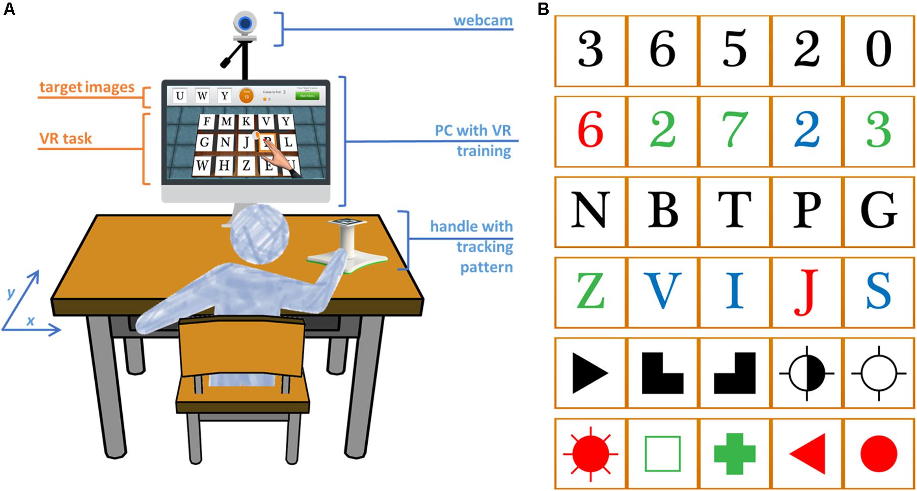
    Stroke is one of the most common causes of acquired disability, leaving numerous adults with cognitive and motor impairments, and affecting patients’ capability to live independently. Virtual Reality (VR) based methods for stroke rehabilitation have mainly focused on motor rehabilitation but there is increasing interest toward the integration of cognitive training for providing more effective solutions. Here we investigate the feasibility for stroke recovery of a virtual cognitive-motor task, the Reh@Task, which combines adapted arm reaching, and attention and memory training. 24 participants in the chronic stage of stroke, with cognitive and motor deficits, were allocated to one of two groups (VR, Control). Both groups were enrolled in conventional occupational therapy, which mostly involves motor training. Additionally, the VR group underwent training with the Reh@Task and the control group performed time-matched conventional occupational therapy. Motor and cognitive competences were assessed at baseline, end of treatment (1 month) and at a 1-month follow-up through the Montreal Cognitive Assessment, Single Letter Cancelation, Digit Cancelation, Bells Test, Fugl-Meyer Assessment Test, Chedoke Arm and Hand Activity Inventory, Modified Ashworth Scale, and Barthel Index. Our results show that both groups improved in motor function over time, but the Reh@Task group displayed significantly higher between-group outcomes in the arm subpart of the Fugl-Meyer Assessment Test. Improvements in cognitive function were significant and similar in both groups. Overall, these results are supportive of the viability of VR tools that combine motor and cognitive training, such as the Reh@Task. Trial Registration:This trial was not registered because it is a small clinical study that addresses the feasibility of a prototype device.

    Introduction

    Stroke is one of the most common causes of adult disability and its prevalence is likely to increase with an aging population (WHO, 2015). It is estimated that 33–42% of stroke survivors require assistance for daily living activities 3–6 months post-stroke and 36% continue to be disabled 5 years later (Teasell et al., 2012). Loss of motor control and muscle strength of the upper extremity are the most prevalent deficits and are those that have a greater impact on functional capacity (Saposnik, 2016). Hence, its recovery is fundamental for minimizing long-term disability and improving quality of life. In fact, most rehabilitation interventions focus on facilitating recovery through motor learning principles (Kleim and Jones, 2008). However, learning engages also cognitive processes such as attention, memory and executive functioning, all of which may be affected by stroke (Cumming et al., 2013). Still, conventional rehabilitation methodologies are mostly motor focused, although 70% of patients experience some degree of cognitive decline (Gottesman and Hillis, 2010), which also affects their capability to live independently (Langhorne et al., 2011).[…]


    Continue —> Frontiers | Combined Cognitive-Motor Rehabilitation in Virtual Reality Improves Motor Outcomes in Chronic Stroke – A Pilot Study | Psychology

    FIGURE 1. Experimental setup and VR task. (A) The user works on a tabletop and arm movements are captured by augmented reality pattern tracking. These movements are mapped onto the movements of a virtual arm on the screen for the execution of the cancelation task. (B) The target stimuli can be letters, numbers, and symbols in black or different colors. The target stimuli in this picture are ordered by increasing complexity.

    , , , , , , ,

    Leave a comment