Archive for April, 2019

[BOOK] The Comorbidities of Epilepsy – Google Books

The Comorbidities of Epilepsy

Front Cover
Marco Mula
Academic PressApr 20, 2019 – Medical – 413 pages

Epilepsy is one of most frequent neurological disorders affecting about 50 million people worldwide and 50% of them have at least another medical problem in comorbidity; sometimes this is a the cause of the epilepsy itself or it is due to shared neurobiological links between epilepsy and other medical conditions; other times it is a long-term consequence of the antiepileptic drug treatment.

The Comorbidities of Epilepsy offers an up-to-date, comprehensive overview of all comorbidities of epilepsy (somatic, neurological and behavioral), by international authorities in the field of clinical epileptology, with an emphasis on epidemiology, pathophysiology, diagnosis and management. This book includes also a critical appraisal of the methodological aspects and limitations of current research on this field. Pharmacological issues in the management of comorbidities are discussed, providing information on drug dosages, side effects and interactions, in order to enable the reader to manage these patients safely.

The Comorbidities of Epilepsy is aimed at all health professionals dealing with people with epilepsy including neurologists, epileptologists, psychiatrists, clinical psychologists, epilepsy specialist nurses and clinical researchers.

  • Provides a comprehensive overview of somatic, neurological and behavioral co-morbidities of epilepsy
  • Discusses up-to-date management of comorbidities of epilepsy
  • Written by a group of international experts in the field


via The Comorbidities of Epilepsy – Google Books

, , , , ,

Leave a comment

[NEWS] Brain stimulation improves depression symptoms, restores brain waves in clinical study — ScienceDaily

Date: March 11, 2019

Source: University of North Carolina Health Care

Summary: With a weak alternating electrical current sent through electrodes attached to the scalp, researchers successfully targeted a naturally occurring electrical pattern in a specific part of the brain and markedly improved depression symptoms in about 70 percent of participants in a clinical study.

FULL STORY

With a weak alternating electrical current sent through electrodes attached to the scalp, UNC School of Medicine researchers successfully targeted a naturally occurring electrical pattern in a specific part of the brain and markedly improved depression symptoms in about 70 percent of participants in a clinical study.

The research, published in Translational Psychiatry, lays the groundwork for larger research studies to use a specific kind of electrical brain stimulation called transcranial alternating current stimulation (tACS) to treat people diagnosed with major depression.

“We conducted a small study of 32 people because this sort of approach had never been done before,” said senior author Flavio Frohlich, PhD, associate professor of psychiatry and director of the Carolina Center for Neurostimulation. “Now that we’ve documented how this kind of tACS can reduce depression symptoms, we can fine tune our approach to help many people in a relatively inexpensive, noninvasive way.”

Frohlich, who joined the UNC School of Medicine in 2011, is a leading pioneer in this field who also published the first clinical trials of tACS in schizophrenia and chronic pain.

His tACS approach is unlike the more common brain stimulation technique called transcranial direct stimulation (tDCS), which sends a steady stream of weak electricity through electrodes attached to various parts of the brain. That approach has had mixed results in treating various conditions, including depression. Frohlich’s tACS paradigm is newer and has not been investigated as thoroughly as tDCS. Frohlich’s approach focuses on each individual’s specific alpha oscillations, which appear as waves between 8 and 12 Hertz on an electroencephalogram (EEG). The waves in this range rise in predominance when we close our eyes and daydream, meditate, or conjure ideas — essentially when our brains shut out sensory stimuli, such as what we see, feel, and hear.

Previous research showed that people with depression featured imbalanced alpha oscillations; the waves were overactive in the left frontal cortex. Frohlich thought his team could target these oscillations to bring them back in synch with the alpha oscillations in the right frontal cortex. And if Frohlich’s team could achieve that, then maybe depression symptoms would be decreased.

His lab recruited 32 people diagnosed with depression and surveyed each participant before the study, according to the Montgomery-Åsberg Depression Rating Scale (MADRS), a standard measure of depression.

The participants were then separated into three groups. One group received the sham placebo stimulation — a brief electrical stimulus to mimic the sensation at the beginning of a tACS session. A control group received a 40-Hertz tACS intervention, well outside the range that the researchers thought would affect alpha oscillations. A third group received the treatment intervention — a 10-Hertz tACS electrical current that targeted each individual’s naturally occurring alpha waves. Each person underwent their invention for 40 minutes on five consecutive days. None of the participants knew which group they were in, and neither did the researchers, making this a randomized double-blinded clinical study — the gold standard in biomedical research. Each participant took the MADRS immediately following the five-day regimen, at two weeks, and again at four weeks.

Prior to the study, Frohlich set the primary outcome at four weeks, meaning that the main goal of the study was to assess whether tACS could bring each individual’s alpha waves back into balance and decrease symptoms of depression four weeks after the five-day intervention. He set this primary outcome because scientific literature on the study of tDCS also used the four-week mark.

Frohlich’s team found that participants in the 10-Hertz tACS group featured a decrease in alpha oscillations in the left frontal cortex; they were brought back in synch with the right side of the frontal cortex. But the researchers did not find a statistically significant decrease in depression symptoms in the 10-Hertz tACS group, as opposed to the sham or control groups at four weeks.

But when Frohlich’s team looked at data from two weeks after treatment, they found that 70 percent of people in the treatment group reported at least a 50 percent reduction of depression symptoms, according to their MADRS scores. This response rate was significantly higher than the one for the two other control groups. A few of the participants had such dramatic decreases that Frohlich’s team is currently writing case-studies on them. Participants in the placebo and control groups experienced no such reduction in symptoms.

“It’s important to note that this is a first-of-its kind study,” Frohlich said. “When we started this research with computer simulations and preclinical studies, it was unclear if we would see an effect in people days after tACS treatment — let alone if tACS could become a treatment for psychiatric illnesses. It was unclear what would happen if we treated people several days in a row or what effect we might see weeks later. So, the fact that we’ve seen such positive results from this study gives me confidence our approach could help many people with depression.”

Frohlich’s lab is currently recruiting for two similar follow-up studies.

Other authors of the Translational Psychiatry paper are co-first authors Morgan Alexander, study coordinator and graduate student, and Sankaraleengam Alagapan, PhD, a postdoctoral fellow, both in the department of psychiatry at UNC-Chapel Hill; David Rubinow, MD, the Assad Meymandi Distinguished Professor and Chair of Psychiatry at the UNC School of Medicine; former UNC postdoctoral fellow Caroline Lustenberger, PhD; and Courtney Lugo and Juliann Mellin, both study coordinators at the UNC School of Medicine.

This research was funded through grants from the Brain Behavior Research Foundation, National Institutes of Health, the BRAIN Initiative, and the Foundation of Hope.

Frohlich holds joint appointments at UNC-Chapel Hill in the department of cell biology and physiology and the Joint UNC-NC State Department of Biomedical Engineering. He is also a member of the UNC Neuroscience Center.

Story Source:

Materials provided by University of North Carolina Health CareNote: Content may be edited for style and length.


Journal Reference:

  1. Morgan L. Alexander, Sankaraleengam Alagapan, Courtney E. Lugo, Juliann M. Mellin, Caroline Lustenberger, David R. Rubinow, Flavio Fröhlich. Double-blind, randomized pilot clinical trial targeting alpha oscillations with transcranial alternating current stimulation (tACS) for the treatment of major depressive disorder (MDD)Translational Psychiatry, 2019; 9 (1) DOI: 10.1038/s41398-019-0439-0


via Brain stimulation improves depression symptoms, restores brain waves in clinical study — ScienceDaily

, , , ,

Leave a comment

[REVIEW ARTICLE] Blood Biomarkers for Traumatic Brain Injury: A Quantitative Assessment of Diagnostic and Prognostic Accuracy – Full Text

Blood biomarkers have been explored for their potential to provide objective measures in the assessment of traumatic brain injury (TBI). However, it is not clear which biomarkers are best for diagnosis and prognosis in different severities of TBI. Here, we compare existing studies on the discriminative abilities of serum biomarkers for four commonly studied clinical situations: detecting concussion, predicting intracranial damage after mild TBI (mTBI), predicting delayed recovery after mTBI, and predicting adverse outcome after severe TBI (sTBI). We conducted a literature search of publications on biomarkers in TBI published up until July 2018. Operating characteristics were pooled for each biomarker for comparison. For detecting concussion, 4 biomarker panels and creatine kinase B type had excellent discriminative ability. For detecting intracranial injury and the need for a head CT scan after mTBI, 2 biomarker panels, and hyperphosphorylated tau had excellent operating characteristics. For predicting delayed recovery after mTBI, top candidates included calpain-derived αII-spectrin N-terminal fragment, tau A, neurofilament light, and ghrelin. For predicting adverse outcome following sTBI, no biomarker had excellent performance, but several had good performance, including markers of coagulation and inflammation, structural proteins in the brain, and proteins involved in homeostasis. The highest-performing biomarkers in each of these categories may provide insight into the pathophysiologies underlying mild and severe TBI. With further study, these biomarkers have the potential to be used alongside clinical and radiological data to improve TBI diagnostics, prognostics, and evidence-based medical management.

Introduction

Traumatic brain injury (TBI) is a common cause of disability and mortality in the US (1) and worldwide (2). Pathological responses to TBI in the CNS include structural and metabolic changes, as well as excitotoxicity, neuroinflammation, and cell death (34). Fluid biomarkers that may track these injury and inflammatory processes have been explored for their potential to provide objective measures in TBI assessment. However, at present there are limited clinical guidelines available regarding the use of biomarkers in both the diagnosis of TBI and outcome prediction following TBI. To inform future guideline formulation, it is critical to distinguish between different clinical situations for biomarker use in TBI, such as detection of concussion, prediction of positive and negative head computed tomography (CT) findings, and prediction of outcome for different TBI severities. This allows for comparisons to determine which biomarkers may be used most appropriately to characterize different aspects of TBI.

The identification of TBI severity has become a contentious issue. Currently, inclusion in TBI clinical trials is primarily based on the Glasgow Coma Scale (GCS), which stratifies patients into categories of mild, moderate, and severe TBI. The GCS assesses consciousness and provides prognostic information, but it does not inform the underlying pathologies that may be targeted for therapy (56). Furthermore, brain damage and persistent neurological symptoms can occur across the spectrum of TBI severity, limiting the use of GCS-determined injury severity to inform clinical management. Biomarkers in TBI have the potential to provide objective and quantitative information regarding the pathophysiologic mechanisms underlying observed neurological deficits. Such information may be more appropriate for guiding management than initial assessments of severity alone. Since the existing literature primarily focuses on applications of biomarkers in either suspected concussion, mild TBI (mTBI), or severe TBI (sTBI), we will discuss biomarker usage in these contexts.

Concussion is a clinical syndrome involving alteration in mental function induced by head rotational acceleration. This may be due to direct impact or unrestrained rapid head movements, such as in automotive crashes. Although there are over 30 official definitions of concussion, none include the underlying pathology. Missing from the literature have been objective measures to not only identify the underlying pathology associated with the given clinical symptoms, but also to indicate prognosis in long-term survival. Indeed, current practices in forming an opinion of concussion involve symptom reports, neurocognitive testing, and balance testing, all of which have elements of subjectivity and questionable reliability (7). While such information generally reflects functional status, it does not identify any underlying processes that may have prognostic or therapeutic consequences. Furthermore, because patients with concussion typically present with negative head CT findings, there is a potential role for blood-based biomarkers to provide objective information regarding the presence of concussion, based on an underlying pathology. This information could inform management decisions regarding resumption of activities for both athletes and non-athletes alike.

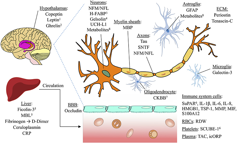
Blood-based biomarkers have utility far beyond a simple detection of concussion by elucidating specific aspects of the injury that could drive individual patient management. For example, biomarkers may aid in determining whether a mTBI patient presenting to the emergency department requires a CT scan to identify intracranial pathology. The clinical outcome for a missed epidural hematoma in which the patient is either discharged or admitted for routine observation is catastrophic; 25% are left severely impaired or dead (8). The Canadian CT Head Rule (9) and related clinical decision instruments achieve high sensitivities in predicting the need for CT scans in mild TBI cases. However, they do this at specificities of only 30–50% (10). Adding a blood biomarker to clinical evaluation may be useful to improve specificity without sacrificing sensitivity, as recently suggested (11). In addition, given concern about radiation exposure from head CT scans in concussion cases, particularly in pediatric populations, identification of patients who would be best assessed with neuroimaging is crucial. Thus, the use of both sensitive and specific biomarkers may serve as cost-effective tools to aid in acute assessment, especially in the absence of risk factors for intracranial injury (12). S-100B, an astroglial protein, has been the most extensively studied biomarker for TBI thus far and has been incorporated into some clinical guidelines for CT scans (1314). However, S-100B is not CNS-specific (1516) and has shown inconsistent predictive capacity in the outcome of mild TBI (1718). Given that several other promising biomarkers have also been investigated in this context, it is important to evaluate and compare the discriminative abilities of S-100B with other candidate blood-based biomarkers for future use.

Blood biomarkers also have the potential to help predict unfavorable outcomes across the spectrum of TBI severity. Outcome predication is difficult; in mTBI, existing prognostic models performed poorly in an external validation study (19). Identifying biomarkers that best predict delayed recovery or persistent neurological symptoms following mTBI would help with the direction of resources toward patients who may benefit most from additional rehabilitation or prolonged observation. In sTBI, poorer outcome has often been associated with a low GCS score (20). However, factors such as intoxication or endotracheal intubation may make it difficult to assess GCS reliably in the acute setting (2122). The addition of laboratory parameters to head CT and admission characteristics have improved prognostic models (23). Thus, prognostic biomarkers in sTBI could help determine whether patients are likely to benefit from intensive treatment. Several candidate biomarkers that correlate with various pathologies of mild and severe TBI have been studied (24), but their relative prognostic abilities remain unclear.

Existing reviews on biomarkers in TBI have provided valuable insight into the pathologic correlates of biomarkers, as well as how biomarkers may be used for diagnosis and prognosis (2531). However, there has been no previous quantitative comparison of the literature regarding biomarkers’ discriminative abilities in specific clinical situations. Here, we compare existing studies on the discriminative abilities of serum biomarkers for four commonly studied clinical situations: detecting concussion, predicting intracranial damage after mTBI, predicting delayed recovery after mTBI, and predicting adverse outcome after sTBI.[…]


Continue —-> Frontiers | Blood Biomarkers for Traumatic Brain Injury: A Quantitative Assessment of Diagnostic and Prognostic Accuracy | Neurology

Figure 2. Anatomical locations of potential TBI biomarkers. The biomarkers included in this schematic all rated as “good” (AUC=0.800.89) or better for any of the four clinical situations studied (detecting concussion, predicting intracranial damage after concussion, predicting delayed recovery after concussion, and predicting adverse outcome after severe TBI). Biomarkers with a pooled AUC <0.8 are not shown. 1Also found in adipose tissue; 2synthesized in cells of stomach and pancreas; may regulate HPA axis; 3found mostly in pons; 4also found extracellularly; 5lectin pathway of the complement system; 6also found in endothelial cells. BBB, blood brain barrier. ECM, Extracellular matrix. Image licensed under Creative Commons Attribution-ShareAlike 4.0 International license. https://creativecommons.org/licenses/by-sa/4.0/deed.en. See Supplementary Material for image credits and licensing.

, , , , , , ,

Leave a comment

[REVIEW ARTICLE] Robot-Assisted Therapy in Upper Extremity Hemiparesis: Overview of an Evidence-Based Approach – Full Text

Robot-mediated therapy is an innovative form of rehabilitation that enables highly repetitive, intensive, adaptive, and quantifiable physical training. It has been increasingly used to restore loss of motor function, mainly in stroke survivors suffering from an upper limb paresis. Multiple studies collated in a growing number of review articles showed the positive effects on motor impairment, less clearly on functional limitations. After describing the current status of robotic therapy after upper limb paresis due to stroke, this overview addresses basic principles related to robotic therapy applied to upper limb paresis. We demonstrate how this innovation is an evidence-based approach in that it meets both the improved clinical and more fundamental knowledge-base about regaining effective motor function after stroke and the need of more objective, flexible and controlled therapeutic paradigms.

Introduction

Robot-mediated rehabilitation is an innovative exercise-based therapy using robotic devices that enable the implementation of highly repetitive, intensive, adaptive, and quantifiable physical training. Since the first clinical studies with the MIT-Manus robot (1), robotic applications have been increasingly used to restore loss of motor function, mainly in stroke survivors suffering from an upper limb paresis but also in cerebral palsy (2), multiple sclerosis (3), spinal cord injury (4), and other disease types. Thus, multiple studies suggested that robot-assisted training, integrated into a multidisciplinary program, resulted in an additional reduction of motor impairments in comparison to usual care alone in different stages of stroke recovery: namely, acute (57), subacute (18), and chronic phases after the stroke onset (911). Typically, patients engaged in the robotic therapy showed an impairment reduction of 5 points or more in the Fugl-Meyer assessment as compared to usual care. Of notice, rehabilitation studies conducted during the chronic stroke phase suggest that a 5-point differential represents the minimum clinically important difference (MCID), i.e., the magnitude of change that is necessary to produce real-world benefits for patients (12). These results were collated in multiple review articles and meta-analyses (1317). In contrast, the advantage of robotic training over usual care in terms of functional benefit is less clear, but there are recent results that suggest how best to organize training to achieve superior results in terms of both impairment and function (18). Indeed, the use of the robotic tool has allowed us the parse and study the ingredients that should form an efficacious and efficient rehabilitation program. The aim of this paper is to provide a general overview of the current state of robotic training in upper limb rehabilitation after stroke, to analyze the rationale behind its use, and to discuss our working model on how to more effectively employ robotics to promote motor recovery after stroke.

Upper Extremity Robotic Therapy: Current Status

Robotic systems used in the field of neurorehabilitation can be organized under two basic categories: exoskeleton and end-effector type robots. Exoskeleton robotic systems allow us to accurately determine the kinematic configuration of human joints, while end-effector type robots exert forces only in the most distal part of the affected limb. A growing number of commercial robotic devices have been developed employing either configuration. Examples of exoskeleton type include the Armeo®Spring, Armeo®Power, and Myomo® and of end-effector type include the InMotion™, Burt®, Kinarm™ and REAplan®. Both categories enable the implementation of intensive training and there are many other devices in different stages of development or commercialization (1920).

The last decade has seen an exponential growth in both the number of devices as well as clinical trials. The results coalesced in a set of systematic reviews, meta-analyses (1317) and guidelines such as those published by the American Heart Association and the Veterans Administration (AHA and VA) (21). There is a clear consensus that upper limb therapy using robotic devices over 30–60-min sessions, is safe despite the larger number of movement repetitions (14).

This technic is feasible and showed a high rate of eligibility; in the VA ROBOTICS (911) study, nearly two thirds of interviewed stroke survivors were enrolled in the study. As a comparison the EXCITE cohort of constraint-induced movement therapy enrolled only 6% of the screened patients participated (22). On that issue, it is relevant to notice the admission criteria of both chronic stroke studies. ROBOTICS enrolled subjects with Fugl-Meyer assessment (FMA) of 38 or lower (out of 66) while EXCITE typically enrolled subjects with an FMA of 42 or higher. Duret and colleagues demonstrated that the target population, based on motor impairments, seems to be broader in the robotic intervention which includes patients with severe motor impairments, a group that typically has not seen much benefit from usual care (23). Indeed, Duret found that more severely impaired patients benefited more from robot-assisted training and that co-factors such as age, aphasia, and neglect had no impact on the amount of repetitive movements performed and were not contraindicated. Furthermore, all patients enrolled in robotic training were satisfied with the intervention. This result is consistent with the literature (24).

The main outcome result is that robotic therapy led to significantly more improvement in impairment as compared to conventional usual care, but only slightly more on motor function of the limb segments targeted by the robotic device (16). For example, Bertani et al. (15) and Zhang et al. (17) found that robotic training was more effective in reducing motor impairment than conventional usual care therapy in patients with chronic stroke, and further meta-analyses suggested that using robotic therapy as an adjunct to conventional usual care treatment is more effective than robotic training alone (1317). Other examples of disproven beliefs: many rehabilitation professionals mistakenly expected significant increase of muscle hyperactivity and shoulder pain due to the intensive training. Most studies showed just the opposite, i.e., that intensive robotic training was associated with tone reduction as compared to the usual care groups (92526). These results are shattering the resistance to the widespread adoption of robotic therapy as a therapeutic modality post-stroke.

That said, not all is rosy. Superior changes in functional outcomes were more controversial until the very last years as most studies and reviews concluded that robotic therapy did not improve activities of daily living beyond traditional care. One first step was reached in 2015 with Mehrholz et al. (14), who found that robotic therapy can provide more functional benefits when compared to other interventions however with a quality of evidence low to very low. 2018 may have seen a decisive step in favor of robotic as the latest meta-analysis conducted by Mehrholz et al. (27) concluded that robot-assisted arm training may improve activities of daily living in the acute phase after stroke with a high quality of evidence However, the results must be interpreted with caution because of the high variability in trial designs as evidenced by the multicenter study (28) in which robotic rehabilitation using the Armeo®Spring, a non-motorized device, was compared to self-management with negative results on motor impairments and potential functional benefits in the robotic group.

The Robot Assisted Training for the Upper Limb after Stroke (RATULS) study (29) might clarify things and put everyone in agreement on the topic. Of notice, RATULS goes beyond the Veterans Administration ROBOTICS with chronic stroke or the French REM_AVC study with subacute stroke. RATULS included 770 stroke patients and covered all stroke phases, from acute to chronic, and it included a positive meaningful control in addition to usual care.[…]


Continue —->  Frontiers | Robot-Assisted Therapy in Upper Extremity Hemiparesis: Overview of an Evidence-Based Approach | Neurology

, , , , , , , , , , ,

Leave a comment

[WEB SITE] How to Help Patients in Wheelchairs Express Their Personal Style – Rehab Managment

Published on 

disabled-wheelchair

By Rae Steinbach

A physical therapist doesn’t merely help someone recover from an injury or accident by improving their mobility. Physical therapists also pay attention to the emotional experiences of those with whom they work.

For instance, people who have recently begun to use wheelchairs often struggle with depression. Their inability to live a fully independent life can result in negative self-talk. Ideally, a physical therapist working with such a patient would notice their mood and identify ways to provide support.

Marla Ranieri of BetterPT further emphasizes this point, sharing that, “Being able to integrate a person back to their community and social activities physically and mentally is one of the most rewarding aspects of being a physical therapist. We provide them the tools they need to enjoy life again.”

Offering fashion tips such as these is one way they can help. Liking what they see in the mirror can help those in wheelchairs start to feel more confident in themselves and their appearance. The problem is, a person who isn’t accustomed to using a wheelchair may struggle to dress well if this experience is new to them.

That doesn’t need to be the case. If you’re a physical therapist working with patients in wheelchairs, help them be more fashionable by offering this key advice:

Wear a Stylish Belt

Long dresses or jackets that look impressive when a person wearing them is standing can get bunched up when a person is sitting down. Luckily, someone in a wheelchair can still enjoy these types of garments. They simply need to add a belt or waistband to the outfit. This smooths out tops and creates a more tailored look.

Choose Form-Fitting Clothing

Again, wheelchair users can wear longer dresses and jackets if they wish. It may even be advisable. Longer tops create the illusion of a longer torso.

That said, wheelchair users need to make sure their tops aren’t too loose. Too much fabric will look boxy when the person wearing such garments is seated. It’s smarter to choose form-fitting clothing. Additionally, excess fabric can get caught in wheelchair components, making it difficult for a person to easily use their wheelchair.

Display Your Shoulders

Dressing to impress is a lot easier for wheelchair users than some may realize. It’s often as simple as wearing a shoulder-baring top. This is a subtle but effective way to make an outfit a bit more daring.

Don’t Overlook Accessories

Learning how to adjust your personal style appropriately if you’ve recently begun using a wheelchair is a process. Although the advice you give patients will help, there are still likely to be instances when they’re not happy with the way a certain outfit looks.

Encourage them to accessorize when this happens. Adding the right accessories can transform a dull outfit into something much more remarkable. Consider recommending a summer subscription box or one that coincides with the approaching season. That way, your patient is sure to have new accessories and styling pieces that excite them throughout the year.

Again, these are important points for physical therapists to keep in mind. You have the chance to help people in wheelchairs feel much more confident. Providing this type of advice will help.

Rae Steinbach is a graduate of Tufts University with a combined International Relations and Chinese degree. Rae is passionate about travel, food, and writing for Jetty.


via How to Help Patients in Wheelchairs Express Their Personal Style – Rehab Managment

, ,

Leave a comment

[Abstract] The effectiveness of somatosensory retraining for improving sensory function in the arm following stroke: a systematic review

The aim of this study was to evaluate if somatosensory retraining programmes assist people to improve somatosensory discrimination skills and arm functioning after stroke.

Nine databases were systematically searched: Medline, Cumulative Index to Nursing and Allied Health Literature, PsychInfo, Embase, Amed, Web of Science, Physiotherapy Evidence Database, OT seeker, and Cochrane Library.

Studies were included for review if they involved (1) adult participants who had somatosensory impairment in the arm after stroke, (2) a programme targeted at retraining somatosensation, (3) a primary measure of somatosensory discrimination skills in the arm, and (4) an intervention study design (e.g. randomized or non-randomized control designs).

A total of 6779 articles were screened. Five group trials and five single case experimental designs were included (N = 199 stroke survivors). Six studies focused exclusively on retraining somatosensation and four studies focused on somatosensation and motor retraining. Standardized somatosensory measures were typically used for tactile, proprioception, and haptic object recognition modalities. Sensory intervention effect sizes ranged from 0.3 to 2.2, with an average effect size of 0.85 across somatosensory modalities. A majority of effect sizes for proprioception and tactile somatosensory domains were greater than 0.5, and all but one of the intervention effect sizes were larger than the control effect sizes, at least as point estimates. Six studies measured motor and/or functional arm outcomes (n = 89 participants), with narrative analysis suggesting a trend towards improvement in arm use after somatosensory retraining.

Somatosensory retraining may assist people to regain somatosensory discrimination skills in the arm after stroke.

via The effectiveness of somatosensory retraining for improving sensory function in the arm following stroke: a systematic review – Megan L Turville, Liana S Cahill, Thomas A Matyas, Jannette M Blennerhassett, Leeanne M Carey, 2019

, , , , , ,

Leave a comment

[ARTICLE] Social cognition and emotion regulation: a multifaceted treatment (T-ScEmo) for patients with traumatic brain injury – Full Text

Many patients with moderate to severe traumatic brain injury have deficits in social cognition. Social cognition refers to the ability to perceive, interpret, and act upon social information. Few studies have investigated the effectiveness of treatment for impairments of social cognition in patients with traumatic brain injury. Moreover, these studies have targeted only a single aspect of the problem. They all reported improvements, but evidence for transfer of learned skills to daily life was scarce. We evaluated a multifaceted treatment protocol for poor social cognition and emotion regulation impairments (called T-ScEmo) in patients with traumatic brain injury and found evidence for transfer to participation and quality of life.

In the current paper, we describe the theoretical underpinning, the design, and the content of our treatment of social cognition and emotion regulation (T-ScEmo).

The multifaceted treatment that we describe is aimed at improving social cognition, regulation of social behavior and participation in everyday life. Some of the methods taught were already evidence-based and derived from existing studies. They were combined, modified, or extended with newly developed material.

T-ScEmo consists of 20 one-hour individual sessions and incorporates three modules: (1) emotion perception, (2) perspective taking and theory of mind, and (3) regulation of social behavior. It includes goal-setting, psycho-education, function training, compensatory strategy training, self-monitoring, role-play with participation of a significant other, and homework assignments.

It is strongly recommended to offer all three modules, as they build upon each other. However, therapists can vary the time spent per module, in line with the patients’ individual needs and goals. In future, development of e-learning modules and virtual reality sessions might shorten the treatment.

Traumatic brain injury refers to a brain lesion caused by an external mechanical force, leading not only to physical impairments and cognitive deficits, but also to changes in behavior and personality.1,2 Especially after damage to orbitofrontal and ventromedial prefrontal brain areas, deficits in social cognition can occur.3,4

According to Adolphs,5 social cognition consists of three stages: (1) the ability to perceive social information (i.e. emotional facial expressions, bodily language), (2) the capacity to process and interpret social information (i.e. theory of mind, perspective taking), and (3) the ability to adapt behavior in accordance with the situation. Babbage et al.6 estimated that 13%–39% of individuals with moderate to severe traumatic brain injury experienced emotion perception deficits and up to 70% reported low empathy.79

Deficits in social cognition often appear in the shape of socially inadequate behavior, such as disinhibited or indifferent emotional behavior.1012 Such behaviors have detrimental consequences for the ability of patients to establish and maintain social relationships, to hold jobs, and to participate in society.1,13,14 It has been found that poor theory of mind and behavioral problems significantly predict poor participation and community integration.15,16For all these reasons, it is important to provide a tailored rehabilitation treatment, in order to prevent an unfavorable outcome.

In their review of cognitive rehabilitation, Cicerone et al.17 stressed the need to provide detailed information about the theoretical base, the protocol design, and the ingredients of a treatment, as a prerequisite to analyze its effectiveness. In the current paper, we give a comprehensive description of the treatment of social cognition and emotion regulation protocol (T-ScEmo). The effectiveness of T-ScEmo was evaluated in 59 patients with traumatic brain injury. It was compared with a computerized control treatment in a randomized controlled trial.18 Compared to the control treatment, T-ScEmo resulted in significant improvements in emotion recognition, theory of mind, emphatic behavior, quality of life partner relationship, quality of life and societal participation, up to five months posttreatment. Patients with traumatic brain injury as well as their life partners were satisfied with the treatment.18 A detailed description of the T-ScEmo protocol is relevant for researchers and clinical therapists; they can use, replicate, or expand this newly developed treatment.[…]


Continue —-> Social cognition and emotion regulation: a multifaceted treatment (T-ScEmo) for patients with traumatic brain injury – Herma J Westerhof-Evers, Annemarie C Visser-Keizer, Luciano Fasotti, Jacoba M Spikman, 2019


                        figure

Figure 1. Thoughts–feelings–behavior scheme (module 2).

, , , , ,

Leave a comment

[Abstract] Attention-controlled assistive wrist rehabilitation using a low-cost EEG Sensor

Abstract

It is essential to make sure patients be actively involved in motor training using robot-assisted rehabilitation to achieve better rehabilitation outcomes. This paper introduces an attention-controlled wrist rehabilitation method using a low-cost EEG sensor. Active rehabilitation training is realized using a threshold of the attention level measured by the low-cost EEG sensor as a switch for a flexible wrist exoskeleton assisting wrist ?exion/extension and radial/ulnar deviation. We present a prototype implementation of this active training method and provide a preliminary evaluation. The feasibility of the attention-based control was proven with the overall actuation success rate of 95%. The experimental results also proved that the visual guidance was helpful for the users to concentrate on the wrist rehabilitation training; two types of visual guidance, namely looking at the hand motion shown on a video and looking at the user’s own hand, had no significant performance difference; a general threshold of a certain group of users can be utilized in the wrist robot control rather than a customized threshold to simplify the procedure.

via Attention-controlled assistive wrist rehabilitation using a low-cost EEG Sensor – IEEE Journals & Magazine

, , , , , , , ,

Leave a comment

[WEB PAGE] When Will There Ever be a Cure for Epilepsy?

The three-pound organ that serves as command central for the human organism is certainly a marvel, just by virtue of the fact that the brain can appreciate its own awesomeness, even if it hasn’t quite perfected the flying car or even self-driving cars. Yet. Companies developing brain-computer interface technology are enabling humans to do things like send commands to computers by just flexing a bit of muscle. Still, there is much we don’t know about ourselves, no matter how much telepsychiatry we do. And that applies especially to medical conditions that affect the brain like epilepsy, a neurological condition for which there is no cure.

What is Epilepsy?

While most of us are probably familiar with some Hollywood-ized version of epilepsy in which someone starts flailing around after being hit by strobe lights on the disco floor, the reality is that epilepsy refers to a large group of neurological disorders that generally involve chronic, spontaneous seizures that vary greatly in how they manifest. The causes of epilepsy are also all over the place, from traumatic brain injuries and stroke to viral and bacterial infections to genetics.

A new set of classifications for epilepsy came out in 2017.

It is considered a brain disorder, according to the U.S. Centers for Disease Control (CDC), though some researchers have suggested it could be classified as a neurodegenerative disease like Parkinson’s or Alzheimer’s. In fact, there is research that suggests a genetic link between epilepsy and neurodegenerative diseases.

Not surprisingly, many of the companies developing therapies for neurodegenerative diseases are also working on treatments for epilepsy and vice versa. For example, a new, well-funded joint venture involving Pfizer (PFE) and Bain Capital called Cerevel, which we profiled in our piece on Parkinson’s disease, is also in advanced clinical trials for an epileptic drug. Its GABA A positive modulator drug candidate targets GABA (Gamma-Aminobutyric Acid) neurotransmitters that block impulses between nerve cells in the brain, helping keep the nervous system chill.

Impacts of Epilepsy

More than 50 million people worldwide have epilepsy, making it one of the most common neurological diseases globally, according to the World Health Organization (WHO). The CDC estimates about 3.4 million Americans live with the condition. Globally, an estimated 2.4 million people are diagnosed with epilepsy each year. Interestingly, the disorder seems to target those who can least afford it: WHO said nearly 80% of people with epilepsy live in low- and middle-income countries.

Impacts of epilepsy graphic

A 2015 study of a bunch of other studies that estimated the cost of epilepsy in the United States found that epilepsy-specific costs probably average out to about $10,000 based on the variety of ranges, which means epilepsy costs the United States healthcare system about $34 billion, though the numbers are widely debated. Conversely, WHO says low-cost treatments are available, with daily medication coming as cheaply as $5 per year, so another win for the U.S. healthcare system.

Treatments for Epilepsy

There are more than 20 antiepileptic drugs used to treat epilepsy, usually to help prevent or slow the occurrence of seizures. Other therapies include surgery and electroceutical treatment in which electrical stimulation is applied, usually to the vagus nerve, the longest cranial nerve in the body. While many find relief from one or more of these options, a third of those who suffer from epilepsy are not able to manage their seizures, according to the U.S. National Institutes of Health (NIH). Below we take a look at a range of innovative therapies designed to detect, stop, or find a cure for epilepsy.

Brain Stimulation Therapies

In our article on electroceutical treatments, we highlighted a London company called LivaNova (LIVN) that offers an implantable Vagus Nerve Stimulation (VNS) therapy that has been approved by the U.S. Food and Drug Administration (FDA) to help treat those with partial seizures who do not respond to seizure medications. A medical device company with a lengthy track record of returning value to investors, Medtronic (MDT) got FDA pre-market approval last year for its Deep Brain Stimulation (DBS) therapy for use in reducing partial-onset seizure for those who have proven to not respond to three or more antiepileptic medications. DBS therapy delivers controlled electrical pulses to an area in the brain called the anterior nucleus of the thalamus, which is part of a network involved in seizures. Yet another company offering a variation of brain stimulation therapy is NeuroPace, which markets its responsive neurostimulation device, or RNS system, as “the first and only brain-responsive neurostimulation system designed to prevent epileptic seizures at their source.”

Artificial Intelligence to Detect, Predict, and Control Epilepsy

The NIH is funding further research into implantable devices that can detect, predict, and stop a seizure before it happens, “working closely with industry partners to develop pattern-recognition algorithms,” which sounds an awful lot like artificial intelligence and machine learning will be at the forefront of some future diagnostics and treatment. AI in healthcare has been an ongoing theme around here, with a recent dive into AI and mental health. Back to AI and epilepsy: A group of neurologists at the Medical University of South Carolina developed a new method based on artificial intelligence to predict which patients will see success with surgical procedures designed to stop seizures. Sounds like a great idea to learn beforehand if it’s necessary to crack open your skull.

Click for company websiteA Boston area startup called Empatica, spun out from MIT in 2011, has raised $7.8 million for a smartwatch that detects possible seizures by monitoring subtle electrical changes across the surface of the skin. Other methods normally rely on electrical activity in the brain that tracks and records brain wave patterns called an electroencephalogram. Empatica’s seizure detection algorithm, on the other hand, can detect complex physiological patterns from electrodermal activity that is most likely to accompany a convulsive seizure. Psychology Today reportedthat the device, Embrace Watch, received FDA approval earlier this year for seizure control in children after getting the green light for the technology for adults in 2018.

The Empatica smartwatch can detect electrical currents in the skin to predict the onset of an epileptic seizure.

Click for company websiteAI and drug discovery for better epileptic drug candidates is yet another application that we would expect to see grow in the coming years. Silicon Valley-based startup System1 Biosciences raised $25 million last year, which included Pfizer among its dozen investors. System1 builds a sort of brain model for testing drug candidates using stem cell lines derived from patients with brain disease. The company uses robotic automation to develop these three-dimensional cerebral organoids, allowing it to generate huge datasets in a relatively short amount of time, then applying “advanced data analysis” (also AI?) to detect patterns that might match the characteristics of a neurological disease (what it refers to as deep phenotypes) such as epilepsy with novel treatments.

Cannabis for Controlling Seizures

We’ve written extensively about the suddenly booming hemp CBD market, noting that the FDA approved a CBD-based drug for epilepsy last year in our recent article on the best certified CBD oils on the market. However, we’ve only briefly profiled the company behind Epidiolex for treating rare forms of epilepsy, GW Pharmaceuticals (GWPH). Sporting a market cap just south of $5 billion, GW Pharmaceuticals has taken in about $300 million in post-IPO equity since our article, bringing total post-IPO equity funding to about $568 million. Aside from its successful epileptic drug, GW also developed the world’s first cannabis-based prescription medicine for the treatment of spasticity due to multiple sclerosis that is available in 25 countries outside the United States.

The forms of epilepsy that GW Pharmaceuticals can treat or can potentially treat.

Back on the epilepsy side, Epidiolex has been approved for two rare forms of epilepsy, with clinical trials underway for two more rare neurological disorders associated with seizures – tuberous sclerosis complex and Rett syndrome. Also in the pipeline is a drug dubbed CBDV (GWP42006) that’s also for treating epileptic seizures, though the results of a trial last year were not encouraging. The same compound is also being investigated for autism. Be sure to check out our article on Charlotte’s Web, a CBD company that came about because of epilepsy.

Helping Cells Get Their Vitamin K

Click for company websiteNeuroene Therapeutics is a small startup spun out of the Medical University of South Carolina that recently picked up $1.5 million in funding to tests its lead drug compounds, which are analogous to the naturally occurring form of vitamin K that is essential for brain health. In particular, the lab-developed vitamin K protects the integrity of the cell’s mitochondria, which serves as a sort of power plant for brain cells, helping the neural circuit fire better. Unfortunately, you can’t get the effect from simply eating a bowl of Special K each morning covered in an organic sugar substitute, so the company is developing a method to deliver the effects directly to the brain.

A Nasal Spray to Stop Seizures

Click for company websiteFounded in 2007 near San Diego, Neurelis licenses, develops, and commercializes treatments for epilepsy and other neurological diseases. It has raised $44.8 million in disclosed funding, most coming in a $40.5 million venture round last November. The company’s flagship product is called Valtoco, a formulation that incorporates diazepam, an existing drug used to control seizures and alcohol withdrawal, with a vitamin E-based solution that is delivered using a nasal spray when a sudden seizure episode occurs. The product uses an absorption enhancement technology called Intravail developed by another San Diego area company called Aegis Therapeutics that Neurelis acquired in December last year. Neurelis submitted Valtoco to the FDA for approval in September.

Conclusions

While many people with epileptic conditions can control their seizures with many of the current medications or other therapies available now, there’s a big chunk of the population that is living with uncertainty. Considering the strong link between neurological disorders like epilepsy and certain neurodegenerative disorders, expect to see some good synergies in the next five to 10 years, especially as automation and advanced analytics using AI start connecting the dots between genetics, biochemistry, and brain disorders.

via When Will There Ever be a Cure for Epilepsy? – Nanalyze

, , , , , , , , ,

Leave a comment

[NEWS] The brain-computer interface at UCLA – from the 1970s to today

Apr 19, 2019 By UCLA Samueli Newsroom

In 1973, UCLA computer science professor Jacques Vidal published a landmark paper, “Toward direct brain-computer communication” that both coined the term “brain-computer interface” and set the foundation for an emerging field.

“That whole concept of interacting with and sensing the brain – interpreting signals with a computer and controlling the cursor on a computer with the mind – that paper is pretty much the essence of it,” said Dejan Markovic, a professor of electrical and computer engineering and leader of the Parallel Data Architectures Laboratory. “The real question is: Can we build technologies that enable those types of things that are clinically sustainable, efficacious, and attractive to patients?”

Looking to answer that question, Markovic carries on the legacy of brain-computer interface research at the UCLA Samueli School of Engineering. For nearly a decade, he has been leading the development of a device that would be implanted in the brain to help people with a range of neurological conditions, such as anxiety, depression, or post-traumatic stress disorder.  And he’s been working closely with doctors and scientists at UCLA and UC San Francisco who study the brain.

“The concepts laid out in 1973 by Vidal haven’t changed too much,” he added. “The brain and a computer can ‘talk’ to each other through electrical signals. The big thing that we are trying to change is to be able to quantify what those signals are, and affect functional networks of the brain.”

Markovic’s prototype is a small implantable device with sixty-four electrodes that fan out onto the brain’s surface. With four modules for each electrode, it constitutes a 256-channel system. The system measures tiny electric signals that tell what’s happening in the brain. The device then interprets that data, and responds with electrical pulses, which research has shown can alter mood.

In several ways, it is leaps and bounds more advanced than implants that have come before it. It’s much smaller for one. In fact it’s not immediately noticeable, unless someone’s really looking for it. It has a tiny battery than can be wirelessly charged. The device is also much more sensitive, able to detect and decipher very faint signals from the brain.

Finally, it’s a closed loop system – meaning that while still picking up the brain’s signals, it can modify the frequency and amplitude of the stimulating signal. The system brings much more data into the loop, giving  doctors and scientists more information about what’s happening in real time . Other devices only deliver a constant electric signal, while this new system offers a therapy  that can be more personalized to a particular patient

“Our technology could revolutionize non-pharmacological treatment of brain disorders,” Markovic said. “We want to be able to understand how various indications are expressed in the actual time waveforms, from specific points inside the brain.”

Markovic and UC San Francisco colleagues saw a major breakthrough in an experiment, which was funded by the Defense Advanced Research Projects Agency. A patient with severe anxiety was recorded before and after electrical stimulation was applied. The change in mood following stimulation was immediate and striking.

“For a person to say, ‘now I feel normal, this is me,’ that was the biggest impact point,” he said.

With a series of successful demonstrations, Markovic is now looking to commercialize the technology.  This includes miniaturizing the external device down to just four cubic centimeters. But first, why go with a brain implant in the first place?

“The brain is an electrochemical organ and the vast majority of our treatments for neurological and psychiatric diseases focus on the chemical part,” explained Dr. Nader Pouratian, a UCLA neurosurgeon working with Markovic. “The goal with devices like the one that Dr. Markovic is creating is to target the electrical abnormalities that occur in the brain as a result of neurological and psychiatric disease.”

Added Markovic, “We are looking into patients that have tried pharmaceuticals. In some people, pharmaceuticals have some effect, but there are a sizeable amount of people where pharmaceuticals do not help.”

On a parallel track, Markovic’s technology also offers scientists a powerful magnifying glass into the inner workings of the brain. One of his collaborators is Nanthia Suthana, a UCLA assistant professor at the Jane and Terry Semel Institute for Neuroscience and Human Behavior who studies neuromodulation and neuroimaging.

“The research potential is really endless with such a device,” Suthana said. “Relevant to my own research field, we will be able to investigate the role of single neuron and local field potential activity in freely moving human behaviors such as in spatial navigation, learning and memory.”

“These newer details will allow us to better understand the neuronal mechanisms that support typical human brain functions as well as abnormalities that may occur in neurologic and psychiatric disorders such epilepsy,” she added.


via The brain-computer interface at UCLA – from the 1970s to today | UCLA Samueli School Of Engineering

,

Leave a comment