Archive for November, 2017

[BOOK Chapter] Hand Rehabilitation after Chronic Brain Damage: Effectiveness, Usability and Acceptance of Technological Devices: A Pilot Study – Full Text

By Marta Rodríguez-Hernández, Carmen Fernández-Panadero, Olga López-Martín and Begoña Polonio-López
DOI: 10.5772/67532

READ CHAPTER

Download as PDF

via Hand Rehabilitation after Chronic Brain Damage: Effectiveness, Usability and Acceptance of Technological Devices: A Pilot Study | InTechOpen


, , , , , , , ,

Leave a comment

[ARTICLE] The Optimal Speed for Cortical Activation of Passive Wrist Movements Performed by a Rehabilitation Robot: A Functional NIRS Study – Full Text

Objectives: To advance development of rehabilitation robots, the conditions to induce appropriate brain activation during rehabilitation performed by robots should be optimized, based on the concept of brain plasticity. In this study, we examined differences in cortical activation according to the speed of passive wrist movements performed by a rehabilitation robot.

Methods: Twenty three normal subjects participated in this study. Passive movements of the right wrist were performed by the wrist rehabilitation robot at three different speeds: 0.25 Hz; slow, 0.5 Hz; moderate and 0.75 Hz; fast. We used functional near-infrared spectroscopy to measure the brain activity accompanying the passive movements performed by a robot. The relative changes in oxy-hemoglobin (HbO) were measured in two regions of interest (ROI): the primary sensory-motor cortex (SM1) and premotor area (PMA).

Results: In the left SM1 the HbO value was significantly higher at 0.5 Hz, compared with movements performed at 0.25 Hz and 0.75 Hz (p < 0.05), while no significant differences were observed in the left PMA (p > 0.05). In the group analysis, the left SM1 was activated during passive movements at three speeds (uncorrected p < 0.05) and the greatest activation in the SM1 was observed at 0.5 Hz.

Conclusions: In conclusion, the contralateral SM1 showed the greatest activation by a moderate speed (0.5 Hz) rather than slow (0.25 Hz) and fast (0.75 Hz) speed. Our results suggest an ideal speed for execution of the wrist rehabilitation robot. Therefore, our results might provide useful data for more effective and empirically-based robot rehabilitation therapy.

Introduction

A number of rehabilitation robots have been developed in the past two decades to aid functional recovery of impaired limbs in patients with brain injury (Volpe et al., 2000Hesse et al., 2005Kahn et al., 2006Lum et al., 2006Masiero et al., 2007Nef et al., 2007Coote et al., 2008Housman et al., 2009Chang et al., 2014). In the field of rehabilitation, high intensive, task-oriented and repetitive execution of movements is effective for functional recovery of impaired upper limbs following brain injury (Bütefisch et al., 1995Kwakkel et al., 2004Schaechter, 2004Levin et al., 2008Murphy and Corbett, 2009Oujamaa et al., 2009). Rehabilitation robots can easily and precisely provide these labor-intensive rehabilitative treatments, and the effect of rehabilitation robots on functional recovery in patients with brain injury has been demonstrated in many studies (Volpe et al., 2000Hesse et al., 2005Lum et al., 2006Masiero et al., 2007Coote et al., 2008Norouzi-Gheidari et al., 2012). Compared to conventional therapy (CT) provided by a therapist, the effectiveness of robot assisted therapy (RT) is questionable (Masiero et al., 2011Norouzi-Gheidari et al., 2012). There is no difference between RT and intensive CT of the same duration/intensity condition, and extra sessions of RT in addition to CT bring better motor recovery of the shoulder and elbow (not for hand and wrist) compared with CT (Norouzi-Gheidari et al., 2012). To make the best use of robot for upper limb rehabilitation, increased efficacy of robotic rehabilitation is necessary. However, research on the optimal conditions to maximize the rehabilitative effect during treatment with a rehabilitation robot has been limited (Reinkensmeyer et al., 2007).

Brain plasticity, the ability of our brain system to reorganize its structure and function, is the basic mechanism underlying functional recovery in patients with brain injury (Schaechter, 2004Murphy and Corbett, 2009). The underlying principle of rehabilitation in terms of brain plasticity is based on the modulation of cortical activation induced by the manipulation of external stimuli (Kaplan, 1988). Little is known about the cortical effects resulting from rehabilitation robot treatment (Li et al., 2013Chang et al., 2014Jang et al., 2015).

Functional neuroimaging techniques, including functional MRI (fMRI), Positron Emission Tomography (PET) and functional Near Infrared Spectroscopy (fNIRS) provide important information about the activation of the brain by external stimuli (Frahm et al., 1993Willer et al., 1993Miyai et al., 2001Fujii and Nakada, 2003Perrey, 2008Kim et al., 2011Leff et al., 2011Gagnon et al., 2012). Of these, fNIRS provides a non-invasive method for measurement of the hemodynamic responses associated with activation of the cerebral cortex based on the intrinsic optical absorption of blood (Arenth et al., 2007Irani et al., 2007Perrey, 2008Ye et al., 2009Leff et al., 2011). Compared with other functional neuroimaging techniques, fNIRS has a unique advantage of less sensitivity to motion artifact and metallic material. Therefore, fNIRS appears suitable for the study of brain response during treatment with rehabilitation robots (Perrey, 2008Mihara et al., 2010Leff et al., 2011Li et al., 2013Chang et al., 2014).

In this study, we hypothesized that there exists optimal conditions for robotic rehabilitation to enhance the rehabilitative effect. The speed of movement performed by rehabilitation robot could be a unique aspect of robot rehabilitation, because varied speed can be provided consistently only with the robot. To confirm our hypothesis, using fNIRS, we examined the optimal speed of passive wrist movements performed by a rehabilitation robot that induces cortical activation through proprioceptive input by passive movements (Radovanovic et al., 2002Francis et al., 2009Lee et al., 2012). As a part of upper limb, the wrist enhances the usefulness of the hand by allowing it to take different orientations with respect to the elbow (van der Lee, 2001). If there exists an optimal speed that offers the greatest cortical activation, it could be applicable for robotic rehabilitation and research for other optimal conditions such as duration.

Subjects and Methods

Subjects

Healthy right-handed subjects (15 males, 8 females; mean age 26.5, range 21–30) with no history of neurological, psychiatric, or physical illness were recruited for this study. Handedness was evaluated using the Edinburg Handedness Inventory (Oldfield, 1971). All subjects were fully informed about the purpose of the research and provided written, informed consent prior to participation in this study. The study protocol was approved by the Institutional Review Board of the Daegu Gyeongbuk Institute of Science and Technology (DGIST). Data from two subjects were excluded because the subjects did not follow the required instructions during the data collection.

Methods

Robot

Regarding flexion and extension only, the human wrist can be simplified as a one degree of freedom (DOF) kinematic model with one revolute joint (Zatsiorsky, 2002). As mentioned above, the wrist rehabilitation robot was designed and manufactured as a simplified kinematic model of the wrist. The robot used for wrist rehabilitation has three parts: hand, wrist joint and forearm, and provides passive movement of flexion and extension (Figure 1). It has a gear driven mechanism using a single motor. The actuation system for the wrist part is composed of DC, a brushless motor with encoder (EC-i 40, Maxon motor), harmonic drive (CSF-11-50, Sam-ik THK, gear ratio 50:1), and force-torque sensor (Mini 45, ATI). In house developed software was used to control the robot. For the real-time control, Linux Fedora 11 and the Real Time Application Interface for Linux (RTAI) Ver 3.8 systems were mounted. Real-time sensing control was achieved using an encoder and Sensoray s626 board, in which time delay control (TDC) was used for precise position control. The robot showed a position error of 0.1°–1° during the experiment.

Enter Figure 1. (A) The wrist rehabilitation robot. Lateral view of the wrist rehabilitation robot, the hand part (dotted line), wrist part (solid line) and forearm part (dashed line). (B) A front view of robot and subjects with the trunk strap and near infrared spectroscopy (NIRS) optodes. (C) Wrist flexion of the robot. (D) Wrist extension of the robot.a caption

 When using the robot for wrist rehabilitation, the hand and forearm must be fixed to the robot in order to perform the passive wrist movement. First, the subjects placed their forearm on the armrest made of foam covered with a soft cloth. They were instructed to place their hand on the support bar under the hand part of the robot before fixing all fingers to the finger holder with velcro straps. The robot performs the passive wrist exercise using a rotary motion of a gear driven by a motor and realizes a full range of motion (ROM) from 80° (flexion) to 75° (extension) when the degree of neutral wrist position is 0°, with the wrist in a flat position, with velocity of the wrist motions up to 2 Hz.[…]


Continue —> Frontiers | The Optimal Speed for Cortical Activation of Passive Wrist Movements Performed by a Rehabilitation Robot: A Functional NIRS Study | Frontiers in Human Neuroscience

, , , ,

Leave a comment

[WEB SITE] 17 Ways To Help Stroke Survivors Recover Faster – Saebo

If you or a loved one has suffered from a stroke, there are many difficulties that can develop as a result. Primarily, these effects are physical, emotional, and cognitive.

Below, we provide tips on how to overcome these common post-stroke conditions. Keep in mind that dealing with the aftermath of a stroke can be frustrating, but with patience and consistent effort, considerable progress can be made.


Tip 1. Recognize Symptoms of Stroke

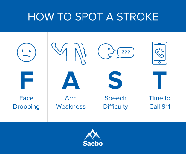
One of the most important ways to successfully recover from stroke, is by taking preventative measures such as knowing and recognizing the symptoms of a stroke because immediate treatment can be life saving and greatly affects the chances for a full recovery. Unfortunately the chances of a second stroke occurring increases in stroke survivors. According to The National Stroke Association, about 25% of stroke survivors will experience a second stroke. Within the first 5 years after the first stroke, risk of a second stroke is about 40% higher. Fortunately it is estimated that of all secondary strokes, about 80% of them are preventable with lifestyle changes and medical intervention. Read more about recognizing the symptoms of stroke in men and in women to better prepare you to act FAST.


Tip 2. Walking Again and Foot Drop

Foot drop is the difficulty or inability to lift the front part of the foot because of fatigue or damage affecting the muscles and nerves that aid in its movement. To combat this, using a brace or Ankle-foot Orthoses (AFO) has proven to be a major aid in rehabilitation. These devices prevent the front of the foot from dipping down and disrupting walking movements.

Leg exercises described in this supplementary post after experiencing a stroke are crucial for recovery. While each patient should have a custom exercise routine, personalized for you, there are several exercises that should be included in most every stroke survivor’s regimen. These low-impact strength and stretching leg exercises for stroke recovery are a good complement to use in conjunction with the Saebo MyoTrac Infiniti biofeedback system.

Richard Sealy, director of The Rehab Practice, a private neuro-therapy rehabilitation program in the United Kingdom, regularly works with individuals, families, and caregivers to establish custom exercise routines to aid in recovery from long-term neurological problems, like the damage caused by stroke. While he acknowledges that each patient should have a custom exercise routine specific and personal to their struggles, he recommends a series of exercises for anyone working to strengthen their legs and improve range of motion during stroke recovery.

Rehabilitation of the legs and feet can occur at a faster rate with a combination of the aforementioned exercises and orthopedic aids such as the SaeboStep.The SaeboStep is a unique foot drop brace worn on the outside of the shoe that assists with lifting the toes when walking. It is made to eliminate cumbersome, unreliable splints and braces placed within the shoe.


Tip 3. Dealing with Curled Toes

Often referred to as “curled toes” or “claw toe,” this symptom is caused by a miscommunication between the brain and muscles within the foot. This misfiring of signals causes an issue with controlling muscular movements, leading to over-contracting of the toes and spasticity, a condition where there is a miscommunication between the brain and the muscles in the toes, causing them to over contract.

The best way to regain strength and movement while dealing with this condition is to create a routine with a variety of exercises—toe taps, floor grips, finger squeezes, and toe-extensor strengthening. With effort and repetition, these workouts can make a huge difference in recovery.


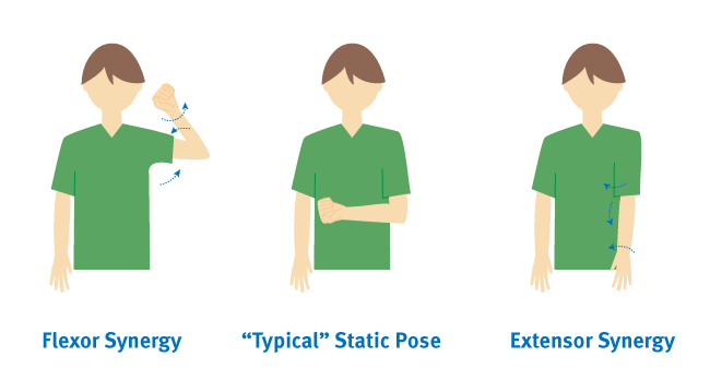
Tip 4. Lack of Arm Function

One of the most common deficiencies following a stroke is the impairment of the arm and hand. This typically results in decreased strength, coordination, and range of motion. Those affected are often unable to support their own arms in order to perform rehabilitation exercises. When this occurs it is crucial that you include additional arm support during rehabilitation to avoid the arms becoming weaker due to learned non-use.

Learned non-use occurs when a stroke survivor prefers to use their strong arm because it is easier to move. This tendency makes it even more difficult for a stroke survivor to recover, because challenging the weakened arm with these exercises plays a crucial role in regaining arm function. Often physical therapists and occupational therapists use a technique known as Constraint-Induced Movement Therapy (or CIMT) to challenge a weakened shoulder and make further exercises and drills possible. Mobile arm supports such as the SaeboMAS and SaeboMAS mini help support the weight of the arm, allowing the user to do a much wider range of exercises. For more information about the SaeboMAS and how it can aid in stroke recovery click here.

As with rehabilitating any part of the body with reduced function after a stroke, it is important to consistently repeat the exercises and stretches to strengthen the brain-muscle connections. It is also important to stay positive and try to have fun with your rehab. Here are 35 fun rehab activities for stroke patients to help keep you motivated.


Tip 5. Hand Paralysis

Paralysis is the inability of a muscle to move voluntarily. The National Stroke Association sites as many as 9 out of 10 stroke survivors have some degree of paralysis following a stroke. Rehabilitation and therapy can help to regain voluntary movement, even several years after the stroke takes place.

The primary symptoms of hand paralysis are spasticity (stiff muscles), weakness, and lack of coordination. Fortunately, there are several methods of treatment in addition to therapy to help manage and recover from spasticity. Additional treatments include medications to relax muscles, botox injections (relaxes muscles temporarily), stretching exercises, anti-spasticity orthotics, and functional orthoses. Surgery is another option in the most severe cases.

The least invasive and most permanent treatment for hand paralysis is therapy to rehabilitate the connection between your brain and muscles using neuroplasticity. To make these exercises even more effective and to increase your rate of recovery, it is important to repeat your hand exercises often. By performing exercises repeatedly, you are strengthening that brain-muscle connection.


Tip 6. Difficulty Speaking and Communicating

Another common side effect of stroke is aphasia, which is the inability to speak or understand speech. This is one of the most frustrating side effects for survivors to deal with. It’s estimated that 25 to 40 percent of people who suffer from a stroke develop aphasia, though this condition is not limited to stroke survivors. Aphasia occurs when there is damage to the brain, specifically the left side that deals with language. There are two primary forms of aphasia: receptive aphasia and expressive aphasia. Receptive aphasia is when the individual has trouble understanding what is being said to them. Expressive aphasia is when the individual is having difficulty expressing what they want to say.

When communicating with someone with receptive aphasia, try not to use long complex sentences. When communicating with someone with expressive aphasia, it is important to be patient and remember that the person’s intelligence has not been affected by the stroke, just their ability to speak.

For those with aphasia, the most important thing you can do to improve your communication is to take a deep breath and try to relax. If you can remain relaxed and focus on what you are trying to say you will have much greater success. It is easy to get flustered or feel self conscious, but you shouldn’t. Create tools that you can use to make communication easier such as a book of words, pictures, phrases, or symbols that can help you get your message across. If you are going out and know you will not be around friends or family, it may also be helpful to carry a card or piece of paper that indicates that you have aphasia and explains what it is, just in case you find yourself needing to explain your condition.

Once these tools are set in place, seeking the help of a speech-language pathologist (SLP) can greatly increase one’s ability to regain normal speech behavior. SLPs can assist in rehabilitating all types of physical speech behavior and offer support and proper guidance for you or a loved one. Read more about aphasia and recovery here.


Tip 7: Coping with PTSD After Stroke

Following a stroke, it is fairly common for a survivor to experience PTSD, or Post-Traumatic Stress Disorder. This condition is usually associated with combat veterans and sexual-assault survivors; however, according to a study published in the journal PLoS One, almost a quarter of stroke survivors experience some form of PTSD.

Common symptoms of PTSD include the victim experiencing the traumatic event over and over in their head or in the form of nightmares. This replaying of the event is typically accompanied by the individual’s unyielding anxiety and feelings of self doubt or misplaced guilt over their condition. Some experience a state of hyperarousal or feelings of being overly alert.

The two main treatments for PTSD include medications such as antidepressants, anti-anxiety medications or psychotherapy. If you are experiencing PTSD, it is important that you communicate how you feel with your doctor, family, and friends, as a strong support system can help you find the relief from psychological pain that you deserve.


Tip 8: Understanding Fatigue

Feeling tired is a normal part of life for everyone, but for stroke survivor, fatigue is a very common symptom that can be frustrating to deal with. About 40 to 70 percent of stroke survivors experience fatigue, which can make recovering feel even more difficult. Post-stroke fatigue is draining both physically and emotionally/mentally, and rest may not be the only solution.

It is important to discuss the fatigue with a doctor so they can rule out potential medical causes or determine if fatigue might stem from current medications. By speaking with the proper medical professionals and taking time to squeeze in a nap or rest as often as possible—and by maintaining a positive mindset—you can help yourself or a loved one combat the constant drowsiness of fatigue and work on returning to pre-stroke energy levels. The key thing to realize is that some level of post-stroke fatigue is normal and that survivors need to be proactive about treating and working around it.


Tip 9: Counteract Learned Non-Use

If the limbs weakened after stroke are not consistently exercised over time, muscles have the potential to atrophy—waste away due to cell degeneration. This often occurs when the person tries to compensate for their weak limb by using their stronger limb more often. Daily attempts to move the affected limbs are necessary to maintain and improve functionality.One method is the use of Constraint-Induced Movement Therapy (CIMT). CIMT is a form of therapy that prevents the unaffected limbs from moving while trying to exercise the affected ones.


Tip 10: Reduce Inflammation and Stress

Inflammation in the body can cause other issues to arise, which is why it’s important to stay stress free whenever possible. When stress does begin to take hold, a hormone called cortisol floods the body, causing pH levels to become imbalanced with acidity. High acidity levels—after an extended period of time—can kill good bacteria in the body while giving rise to bad bacteria, ultimately weakening the immune system.

With that in mind, a natural probiotic like yogurt is a great way to boost good bacteria in the body. Supplemental drinks can also improve the immune system significantly. In addition to pH balance, adopting stress management exercises such as yoga, deep breathing, tai chi, qi gong, and meditation, can limit one’s cortisol levels, promoting overall health.


Tip 11: Coping with Emotional Effects

Experiencing a stroke is not only a major hardship to overcome physically; it can also take a huge toll on a survivor’s emotions in many ways.

If the area of your brain that controls personality or emotion is affected, you may be susceptible to changes in your emotional response or everyday behavior. Strokes may also cause emotional distress due to the suddenness of their occurrence. As with any traumatic life experience, it may take time for you or your loved one to accept and adapt to the emotional trauma of having experienced a stroke.

Some common emotional changes strokes may cause are PseudoBulbar Affect, depression, and anxiety. Thankfully, there are several methods for treating the emotional changes associated with a stroke, with the first step being to discuss how you or your loved one is feeling with a doctor. Treatment may consist of one, or a combination, of the following: one-on-one counseling, group counseling, medication, diet, and exercise. The most effective treatment is different for everyone, so it is important to discuss and explore which combination works best for your or your loved one.


PseudoBulbar Affect

Sometimes referred to as “reflex crying,” “emotional lability,” or “labile mood,” PseudoBulbar Affect (PBA) is a symptom of damage to the area of the brain that controls expression of emotions, and it is one of the most frequently reported post-stroke behaviors. Characteristics of the disorder include rapid changes in mood, such as suddenly bursting into tears and stopping just as suddenly or even beginning to laugh at inappropriate times.


Depression

Survivors have a one in four chance of developing serious depression as a side effect of stroke. If you are feeling sad, hopeless, or helpless after having suffered a stroke, you may be experiencing this. Other symptoms of depression may include irritability or changes to your eating and sleeping habits. Talk to your doctor if you are experiencing any of these symptoms, as it may be necessary to treat with prescription antidepressants or therapy to avoid it becoming a road block to your recovery.

Along with medication and therapy, a lot of research shows that a few simple lifestyle changes help relieve the symptoms of depression. If you or a loved one is having a difficult time coping with the emotional repercussions of a stroke, here are tips on how to implement positivity and resilience:

  • Attend a support group. Talking about your struggles with people in the same situation makes you feel less lonely and can offer helpful insight or different approaches to dealing with difficulties.
  • Eat healthy food. A good diet is important for your general health and your recovery from stroke and can also improve your mental health.
  • Remain socially active. Although you may not be able to do as much as you used to, it’s crucial to stay in touch with family and friends and take part in social activities.
  • Be as independent as possible. Humans need to feel independent and competent. Stroke recovery may require the help of caregivers, but if there are things that you can safely do by yourself, insist on it.
  • Exercise regularly. Physical activity, especially a low-impact one like walking, is proven to boost mental health and will also contribute to your recovery.


Tip 12: Set Recovery Goals with Your Therapist

Setting specific and meaningful goals can help keep one focused and motivated once they are achieved, and these goals can range from simple tasks to long-term accomplishments. By establishing a list consisting of difficulties and goals, overcoming obstacles can be put within reach.

When setting these goals, working with a therapist, doctor, or close friend can be a good way to find encouragement, as well as assistance in creating a list that places goals into an appropriate timeframe. Overall, a therapist will be familiar with your case, understanding the issues and complications, and will be able to offer sound advice in all aspects of recovery.


Tip 13: Stay Motivated

Since apathy is common during stroke recovery, staying motivated can be a challenge. Combining one’s interests with a solid rehabilitation regimen can effectively eradicate feelings of lethargy and depression. The best thing to do is to focus on a reason for recovery and to associate it with your plan of action. This can be done by implementing sentimental items into daily routines, thus giving you personal and motivational support at all times. For example, if one of your routines is to write a list of things to do for the day, try writing it on the back of a special photo. That way, as you’re checking things off, you’ll have a little reminder to keep you motivated.


Tip 14: Watch Out For The Recovery Plateau Stage

The recovery plateau stage refers to the point at which a stroke survivor begins to see a slow down or stop in the progression in their recovery. Some of the most significant improvements often occur in the subacute phase, which is usually the first three to six months after the stroke  (though there is anecdotal evidence of people making significant stroke recovery progress outside of that zone.)

Seeing improvement in the early days of a survivor’s recovery can make it a lot easier for them to stay motivated and continue working hard in therapy. Research shows that further recovery is still very possible after the plateau stage though, which is why it is so important to have a strong support system to encourage you to continue with therapy and working on recovery.


Tip 15: Working After Stroke

Since the brain is a major organ affected when it comes to strokes, chances are that some of its functions may have trouble performing like they did before. After a stroke, learning new things, or even just recalling information can be a challenge, and working through thoughts may suddenly be difficult.

After rehabilitation, many stroke survivors do find themselves able to return to work, but preparing for this transition can come with a lot of questions. Are you physically going to be able to perform your job? Will your disability benefits lapse? What do you need to communicate with your employer? These can be tough questions, but they do have answers. Some may not ever be able to go back to the same work, but for others, just a little assistance is needed.

When you are ready to return to work, it is important to know your rights and what your employer is, and is not, legally required to provide to employees with disabilities. Keep in mind that if you are unable to perform the essential functions of your job even with reasonable accommodation, your employer is not obligated to offer you a different position or create a new role for you. They might be willing to anyway, but it is not a requirement.


Tip 16: Understand and Combat Memory Loss

Not only is it common for stroke survivors to experience, but memory loss can affect a wide range of people through multiple factors. Age, physical trauma, and emotional stress have the potential to cause memory decline, but rebuilding memory’s strength is highly possible and can be fun.

Specifically, incorporating technology into daily rehabilitation exercises is a great way to show quick improvements. There are numerous apps for smartphones and tablets that use different techniques to significantly improve memory, and they have the ability to set reminders, schedule appointments, and oversee other illnesses.


Tip 17: Be Aware of Vascular Dementia

A common problem among stroke survivors, this symptom disrupts cognitive functions, which can make it challenging for one to sort out information.

Due to the damage of blood vessels from a stroke, blood pressure, cholesterol, and blood sugar must be maintained at healthy levels to ensure good blood flow throughout the body. If you are diabetic, it is crucial that you are paying careful attention to your blood sugar and insulin levels. Studies have shown that by managing these three components, vascular dementia can be decreased or prevented.

Helping Stroke Survivors Help Themselves

The process of stroke recovery is long and full of ups, downs, twists, and turns. It takes hard work and dedication to regain mental and physical function after a stroke. The information and tips above will help you to identify and overcome the many challenges that come with recovery.

To read our answers to the most common stroke recovery questions, click here. And remember, at the end of the day, there are dozens of approaches you can take to improve the speed of stroke recovery.


All content provided on this blog is for informational purposes only and is not intended to be a substitute for professional medical advice, diagnosis, or treatment. Always seek the advice of your physician or other qualified health provider with any questions you may have regarding a medical condition. If you think you may have a medical emergency, call your doctor or 911 immediately. Reliance on any information provided by the Saebo website is solely at your own risk.

via 17 Ways To Help Stroke Survivors Recover Faster | Saebo

, , , , , , , , , , ,

Leave a comment

[WEB SITE] The Brunnstrom Stages of Stroke Recovery – Saebo


The Brunnstrom Stages of Stroke Recovery


Life after a stroke can be challenging. Many patients wonder if they will ever fully recover their muscle coordination, or how long or difficult the process of recovery may be. Fortunately, the field of occupational and physical therapy has come a long way in developing approaches that help patients regain controlled muscle movements after a stroke.
There are seven recognized stages of stroke recovery through which most patients progress. Also known as the Brunnstrom Approach, the seven stages framework views spastic and involuntary muscle movement as part of the process and uses them to aid in rehabilitation.


What Is The Brunnstrom Approach?

The Brunnstrom Approach was developed in the 1960’s by Signe Brunnstrom, an occupational and physical therapist from Sweden. With seven stages, the Brunnstrom Approach breaks down how motor control can be restored throughout the body after suffering a stroke.

Normally, muscle movements are the result of different muscle groups working together. Researchers have termed this collaboration between muscles as “synergies”. The brain has the delicate task of coordinating these movements, many of which become severely affected after a stroke.

After the stroke has occurred, your muscles become weak due to the lack of coordination between the brain and body. This causes the muscle synergies to move in abnormal patterns. Most treatments offered to stroke patients will focus on trying to inhibit atypical muscle synergies and movements. The Brunnstrom Approach, on the other hand, teaches patients how to use the abnormal synergy patterns to their advantage.
This approach has become a popular choice among both occupational and physical therapists as well as patients since its inception. It can be effective in clinical settings and can dramatically improve voluntary muscle movements after suffering a stroke.

image05

Stage 1: Flaccidity 

The first stage in Brunnstrom’s Approach is the initial period of shock immediately after stroke where flaccid paralysis sets in. Flaccid paralysis (flaccidity) is the medical term for a complete lack of voluntary movement. This paralysis is caused by nerve damage that prevents the muscles from receiving appropriate signals from the brain, whether or not the brain is still capable of moving those muscles.

In the early state of flaccid paralysis, the stroke survivor cannot initiate any muscle movements on the affected side of their body. If this continues for long enough without intervention or physical therapy, the unused muscles become much weaker, and begin to atrophy. Simply put, muscles need to be used in order to retain their tone and definition, and flaccid paralysis prevents muscles from doing this important work.

Stroke recovery stage 1 left side of brain / right side of brain paralysis

The medical term for this loss of muscle tone is hyptonia. Hyptonia causes weakness and sometimes numbness that seriously interferes with a patient’s quality of life. In addition to therapy exercises and treatments that reduce the severity of hypotonia, this Stage 1 condition also requires lifestyle modifications to protect the affected limbs from injury.

Stroke recovery stage 1 - muscle atrophy suffering
Though stroke does serious neurological damage, other healthy brain cells and muscles can help make up for some of this damage. In fact, the patient’s own body is full of tools that reduce complications and increase their likelihood of entering new stages of recovery. It’s never too early to start retraining the body and brain after stroke, even if patients are still experiencing flaccid paralysis and hypotonia.

image07

Stage 2: Dealing with the Appearance of Spasticity

The second stage in stroke recovery marks the redevelopment of some basic limb synergies as certain muscles are stimulated or activated and other muscles in the same system begin to respond. Muscles begin to make small, spastic, and abnormal movements during this stage. While these movements are mostly involuntary, they can be a promising sign during your recovery. Minimal voluntary movements might or might not be present in stage two.

Muscle synergies result from muscles coordinating movements to perform different tasks. These synergies allow common patterns of movement that involve either cooperative or reciprocal activation of muscle. Because the muscles are linked, one activated muscle may lead to partial or complete responses in other muscles. These synergies may limit patient’s muscles to certain movements, preventing them from completing the voluntary movements they want to make. However, as neurological development and cell regrowth occurs after a stroke, some new connections may be formed to impaired muscle tissue.


flexor-and-extensor-synergies during stage 2 of stroke recovery

Two limb synergies determine a patient’s reactions to cell regrowth during Stage 2 of recovery. The first, the flexor synergy, includes the external rotation of the shoulder, flexion of the elbow, and supination of the forearm. The second, the extensor synergy, includes internal rotation of the shoulder with elbow extension and pronation of the forearm. These synergies may produce one or both of the following postures, which indicate varying levels of brain trauma after stroke.

Coupled with the presence of muscle synergies, between 30 and 40 percent of stroke survivors also experience spasticity. This is a velocity-dependent increase in your normal stretch reflexes, and during Stage 2, it presents as aresistance to passive movement. Stage 2 spasticity contributes to the jerky upper body movements characteristic of the flexor and extensor synergies.

Unused limbs still need stimulation to maintain or form connections to neurons. Though the nerves and connections that originally controlled your affected limbs may be damaged too much to create voluntary movements, it could still be possible to regain movement in later stages of recovery. In order to leave this possibility open and prevent the body’s tendency toward learned non-use, it’s important to continue using and moving your affected limbs and muscles as much as possible.


image04

Stage 3: Increased Spasticity

Spasticity in muscles increase during stage three of stroke recovery, reaching its peak. Spasticity is a feeling of unusually stiff, tight, or pulled muscles. It is caused by damage from a stroke to nerve pathways within the brain or spinal cord that control muscle movement. The lack of ability to restrict the brain’s motor neurons causes muscles to contract too often. Spasticity causes an abnormal increase in muscle stiffness and tone that can interfere with movement, speech, or cause discomfort and pain.

During stage 3, synergy patterns also start to emerge and minimal voluntary movements should be expected. The increase in voluntary movement is due to being able to initiate movement in the muscle, but not control it (yet). The appearance of synergy patterns and coordination between muscles facilitate the voluntary movements which become stronger with occupational and physical therapy.

Muscles with severe spasticity, like the ones in stage 3 of stroke recovery, are likely to be more limited in their ability to exercise and may require help to do this. Patients and family/caregivers should be educated about the importance of maintaining range of motion and doing daily exercises. It is important to minimize highly stressful activities this early in training.

motion-excercise-stretch during stage 3 of stroke recovery

Passive exercises, also known as passive range-of-motion (PROM) exercises, should be continued during this stage to improve your range of motion. Treatment includes how far the therapist can move your joints in different directions, like raising your hand over your head or bending your knee toward your chest.

image00

Stage 4: Decreased Spasticity

During stage four of stroke recovery, spastic muscle movement begins to decline. Patients will regain control mostly in the extremities, and they will have a limited ability to move normally. The movements may still be out of sync with muscle synergies, but this will improve quickly over the length of this stage.

The focus during this stage is to strengthen and improve muscle control. Now that you are regaining motor control and can start to make normal, controlled movements on a limited basis, you can start to build strength back in your limbs and continue work on your range of motion. Continuing to stretch out your muscles is still important in this stage.

Physiotherapist helping her patient with arm exercise Rehabilitation concept during stage 4 of stroke recovery


Therapists use active-assisted range of motion (AAROM) exercises when a stroke patient has some ability to move but still needs help to practice the exercises or complete the movement. A therapist may help guide the movement with their own body (hold the limb, for example) or use bands and other exercise equipment to support the patient. Gravity-assisted devices such as the SaeboMAS, are beneficial in helping the patient perform the movements.


Female therapist assisting senior couple with exercises in the medical office during stage 4 of stroke recovery


You can begin active range-of-motion (AROM) exercises once you have regained some muscle control and can perform some exercises without assistance. They often involve moving a limb along its full range of motion, like bending an elbow or rotating a wrist. AROM exercises increase flexibility, muscle strength, and endurance. Range-of-motion exercises should be practiced equally on both the affected and unaffected sides of the body.

Of course, when it comes to building a stage 4 stroke recovery exercise program, you should always consult with a professional physical or occupational therapist. They can help you with exercise specifics, finding the right tools and equipment, and, of course, to provide assistance, especially in the beginning.

image02

Stage 5: Complex Movement Combinations

In stage 5, spasticity continues to decline and synergy patterns within the muscles also become more coordinated, allowing voluntary movements to become more complex. Abnormal movements also start to decline dramatically during stage 5, but some may still be present.

The patient will be able to make more controlled and deliberate movements in the limbs that have been affected by the stroke. Isolated joint movements might also be possible.

All voluntary movements involve the brain, which sends out the motor impulses that control movement. These motor signals are initiated by thought and must also involve a response to sensory stimuli. The sensory stimuli that trigger voluntary responses are dealt with in many parts of the brain.

 stage 5 of stroke recovery - patient ready for complex movement
Voluntary movements are purposeful and goal directed. They are learned movements that improve with repetition or practice and require less attention. Some examples include combing hair, swinging a bat, driving a car, swimming, and using eating utensils.

image06

Stage 6: Spasticity Disappears

At stage six, spasticity in muscle movement disappears completely. You are able to move individual joints, and synergy patterns become much more coordinated. Motor control is almost fully restored, and you can coordinate complex reaching movements in the affected extremities. Abnormal or spastic movements have ceased, and a full recovery may be on the horizon.

image01

Stage 7: Normal Function Returns

The last stage in Brunnstrom’s Approach is when you regain full function in the areas affected by the stroke. You are now able to move your arms, legs, hands, and feet in a controlled and voluntary manner.

Since you have full control over your muscle movements, synergy patterns have also returned to normal. Reaching stage seven is the ultimate goal for therapists and patients alike.


Stroke Recovery In 7 Stages: Spasticity As A Process

With the seven stages of recovery, Brunnstrom effectively changed the way stroke recovery is approached by occupational and physical therapists. She theorized that spastic and primitive muscle movements were a natural part of the recovery process after a stroke. Moreover, she developed an approach that allows patients to use these involuntary movements to their advantage instead of trying to inhibit them.

During each phase, an increasing amount of synergies are available to use. Using the Brunnstrom Approach, occupational and physical therapists will teach you how to use the synergies that are currently available to you. These techniques are used to improve movement and regain motor control.

There is no one approach to stroke recovery, and the stages laid out in these guides may not apply to everyone. Since the Brunnstrom Approach can be effective, however, therapists still use this method to help patients recover after suffering a stroke. Thanks to new medical technology, therapists can use the Brunnstrom Approach in conjunction with tools like the SaeboGloveSaeboReach, and SaeboMAS to help patients reach new levels of independence.

via The Brunnstrom Stages of Stroke Recovery | Saebo

, , , ,

Leave a comment

[ARTICLE] A composite robotic-based measure of upper limb proprioception – Full Text

Abstract

Background

Proprioception is the sense of the position and movement of our limbs, and is vital for executing coordinated movements. Proprioceptive disorders are common following stroke, but clinical tests for measuring impairments in proprioception are simple ordinal scales that are unreliable and relatively crude. We developed and validated specific kinematic parameters to quantify proprioception and compared two common metrics, Euclidean and Mahalanobis distances, to combine these parameters into an overall summary score of proprioception.

Methods

We used the KINARM robotic exoskeleton to assess proprioception of the upper limb in subjects with stroke (N = 285. Mean days post-stroke = 12 ± 15). Two aspects of proprioception (position sense and kinesthetic sense) were tested using two mirror-matching tasks without vision. The tasks produced 12 parameters to quantify position sense and eight to quantify kinesthesia. The Euclidean and Mahalanobis distances of the z-scores for these parameters were computed each for position sense, kinesthetic sense, and overall proprioceptive function (average score of position and kinesthetic sense).

Results

A high proportion of stroke subjects were impaired on position matching (57%), kinesthetic matching (65%), and overall proprioception (62%). Robotic tasks were significantly correlated with clinical measures of upper extremity proprioception, motor impairment, and overall functional independence. Composite scores derived from the Euclidean distance and Mahalanobis distance showed strong content validity as they were highly correlated (r = 0.97–0.99).

Conclusions

We have outlined a composite measure of upper extremity proprioception to provide a single continuous outcome measure of proprioceptive function for use in clinical trials of rehabilitation. Multiple aspects of proprioception including sense of position, direction, speed, and amplitude of movement were incorporated into this measure. Despite similarities in the scores obtained with these two distance metrics, the Mahalanobis distance was preferred.

Background

Stroke is heterogeneous, affecting sensory, motor, and cognitive functions that are required for daily activities. While there are well validated tools to assess motor and speech functions (eg. Fugl-Meyer Assessment (FMA) [1], the National Institute of Health Stroke Scale (NIHSS) [2], Chedoke-McMaster Stroke Assessment Impairment Inventory (CMSA) [3]) the use of high quality, validated assessment tools for measuring sensory function post-stroke (proprioception in particular) is limited [4], and there is still a lack of a gold standard assessment. While the FMA and NIHSS have sensory components to the assessment, they are seldom used as a sole measure of sensory impairment in research studies focused on sensation as they are based on relatively coarse scales. Yet, sensory and proprioceptive impairments have a significant negative impact on functional recovery following stroke [56789]. Individuals with sensory and motor impairments, compared to those with just motor impairments, have longer lengths of hospitalization and fewer discharges home [101112]. Furthermore, it has recently been shown that motor and proprioceptive impairments can occur independently after stroke [13].

Some commonly used clinical assessments of proprioception post-stroke include: 1) simple passive limb movement detection test [14] in which an examiner moves a subject’s limb segment with their eyes closed, and subjects are asked to say which direction the limb was moved; 2) the Revised Nottingham Sensory Assessment [1516] in which the subject is asked to mirror match the movement of a passively moved limb by a therapist; and 3) the Thumb Localizing Test [17] which involves passive movement of a subject’s arm and hand to a random position overhead, and is followed by subjects reaching to grasp their thumb with the opposite (less affected) hand. These assessments are scored crudely as normal, slightly impaired, or absent, and lack the sensitivity to detect smaller changes in proprioceptive function in part due to poor inter- and intrarater reliability [1819]. Therefore, establishing an objective and reproducible method to assess proprioceptive impairments post-stroke is vital to evaluating the efficacy of different treatments.

Other more advanced methods to assess proprioception have been developed [20212223], with many using robotic technology to measure the kinematics of an individual’s movements. Assessment devices can now measure position sense and kinesthetic impairments after stroke using arm contralateral matching [13242526], in which a subject’s affected arm is passively moved by the robot to a position, and the subject mirror-matches the movement/position with their less affected limb. Another paradigm involves passive movement of a subject’s limb to a specified position, returning the limb to the starting position, and then having subjects actively move the same arm to this remembered position [2126]. This method has an advantage in that it does not require interhemispheric transfer of information, but has limited value in assessing people with concurrent motor deficits, or in assessing kinematic aspects of proprioception, such movement speed and amplitude perception. Further, results can be confounded by problems with spatial working memory. Threshold for detection of passive movement paradigms have also been used to assess proprioception [2728]. This paradigm eliminates confounds due to motor impairment and interhemispheric transfer of information but again, little information about the kinematics of movement perception (e.g. speed or direction) are gained from this task, and it typically takes much longer to complete than position/movement matching. Lastly, Carey et al. [20] have developed and validated a wrist position sense test, where a subject’s wrist is moved to a position (wrist flexion or extension) and without vision of the wrist the subject has to use their other arm to move a cursor to the direction the wrist is pointing. This method minimizes confounds due to interhemispheric information transfer and motor deficits, but again does not provide information about kinesthetic impairments.

Many of these assessments are reliable, reproducible, objective, and provide quantitative measures of proprioceptive function in the upper limbs. Dukelow et al. [1324], used a KINARM robot (BKIN Technologies, Kingston, ON), and detailed a contralateral position-matching task for the upper extremities that can measure various aspects of an individual’s position sense including: absolute error, variability in matching positions, systematic shifts in perceived workspace, and perceived contraction or expansion of the workspace. Similarly, Semrau et al. [25] recently detailed a kinesthetic matching task using the KINARM robot that can measure an individual’s ability to mirror-match the speed, direction, and amplitude of a robotically moved limb [825]. These tasks are reliable [24], and provide numerous parameters that describe an individual’s position or kinesthetic sense impairments and can be used to guide a rehabilitation program tailored to the individual. Furthermore, these studies have shown a strong relationship between proprioceptive impairments and functional independence post-stroke, yet proprioceptive impairments are often not addressed in day-to-day therapy. Reliable and quantitative assessment tools are therefore critical for testing the efficacy of rehabilitation treatments, as in clinical rehabilitation trials.

While multiple kinematic parameters can provide a level of exactness around the nature of an individual’s proprioceptive impairments and are helpful for rehabilitation planning, a summary measure is needed for clinical therapeutic trials in rehabilitation. Thus, a single continuous metric of upper limb proprioceptive function that combines all parameters from the position and kinesthetic matching robotic tasks was developed using two common measures of distance, Euclidean distance (EDist) and Mahalanobis distance (MDist) [29]. The EDist was chosen as it is an easily interpretable calculation and considers each parameter independently. It is the square root of the sum of squared distances between data points (i.e. the straight-line distance between two points in three-dimensional space). The MDist is the next measure we used to compare with the EDist. It was chosen because the calculation accounts for correlations between parameters (by using the inverse of the variance-covariance matrix of the data set of interest), therefore preventing the overweighting of correlated parameters in the calculation. It is the distance between a point and the center of a distribution, measured along the major axes of variation (i.e. the standard deviation of an object in more than one dimension) [3031].. Because the kinematic parameters derived from the robotic tasks may demonstrate some degree of correlation with one another [13], the MDist can account for this auto-correlation. Theoretically, it should perform better at identifying stroke subjects who perform abnormally on the tasks and those who have atypical patterns of behavior relative to controls. The MDist is generally preferred over the EDist for multivariable data since it can cope with different structures of data [31].

MDist (or variants of it) has recently been used in other studies when examining reaching movements after stroke [32].. Our primary aim was to examine differences and similarities between two summary scores (EDist and MDist) in their ability to differentiate proprioceptive impairment in individuals with stroke from controls in a large patient sample. We hypothesized that using a composite proprioception score calculated from the Mahalanobis distance would more accurately identify impaired proprioception in individuals with stroke compared to a proprioception score calculated from the Euclidean distance.[…]


Continue —>  A composite robotic-based measure of upper limb proprioception | Journal of NeuroEngineering and Rehabilitation | Full Text


Fig. 1a KINARM robotic exoskeleton (BKIN Technologies, Kingston, ON, Canda). Subjects are seated in the wheelchair base with arms supported by the arm troughs. b Top-down view of the position matching task. The stroke affected arm was positioned by the robot (black targets, green lines) and subjects were required to mirror-match the target positions with their opposite hand (open targets, blue lines). Nine targets were matched to six times each for a total of 54 trials, presented in pseudorandom order. c Top-down view of an exemplar subject performing one trial of the kinesthetic matching task. The stroke affected arm was moved by the robot between two targets (green lines) and subjects were required to mirror match the speed, direction, and amplitude of movement as soon as they felt the robot move their arm (blue lines). The speed versus time profile represents the temporal aspects of the task, by measuring the response latency (time to initiation of the active arm movement) and peak speed ratio (difference between peak speeds of the passive (green) and active (blue) hands)

, , , , , , , , , ,

Leave a comment

[VIDEO] 5 Tips for Surviving the Holidays with an mTBI — How to manage your TBI & still enjoy the holidays – YouTube

Kim & Brie are back! This time they’re here to give you some tips on how to comfortably celebrate the holidays after a TBI. Post Concussion Syndrome can make holidays even more overwhelming than usual, but some forethought and planning can help. The TBI Rockstars guide you through some of their own holiday experiences post brain injury.


, ,

Leave a comment

[Poster] Collaboration of Music and Physical Therapy: Case Study for Treatment of Patient with Chronic Stroke

To evaluate the change in gait speed pre- and post-treatment. To evaluate the change in quality of life pre- and post-treatment. To evaluate the change in outcome measures pre- and post-treatment.

First page of article

via Collaboration of Music and Physical Therapy: Case Study for Treatment of Patient with Chronic Stroke – Archives of Physical Medicine and Rehabilitation

, ,

Leave a comment

[Abstract] Effectiveness of the conductive educational approach added to conventional physiotherapy in the improvement of gait parameters of poststroke patients: randomized-controlled pilot study.

Abstract

Our objective was to assess the benefits of the conductive education (CE) approach added to conventional physiotherapy in gait functions of poststroke, hemiparetic patients. A randomized-controlled trial was designed in a rehabilitation clinic. Late and chronic poststroke patients with gait disturbances (n=17, median age: 55 years, range: 41-72 years) were enrolled in the study. All patients received conventional physiotherapy. However, patients of only one group took part in therapy on the basis of the CE approach. The gait parameters, semiobjective outcome measures, functional independence measure, and International Classification of Functioning, Disability and Health domains were collected. The effectiveness of the CE approach was underlined by those outcome measures that were only significant (P≤0.05) in the conductive group: functional independence measure motor subscale; maintaining body position and walking long distances; and muscle strength in some muscle groups. The results suggest that CE could have an additive effect on gait improvement of stroke patients.

via Effectiveness of the conductive educational approach added to conventional physiotherapy in the improvement of gait parameters of poststroke patien… – PubMed – NCBI

, , , ,

Leave a comment

[Abstract] Feasibility study of a serious game based on Kinect system for functional rehabilitation of the lower limbs

Summary

Introduction

Conventional functional rehabilitation costs time, money and effort for the patients and for the medical staff. Serious games have been used as a new approach to improve the performance as well as to possibly reduce medical cost in the future for cognitive rehabilitation and body balance control. The objective of this present work was to perform a feasibility study on the use of a new real-time serious game system for improving the musculoskeletal rehabilitation of the lower limbs.

Materials and methods

A basic functional rehabilitation exercise database was established with different levels of difficulties. A 3D virtual avatar was created and scaled to represent each subject-specific body. A portable and affordable Kinect sensor was used to capture real-time kinematics during each exercise. A specific data coupling process was developed. An evaluation campaign was established to assess the developed system.

Results

The squats exercise was the hardest challenge. Moreover, the performance of each functional rehabilitation exercise depended on the physiological profile of each participant. Our game system was clear and attractive for all functional rehabilitation exercises. All testing subjects felt motivated and secure when playing the rehabilitation game.

Discussion

The comparison with other systems showed that our system was the first one focusing on the functional rehabilitation exercises of the lower limbs.

Conclusions

Our system showed useful functionalities for a large range of applications (rehabilitation at home, sports training). Looking forward, new in-situation exercises will be investigated for specific musculoskeletal disorders.

via Feasibility study of a serious game based on Kinect system for functional rehabilitation of the lower limbs – ScienceDirect

, , , , , , ,

Leave a comment

[WEB SITE] Brain Injury — Important Facts and Implications for Social Work Practice

Brain Injury — Important Facts and Implications for Social Work Practice
By Jennifer Fleming, MA, LPC, CBIS; Natasha McVey, MSS, LCSW, CBIS; Madeleine Shusterman, LCSW, CBIS; and Eli DeHope, PhD, LCSW, BCD

Roberta is a bright, high-energy, fun-loving, career-oriented woman in her early 30s who worked as a pharmaceutical sales representative. Roberta’s ability to multitask and think quickly and her dedication to working long hours put her on the fast track to project leader of the sales team.

On one winter day while traveling to see a client, Roberta’s car slid on a patch of ice, went off the road, and crashed into a ditch. Roberta sustained minor physical injuries and a bad concussion. She did not lose consciousness. Her family and coworkers believed, as did Roberta, that while this was a serious accident, she would recover fully. The expectation was that she would be back to her usual, highly productive self in a matter of weeks.

After several months of ongoing headaches, fatigue, difficulty concentrating, and decreased motivation to return to work, her family started to wonder what was wrong with Roberta. Why didn’t she just get over the accident and go back to her life? Why was she so unmotivated? What her family didn’t understand was that Roberta’s symptoms were related to her concussion and that Roberta had actually sustained a brain injury.

Brain injury refers to the death of brain cells and the disruption of neural pathways that can change the way a person thinks, feels, and/or actsA brain injury can be caused by an outside force, such as a bump, blow, or jolt to the head related to an accident, fall, or violence, or to a change in air pressure, such as a blast injury. Brain injuries can also result from neurologic diseases, lack of oxygen to the brain, or penetrating wounds like gunshots (Rutland-Brown, Langlois, Thomas, & Xi, 2006).

Brain injury is increasingly recognized as a health concern with lifelong implications; however, it continues to be referred to as the “silent epidemic,” perhaps because awareness about brain injury, although improving, continues to be limited. It is estimated that an average of 1.7 million individuals sustain brain injuries each year (Coronado et al., 2011), which translates to about one person every 23 seconds. Brain injuries are most likely to occur in the very young (under the age of 5), followed by adolescents (ages 15 to 19) and adults over the age of 75 (Coronado et al.). Brain injury has a higher prevalence than HIV, breast cancer, and multiple sclerosis combined. Brain injury is also frequently undiagnosed and underreported (Leibson et al., 2011).

Brain Injury Sequelae
Because the brain is responsible for many functions, once an individual experiences a brain injury, his or her life is often drastically affected. A person with a brain injury can have a range of medical, cognitive, emotional/behavioral, and psychosocial issues. The sequelae listed in this chart are common challenges associated with brain injury; however, this list is not exhaustive:

Clearly, brain injury can impact all aspects of a person’s life. An individual’s identity in his or her family, friendships, and workplace are often affected. Alarmingly, it is estimated that 60% of people with brain injuries are never able to return to their prior employment (van Velzen, van Bennekom, Edelaar, Sluiter, & Frings-Dresen, 2009). Many lose their friendships and spouse or partner. People with a brain injury often experience and report social isolation, feelings of loneliness, and loss of friendships as a primary problem (Temkin, Corrigan, Dikmen, & Machamer, 2009) as well as an inability to participate in their hobbies and leisure activities.

Similarly, family members of these patients often experience significant stress, including the development of significant psychological symptoms as well as the loss of social support (Vangel, Rapport, & Hanks, 2011). Social workers can play a vital role in helping individuals and their families to accept and learn how to adapt to the inevitable changes that result from brain injury.

Making an Impact
All social workers should be equipped to recognize and refer individuals who might be struggling with a brain injury. Knowing who is at risk and what to look for is an important first step.

Recognizing At-Risk Populations and Symptoms
Individuals who have been involved in violence, dangerous occupations or hobbies, or who were athletes, veterans, or substance abusers should be considered as possibly having suffered a brain injury. Via case review, social workers should look for a history of loss/alteration of consciousness or significant events such as falls, motor vehicle accidents, and hospitalizations.

While a history of coma indicates the likelihood of a brain injury and the potential for long-term impairment, short losses of consciousness, or alterations, can also indicate the presence of a brain injury.

There is no medical test that can predict a person’s prognosis based on a specific injury. It is important to understand that even if an MRI or CT scan does not reveal any physical brain changes, the injury may still affect the person. The social worker should take into consideration a client’s history, as there may have been instances of head trauma not documented in the person’s medical record, as well as look for possible symptoms of brain injury.

Screening Questions
If a brain injury is suspected, social workers can ask several questions during an interview that may shed light on the person’s history of head trauma. These questions include the following:

• Have you ever been hit in the head?

• Have you ever lost consciousness?

• Have you ever had a concussion?

• Do you, or did you ever, play contact sports?

• Have you ever been in a car accident?

• Have you ever been in a physical fight or a victim of violence?

• Are you a veteran? Were you ever injured in service?

If an event with the potential to cause head trauma is found, social workers should follow up with questions about the immediate effect of the trauma, including amnesia, disorientation, or confusion, and then with questions about its impact on functioning in the following weeks, months, and even years. The Ohio Valley Center for Brain Injury Prevention and Rehabilitation, in conjunction with BrainLine, has developed a screening tool available at www.brainline.org.

Helpful Referrals
Brain injury rehabilitation services are highly specialized. Knowing where quality rehabilitation services are located in your community is most important for referring clients appropriately. The majority of physicians have little or no experience with brain injury and its short- and long-term impact. So identifying a physiatrist, a rehabilitation doctor, and a neurologist who do have experience treating those with brain injuries can greatly improve an individual’s overall medical care. Major trauma centers and rehabilitation facilities in urban areas are a good place to start.

Each state has a Brain Injury Association, organized by the umbrella organization Brain Injury Association of America (www.biausa.org). These organizations often have online resources that contain provider information, family support services, and general brain injury education.

Children with brain injuries can qualify for specialized services until the age of 21 through their school districts with an individualized education plan. This information is imperative for school social workers to know so they can educate parents, teachers, coaches, colleagues, and students. They can also advocate for needed services and make necessary referrals.

School social workers should also be aware of the special situation concussions can pose for students and schools. While a child can completely recover from a concussion, it is often recommended that a child’s return to school be gradual, following a period of complete rest at home. Social workers can play a critical role in gathering information from family and physicians and ensuring that all school staff are aware of medical recommendations about concussion management and restrictions (e.g., recess, physical education, sports). The Centers for Disease Control and Prevention has many valuable resources on concussion, including free tool kits and checklists (http://www.cdc.gov/concussion).

Finally, social workers should be aware of the adjunct resources in their community that clients may need to access, including assistance with transportation, housing, legal problems, education and/or employment, attendant care, assistive technology, and financial support.

Quick Tips for Working With Individuals With Brain Injury
• Provide simple written information, including summaries of what you are doing and what is next.

• Have a family member present in sessions.

• Speak simply and ask direct questions.

• Avoid long, complicated discussions.

• Check the client’s understanding of the information presented, making sure to allow time for processing.

• Offer breaks.

• Provide appointment reminders.

• Be careful with humor and your personal space.

Additional resources are provided in the table below:

Resources on Brain Injury
BrainLine.org: provides resources for preventing, treating, and living with brain injury
Brain Injury Association of America: a website with links to state affiliates and their resources; also a great source of information on current advocacy efforts in this area
Centers for Disease Control and Prevention – Traumatic Brain Injury: extensive resource on brain injury (mild to severe), including fact sheets, tip cards, and other practical tools
Systematic Approach to Social Work Practice: Working With Clients With Traumatic Brain Injuries: a downloadable 132-page manual completed with support from the National Institute on Disability and Rehabilitation Research
Defense and Veterans Brain Injury Center: provides both information and resources on brain injury in the military for veterans, military families, and other interested parties


Final Thoughts
With brain injury becoming more common, especially with more veterans returning home from war, it is important for social workers to understand this condition and know the best means for helping their clients get the treatment they need to successfully function from day to day. Doing so will prove beneficial not only for the client but for family and friends as well.

— Jennifer Fleming, MA, LPC, CBIS, is a day program specialist at ReMed in Paoli, PA.

— Natasha McVey, MSS, LCSW, CBIS,is a rehabilitation case manager atReMed.

— Madeleine Shusterman, LCSW, CBIS, is a clinical specialist at ReMed.

— Eli DeHope, PhD, LCSW, BCD, is a professor of undergraduate social work at West Chester University of Pennsylvania.


References 
Coronado, V. G., Xu, L., Basavaraju, S. V., McGuire, L. C., Wald, M. W., Faul, M. D., et al. (2011). Surveillance for traumatic brain injury-related deaths—United States, 1997-2007. MMWR Surveill Summ, 60(5), 1-32.

Rutland-Brown, W., Langlois, J. A., Thomas, K. E., & Xi Y. L. (2006). Incident of traumatic brain injury in the United States, 2003. Journal of Head Trauma and Rehabilitiation, 21(6), 544-548.

Leibson, C. L., Brown, A.W., Ransom, J. E., Diehl, N. N., Perkins, P. K., Mandekar, J. et al. (2011). Incidence of traumatic brain injury across the full disease spectrum: A population-based medical record review study. Epidemiology22(6), 836-844.

Temkin, N. R., Corrigan, J. D., Dikmen, S. S., & Machamer, J. (2009). Social functioning after traumatic brain injury. Journal of Head Trauma Rehabilitation, 24(6), 460-467.

Vangel, S. J., Rapport, L. J., & Hanks, R. A. (2011). Effects of family and caregiver psychosocial functioning on outcomes of persons with traumatic brain injury. Journal of Head Trauma Rehabilitation, 26(1), 20-29.

van Velzen, J. M., van Bennekom, C. A., Edelaar, M. J., Sluiter, J. K. & Frings-Dresen, M. H. (2009). How many people return to work after acquired brain injury?: A systematic review. Brain Injury, 23(6), 473-488.

via Brain Injury — Important Facts and Implications for Social Work Practice

,

Leave a comment