Posts Tagged brain activity

[Abstract] Brain–Computer Interface Training Based on Brain Activity Can Induce Motor Recovery in Patients With Stroke: A Meta-Analysis

Abstract

Background

Brain–computer interface (BCI) is a procedure involving brain activity in which neural status is provided to the participants for self-regulation. The current review aims to evaluate the effect sizes of clinical studies investigating the use of BCI-based rehabilitation interventions in restoring upper extremity function and effective methods to detect brain activity for motor recovery.

Methods

A computerized search of MEDLINE, CENTRAL, Web of Science, and PEDro was performed to identify relevant articles. We selected clinical trials that used BCI-based training for post-stroke patients and provided motor assessment scores before and after the intervention. The pooled standardized mean differences of BCI-based training were calculated using the random-effects model.

Results

We initially identified 655 potentially relevant articles; finally, 16 articles fulfilled the inclusion criteria, involving 382 participants. A significant effect of neurofeedback intervention for the paretic upper limb was observed (standardized mean difference = .48, [.16-.80], P = .006). However, the effect estimates were moderately heterogeneous among the studies (I2 = 45%, P = .03). Subgroup analysis of the method of measurement of brain activity indicated the effectiveness of the algorithm focusing on sensorimotor rhythm.

Conclusion

This meta-analysis suggested that BCI-based training was superior to conventional interventions for motor recovery of the upper limbs in patients with stroke. However, the results are not conclusive because of a high risk of bias and a large degree of heterogeneity due to the differences in the BCI interventions and the participants; therefore, further studies involving larger cohorts are required to confirm these results.

Source

, , , , , , , , ,

Leave a comment

[WEB PAGE] Stroke patients now able to boost brain activity at home

by University of South Australia

UniSA researchers have adopted a novel approach to help stroke victims recover greater use of their paralysed limbs, giving patients the chance to re-train their brains at home.

A new project developed by NHMRC Early Career Research Fellow Dr. Brenton Hordacre delivers a home treatment program for chronic stroke patients with hand/arm mobility issues.

The program involves the patient or their carer attaching brain stimulation electrodes to the motor cortex region of the head for 20 minutes each day, followed by a series of physical exercises, lasting an hour in total.

The therapy is designed to increase brain activity and help the brain re-learn the ability to move through a process known as plasticity.

“It’s a two-week treatment that can be delivered in the comfort of a person’s home, without the need to come into a hospital or clinical setting, which can be very stressful for some people, particularly those who are disabled or living in regional and rural areas,” Dr. Hordacre says.

The patient’s rehabilitation over the fortnight is tracked via an iPad with Skype connection to ensure that the treatment is safe and effective. Several advanced brain imaging measures are used to compare brain activity before and after this treatment.

“This is the first time we have performed brain stimulation remotely for stroke patients and it could well be the future direction for this type of treatment,” Dr. Hordacre says.

His study is targeting stroke patients with impaired motor function, but separate treatment projects could be adapted for stroke victims with speech and/or cognitive damage, he adds.

Dr. Hordacre has recruited 21 stroke patients to date, around half of whom have completed the two-week program. People wanting to take part in the project should contact him at Brenton.Hordacre@unisa.edu.au

Source

, , , , , , ,

Leave a comment

[ARTICLE] Increased gait variability during robot-assisted walking is accompanied by increased sensorimotor brain activity in healthy people – Full Text

Abstract

Background

Gait disorders are major symptoms of neurological diseases affecting the quality of life. Interventions that restore walking and allow patients to maintain safe and independent mobility are essential. Robot-assisted gait training (RAGT) proved to be a promising treatment for restoring and improving the ability to walk. Due to heterogenuous study designs and fragmentary knowlegde about the neural correlates associated with RAGT and the relation to motor recovery, guidelines for an individually optimized therapy can hardly be derived. To optimize robotic rehabilitation, it is crucial to understand how robotic assistance affect locomotor control and its underlying brain activity. Thus, this study aimed to investigate the effects of robotic assistance (RA) during treadmill walking (TW) on cortical activity and the relationship between RA-related changes of cortical activity and biomechanical gait characteristics.

Methods

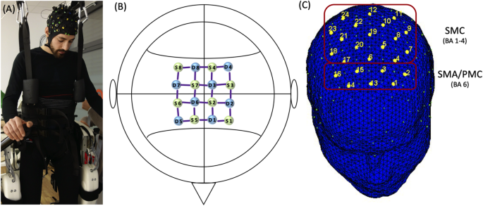
Twelve healthy, right-handed volunteers (9 females; M = 25 ± 4 years) performed unassisted walking (UAW) and robot-assisted walking (RAW) trials on a treadmill, at 2.8 km/h, in a randomized, within-subject design. Ground reaction forces (GRFs) provided information regarding the individual gait patterns, while brain activity was examined by measuring cerebral hemodynamic changes in brain regions associated with the cortical locomotor network, including the sensorimotor cortex (SMC), premotor cortex (PMC) and supplementary motor area (SMA), using functional near-infrared spectroscopy (fNIRS).

Results

A statistically significant increase in brain activity was observed in the SMC compared with the PMC and SMA (p < 0.05), and a classical double bump in the vertical GRF was observed during both UAW and RAW throughout the stance phase. However, intraindividual gait variability increased significantly with RA and was correlated with increased brain activity in the SMC (p = 0.05; r = 0.57).

Conclusions

On the one hand, robotic guidance could generate sensory feedback that promotes active participation, leading to increased gait variability and somatosensory brain activity. On the other hand, changes in brain activity and biomechanical gait characteristics may also be due to the sensory feedback of the robot, which disrupts the cortical network of automated walking in healthy individuals. More comprehensive neurophysiological studies both in laboratory and in clinical settings are necessary to investigate the entire brain network associated with RAW.

Background

Safe and independent locomotion represents a fundamental motor function for humans that is essential for self-contained living and good quality of life [1,2,3,4,5]. Locomotion requires the ability to coordinate a number of different muscles acting on different joints [6,7,8], which are guided by cortical and subcortical brain structures within the locomotor network [9]. Structural and functional changes within the locomotor network are often accompanied by gait and balance impairments which are frequently considered to be the most significant concerns in individuals suffering from brain injuries or neurological diseases [51011]. Reduced walking speeds and step lengths [12] as well as non-optimal amount of gait variability [13,14,15] are common symptoms associated with gait impairments that increase the risk of falling [16].

In addition to manual-assisted therapy, robotic neurorehabilitation has often been applied in recent years [1718] because it provides early, intensive, task-specific and multi-sensory training which is thought to be effective for balance and gait recovery [171920]. Depending on the severity of the disease, movements can be completely guided or assisted, tailored to individual needs [17], using either stationary robotic systems or wearable powered exoskeletons.

Previous studies investigated the effectiveness of robot-assisted gait training (RAGT) in patients suffering from stroke [2122], multiple sclerosis [23,24,25,26], Parkinson’s disease [2728], traumatic brain injury [29] or spinal cord injury [30,31,32]. Positive effects of RAGT on walking speed [3334], leg muscle force [23] step length, and gait symmetry [2935] were reported. However, the results of different studies are difficult to summarize due to the lack of consistency in protocols and settings of robotic-assisted treatments (e.g., amount and frequency of training sessions, amount and type of provided robotic support) as well as fragmentary knowledge of the effects on functional brain reorganization, motor recovery and their relation [3637]. Therefore, it is currently a huge challenge to draw guidelines for robotic rehabilitation protocols [2236,37,38]. To design prologned personalized training protocols in robotic rehabilitation to maximize individual treatment effects [37], it is crucial to increase the understanding of changes in locomotor patterns [39] and brain signals [40] underlying RAGT and how they are related [3641].

A series of studies investigated the effects of robotic assistance (RA) on biomechanical gait patterns in healthy people [3942,43,44]. On one side, altered gait patterns were reported during robot-assisted walking (RAW) compared to unassisted walking (UAW), in particular, substantially higher muscle activity in the quadriceps, gluteus and adductor longus leg muscles and lower muscle activity in the gastrocnemius and tibialis anterior ankle muscles [3942] as well as reduced  lower-body joint angles due to the little medial-lateral hip movements [45,46,47]. On the other side, similar muscle activation patterns were observed during RAW compared to UAW [444849], indicating that robotic devices allow physiological muscle activation patterns during gait [48]. However, it is hypothesized that the ability to execute a physiological gait pattern depends on how the training parameters such as body weight support (BWS), guidance force (GF) or kinematic restrictions in the robotic devices are set [444850]. For example, Aurich-Schuler et al. [48] reported that the movements of the trunk and pelvis are more similar to UAW on a treadmill when the pelvis is not fixed during RAW, indicating that differences in musle activity and kinematic gait characteristics between RAW and UAW are due to the reduction in degrees of freedom that user’s experience while walking in the robotic device [45]. In line with this, a clinical concern that is often raised with respect to RAW is the lack of gait variability [454850]. It is assumed that since the robotic systems are often operated with 100% GF, which means that the devices attempt to force a particular gait pattern regardless of the user’s intentions, the user lacks the ability to vary and adapt his gait patterns [45]. Contrary to this, Hidler et al. [45] observed differences in kinematic gait patterns between subsequent steps during RAW, as demonstrated by variability in relative knee and hip movements. Nevertheless, Gizzi et al. [49] showed that the muscular activity during RAW was clearly more stereotyped and similar among individuals compared to UAW. They concluded that RAW provides a therapeutic approach to restore and improve walking that is more repeatable and standardized than approaches based on exercising during UAW [49].

In addition to biomechanical gait changes, insights into brain activity and intervention-related changes in brain activity that relate to gait responses, will contribute to the optimization of therapy interventions [4151]. Whereas the application of functional magnetic resonance imaging (fMRI), considered as gold standard for the assessment of activity in cortical and subcortical structures, is restricted due to the vulnerability for movement artifacts and the range of motion in the scanner [52], functional near infrared spectroscopy (fNIRS) is affordable and easily implementable in a portable system, less susceptible to motion artifacts, thus facilitation a wider range of application with special cohorts (e.g., children, patients) and in everyday environments (e.g., during a therapeutic session of RAW or UAW) [5354]. Although with lower resolution compared to fMRI [55], fNIRS also relies on the principle of neurovascular coupling and allows the indirect evaluation of cortical activation [5657] based on hemodynamic changes which are analogous to the blood-oxygenation-level-dependent responses measured by fMRI [56]. Despite limited depth sensitivity, which restricts the measurement of brain activity to cortical layers, it is a promising tool to investigate the contribution of cortical areas to the neuromotor control of gross motor skills, such as walking [53]. Regarding the cortical correlates of walking, numerous studies identified either increaesed oxygenated hemoglobin (Hboxy) concentration changes in the sensorimotor cortex (SMC) by using fNIRS [5357,58,59] or suppressed alpha and beta power in sensorimotor areas by using electroencephalography (EEG) [60,61,62] demonstrating that motor cortex and corticospinal tract contribute directly to the muscle activity of locomotion [63]. However, brain activity during RAW [366164,65,66,67,68], especially in patients [6970] or by using fNIRS [6869], is rarely studied [71].

Analyzing the effects of RA on brain activity in healthy volunteers, Knaepen et al. [36] reported significantly suppressed alpha and beta rhythms in the right sensory cortex during UAW compared to RAW with 100% GF and 0% BWS. Thus, significantly larger involvement of the SMC during UAW compared to RAW were concluded [36]. In contrast, increases of Hboxy were observed in motor areas during RAW compared UAW, leading to the conclusion that RA facilitated increased cortical activation within locomotor control systems [68]. Furthermore, Simis et al. [69] demonstrated the feasibility of fNIRS to evaluate the real-time activation of the primary motor cortex (M1) in both hemispheres during RAW in patients suffering from spinal cord injury. Two out of three patients exhibited enhanced M1 activation during RAW compared with standing which indicate the enhanced involvement of motor cortical areas in walking with RA [69].

To summarize, previous studies mostly focused the effects of RA on either gait characteristics or brain activity. Combined measurements investigating the effects of RA on both biomechanical and hemodynamic patterns might help for a better understanding of the neurophysiological mechanisms underlying gait and gait disorders as well as the effectiveness of robotic rehabilitation on motor recovery [3771]. Up to now, no consensus exists regarding how robotic devices should be designed, controlled or adjusted (i.e., device settings, such as the level of support) for synergistic interactions with the human body to achieve optimal neurorehabilitation [3772]. Therefore, further research concerning behavioral and neurophysiological mechanisms underlying RAW as well as the modulatory effect of RAGT on neuroplasticy and gait recovery are required giving the fact that such knowledge is of clinical relevance for the development of gait rehabilitation strategies.

Consequently, the central purpose of this study was to investigate both gait characteristics and hemodynamic activity during RAW to identify RAW-related changes in brain activity and their relationship to gait responses. Assuming that sensorimotor areas play a pivotal role within the cortical network of automatic gait [953] and that RA affects gait and brain patterns in young, healthy volunteers [39424568], we hypothesized that RA result in both altered gait and brain activity patterns. Based on previous studies, more stereotypical gait characteristics with less inter- and intraindividual variability are expected during RAW due to 100% GF and the fixed pelvis compared to UAW [4548], wheares brain activity in SMC can be either decreased [36] or increased [68].

Methods

This study was performed in accordance with the Declaration of Helsinki. Experimental procedures were performed in accordance with the recommendations of the Deutsche Gesellschaft für Psychologie and were approved by the ethical committee of the Medical Association Hessen in Frankfurt (Germany). The participants were informed about all relevant study-related contents and gave their written consent prior to the initiation of the experiment.

Participants

Twelve healthy subjects (9 female, 3 male; aged 25 ± 4 years), without any gait pathologies and free of extremity injuries, were recruited to participate in this study. All participants were right-handed, according to the Edinburg handedness-scale [73], without any neurological or psychological disorders and with normal or corrected-to-normal vision. All participants were requested to disclose pre-existing neurological and psychological conditions, medical conditions, drug intake, and alcohol or caffeine intake during the preceding week.

Experimental equipment

The Lokomat (Hocoma AG, Volketswil, Switzerland) is a robotic gait-orthosis, consisting of a motorized treadmill and a BWS system. Two robotic actuators can guide the knee and hip joints of participants to match pre-programmed gait patterns, which were derived from average joint trajectories of healthy walkers, using a GF ranging from 0 to 100% [7475] (Fig. 1a). Kinematic trajectories can be adjusted to each individual’s size and step preferences [45]. The BWS was adjusted to 30% body weight for each participant, and the control mode was set to provide 100% guidance [64].

figure1

Montage and Setup. a Participant during robot-assisted walking (RAW), with functional near-infrared spectroscopy (fNIRS) montage. b fNIRS montage; S = Sources; D = Detectors c Classification of regions of interest (ROI): supplementary motor area/premotor cortex  (SMA/PMC) and sensorimotor cortex (SMC) 

[…]

Continue —-> Increased gait variability during robot-assisted walking is accompanied by increased sensorimotor brain activity in healthy people | Journal of NeuroEngineering and Rehabilitation | Full Text

, , , , , , , , ,

Leave a comment

[Music] Mozart – Sonata for Two Pianos in D, K. 448 [complete] – YouTube

The Sonata for Two Pianos in D major, K. 448 is a piano work composed in 1781 by Wolfgang Amadeus Mozart, at 25 years of age. It is written in strict sonata-allegro form, with three movements. The sonata was composed for a performance he would give with fellow pianist Josephine von Aurnhammer. Mozart composed this in the galant style, with interlocking melodies and simultaneous cadences. This is one of his only formal compositions written exclusively for two pianos. This sonata was also used in the scientific study that tested the theory of the Mozart Effect, suggesting that classical music increases brain activity more positively than other kinds of music. The sonata is written in three movements, 1. Allegro con spirito 2. Andante and 3. Molto Allegro. The first movement begins in D major, and sets the tonal center with a strong introduction. The two pianos divide the main melody for the exposition, and when the theme is presented both play it simultaneously. Mozart spends little time in the development introducing a new theme unlike most sonata forms, and begins the recapitulation, repeating the first theme. The entire second movement is played Andante, in a very relaxed pace. The melody is played with both pianos, but there is no strong climax in this movement. It is written in a strict ABA form. Molto Allegro begins with a galloping theme. The cadences used in this movement are similar to those in Mozart’s Rondo alla Turca. According to the British Epilepsy Organization, research has suggested that Mozart’s K 448 can have the “Mozart effect”, in that listening to the piano sonata improved spatial reasoning skills and reduce the number of seizures in people with epilepsy. Apart from another Mozart Concerto, K 488, only one other piece of music has been found to have a similar effect, a song by the Greek composer Yanni, entitled “Acroyali/Standing In Motion”, which is featured on his album Yanni Live at the Acropolis. It was determined to have the “Mozart effect”, by the Journal of the Royal Society of Medicine because it was similar to Mozart’s K 448 in tempo, structure, melodic and harmonic consonance and predictability.

via Mozart – Sonata for Two Pianos in D, K. 448 [complete] – YouTube

, , , ,

Leave a comment

[Editorial] Investigating Brain Activity After Acquired and Traumatic Brain Injury: Applications of Functional MRI

  • 1Stroke, Kessler Foundation, West Orange, NJ, United States
  • 2Neuropsychology and Neuroscience, Kessler Foundation, West Orange, NJ, United States
  • 3Traumatic Brain Injury, Kessler Foundation, West Orange, NJ, United States

Editorial on the Research Topic

Investigating Brain Activity After Acquired and Traumatic Brain Injury: Applications of Functional MRI

Every year, approximately 795,000 people in the United States are affected by stroke and 2.8 million lives are impacted by traumatic brain injury (TBI) (1). Stroke and TBI are also major causes of serious long-term disability, reducing mobility, and impacting thinking, memory, sensation, and emotional functioning. Neuroscience holds great promise in addressing the needs of persons with a history of stroke or TBI by improving the current understanding of brain injury and recovery mechanisms. This is the first step in working to inform and improve the available treatments.

While a great many functional neuroimaging methods exist for studying the healthy brain, such methods have not received widespread acceptance in characterizing patient groups. Several methodological barriers may explain why. First, patient populations can be diverse in terms of injury location and stages of recovery. Accurate measurement and interpretation of functional neuroimaging signal in the damaged brain can also pose a challenge, because stroke and TBI can dramatically alter cerebral blood flow, even in areas that are not affected by a structural lesion (23). Finally, correct interpretation of findings in light of impaired and/or changing behavioral function depends on careful experimental design and precise a priorioperational definitions of the anticipated effects.

Despite these challenges, or, perhaps, because of them, functional neuroimaging is a promising area of investigation in TBI and stroke. This Research Topic is a collection of original research and review articles focused on functional neuroimaging in persons with TBI and stroke. Below, we highlight a few of the most notable findings and ideas from this collection of articles. Readers are encouraged to access the full text articles for more details.

In one of the two review articles, Medaglia provides an overview of fMRI methodology, analyses, and the caveats of applying these analyses to the injured brain. This includes methods, such as seed-based and voxel-based functional connectivity, effective connectivity, including psychophysiological interactions, causal connectivity, and graph analyses. Medaglia discusses the concept of functional re-organization. The term is sometimes used to describe a change in the magnitude of activation or of functional connectivity. It is also used to refer to a re-allocation of function to new brain areas following injury. Medaglia suggests that to improve clarity a precise description of the effect should be provided. Formal tests of re-organization should include a search for areas with activity profile closely resembling that of a damaged area, and with corresponding evidence of recovered behavioral function. Distinguishing different innate recovery mechanisms is especially important in intervention studies, because failing to understand which process may be at work when introducing an intervention, may lead to inadvertent interference with endogenous repair mechanisms.

Nair et al. studied brain activation in acute stroke and healthy older controls participants during a covert verbal fluency task. They controlled for the blood oxygen level dependent (BOLD) response variability across participants using resting state fluctuation amplitude (RSFA) (4). RSFA calibration is thought to eliminate any inter-subject variability due to vascular factors and retain any differences due to neuronal activation factors. They found that after scaling, the BOLD response differences between stroke patients and healthy controls were eliminated. This finding suggests that some of the group differences were due to vascular variables. Additionally, some fine-tuning may be required when scaling with RSFA, perhaps scaling by brain region, rather than across the whole brain.

Bernier et al. applied graph theory to a data set of healthy and TBI subjects with moderate/severe TBI. Their aim was to determine if loss of network differentiation accounts for changes in brain connectivity, specifically hyperconnectivity. This hypothesis was examined within the default mode (DMN) and the task positive network. Supporting other results in the field, they observed hyperconnectivity within the DMN and task positive networks. DMN hyperconnectivity was found to be associated with higher scores on the standardized working memory measure. Thus, the work of these authors demonstrates how fMRI and connectivity analyses can inform the cognitive profile observed following TBI.

The second review in the Research Topics explores a common deficit in TBI. Namely, cognitive control, an executive function that is generally necessary for switching between habitual and goal-directed behavior. In his review, Scheibel talks about functional neuroimaging studies of cognitive control in mild TBI (mTBI). The review draws attention to how the fMRI findings are mixed, with reports of decreased as well as increased brain activation in mTBI, and urges for future studies to aim at recruiting more homogenous samples, as the mixed findings might be explained by the presence of comorbidies in TBI samples.

The original research article by Saleh et al. explored how different approaches to rehabilitation of hand function after stroke can alter brain activity across the sensorimotor brain networks and demonstrates network re-organization discussed in the Medaglia review. Both treatment approaches tested in the study improved hand function. However, only the robot-assisted virtual reality group showed reduction of activity and re-lateralization of activation to ipsilesional cortex, a pattern associated with better arm function in this study and with positive recovery in other studies (5).

A contribution by Möller et al. used arterial spin labeling (ASL) fMRI to examine fatigue in mTBI during psychomotor vigilance task performance. The mTBI participants showed different patterns of brain activation compared to healthy controls, in addition to higher self-reported fatigue and reductions in performance as the task progressed (fatigability). Together with the self-reported fatigue ratings and task performance, the ASL results suggested the engagement of disparate functional networks compared in mTBI.

fMRI research in stroke and TBI poses a unique set of challenges to researchers. The articles assembled in this Research Topic address some of these challenges. Using methods designed to work in patients with brain lesions, using appropriate controls, and applying network neuroscience tools are a few of the promising solutions. This topic is an important frontier in neuroscience research today offering tangible benefits for public health and is a potential area of growth in the coming years

References

1. Centers for Disease Control and Prevention (CDC). U. Centers for Disease con [WWW Document]. (2018). Available from: https://www.cdc.gov/ (Accessed: February 22, 2018).

Google Scholar

2. Brumm KP, Perthen JE, Liu TT, Haist F, Ayalon L, Love T. An arterial spin labeling investigation of cerebral blood flow deficits in chronic stroke survivors. Neuroimage (2010) 51:995–1005. doi:10.1016/j.neuroimage.2010.03.008

PubMed Abstract | CrossRef Full Text | Google Scholar

3. Hillis AE. Magnetic resonance perfusion imaging in the study of language. Brain Lang (2007) 102:165–75. doi:10.1016/j.bandl.2006.04.016

PubMed Abstract | CrossRef Full Text | Google Scholar

4. Kannurpatti SS, Motes MA, Rypma B, Biswal BB. Increasing measurement accuracy of age-related BOLD signal change: minimizing vascular contributions by resting-state-fluctuation-of-amplitude scaling. Hum Brain Mapp (2011) 32:1125–40. doi:10.1002/hbm.21097

PubMed Abstract | CrossRef Full Text | Google Scholar

5. Cramer SC. Repairing the human brain after stroke: I. Mechanisms of spontaneous recovery. Ann Neurol (2008) 63:272–87. doi:10.1002/ana.21393

PubMed Abstract | CrossRef Full Text | Google Scholar


via Frontiers | Editorial: Investigating Brain Activity After Acquired and Traumatic Brain Injury: Applications of Functional MRI | Neurology

, , , , ,

Leave a comment

[WEB SITE] There’s a lot of junk fMRI research out there. Here’s what top neuroscientists want you to know.

John Greim/LightRocket via Getty Images

If you’ve followed the latest from the world of neuroscience, you might get the impression that the field is in deep trouble.

In July, a report in the Proceedings of the National Academy of Sciences declared that as many as 40,000 papers using the standard tool in neuroscience research, functional magnetic resonance imaging (fMRI), could suffer from a statistical flaw that rendered their results to be a false positive.

Headlines feared the worst:

These studies aren’t necessarily wrong, per se. It’s that the common statistical software they used is prone to wrongly guess the borders of “lit up” areas in the brain.

But authors of the PNAS paper quickly realized their estimate of 40,000 papers was too high. (The actual number of papers implicated is probably closer to 3,500, one of the authors wrote in a blog post. And PNAS has since amended the paper.)

Still, the results cast some doubt over the validity of fMRI. And they weren’t an anomaly. Over the past few years, doubt about fMRI research has been mounting. Researchers have found brain activity in dead fish. One 2009 paper found an epidemic of “puzzlingly high” correlations in fMRI research.

All this uncertainty has provoked a question: Is fMRI actually a faulty tool that should not be trusted?

Not at all, neuroscientists say. “It’s unfair to blame the tool,” Rebecca Saxe of MIT, who has been using fMRI since its earliest days, told me. “It’s like blaming a telescope when somebody’s wrong about [identifying] a planet. It’s not the telescope’s fault. The general problem is there are lots of ways to fool yourself with fMRI data.”

There are questions fMRI is good at answering and questions it is bad at answering. There are right ways to use fMRI, and there are irresponsible ways to use it.

I asked several experts in the field a simple question: What’s most important to know about fMRI and its limitations? Here’s what I learned.

1) An fMRI measures blood flow, not neural activity

When fMRI first became available as a scientific tool in the 1990s, it was a revelation.

Before then, scientists who wanted to learn about brain function had few options: They could wait for a patient with a brain injury to come along and test her mental abilities. Or they could inject people with radioactive dyes and then scan them with X-rays. The arrival of fMRI meant that a great many more scientists could study the brain. Neuroscience, as a field of research, exploded. In the past 20 years, 40,000 papers have been publishedusing fMRI.

“With fMRI, suddenly you could study the brain of a healthy person,” says Saxe. Better yet, fMRI wasn’t dangerous. You could repeat tests on the same person without fear of harming them. “There was a lot of hope,” she says. Finally, scientists thought they could peer into the brain and find the cause of autism.

And it was all thanks to magnets. An fMRI is conducted in a MRI machine equipped to scan people’s heads — it’s a giant doughnut-shaped device (people are loaded in through the center on a sliding table).The machine’s huge magnets can pick up on small changes in the brain; specifically, they’re looking for the presence of oxygenated blood.

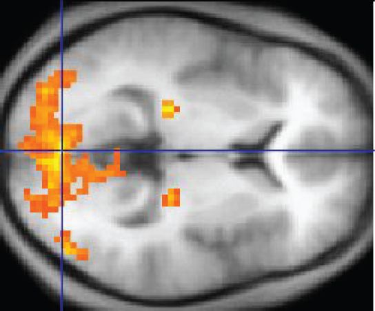
When the brain region is activated, it calls out for more oxygen. The fMRI then follows oxygenated blood as it flows through the brain. On the printout of the scan, these oxygenated brain regions “light up.” Scientists can see areas smaller than a millimeter cubed (also called a voxel, which just means it’s a pixel in three dimensions) That blood flow is a sign that neural activity is happening in a given brain region.
 Wikipedia

From the 1990s onward, the use of the tool exploded. In 2015, there were more than 29,000 academic articles in the PubMed database mentioning “fMRI.”

2) fMRI studies go wrong not just because of the limitations of the tool. They go wrong because science, overall, has flaws.

With fMRI, scientists began to chart an atlas of the brain and its function. They confirmed that there was a specific area of the brain devoted to interpreting faces. And they found wonderfully simple things, like how wiggling of the fingers are controlled by a tiny area near the earlobes. (Alas, fMRI hasn’t revealed the secrets to disorders like autism or schizophrenia.)

But amid the successes, there was a quietly growing concern that labs were using the tool inappropriately and generating false positives with it.

The July PNAS paper was the latest in a string of papers casting doubt fMRI science. Here are two most famous ones:

  1. In 2009, a researcher showed brain activity in a dead salmon. Dead salmon aren’t thinking, and the paper revealed that neuroscientists need to be vigilant about separating out signal from noise. (The neuroscientists I spoke with said the field was largely aware of this problem and could correct for it when this paper came out.)
  2. Also in 2009, a paper in Perspectives in Psychological Science found an epidemic of “puzzlingly high” correlations in papers that tried to associate brain scans with personality types.

“The common thing people would do is say, ‘We found that activation in some area correlates with some aspect of people’s personality,’” Russell Poldrack, a researcher at Stanford’s Center for Reproducible Neuroscience, explains. They’d then focus their analysis on that specific area and find extremely high correlations. “This is problematic because it is basically double dipping.”

The Perspectives paper concluded: “[I]t is quite possible that a considerable number of relationships reported in this literature are entirely illusory.”

Each of these papers highlighted a big concern with fMRI methods, and for the most part, the field has corrected course. But problems sill remain.

There’s currently a bigger “crisis” in psychology and social science where researchers are realizing some of their most celebrated findings don’t replicate under stricter methods. “All the things that are wrong in psychology are clearly wrong in neuroimaging,” Poldrack says.

At Vox, we’ve discussed these problems in science at length. There’s publication bias — the trend that journals only publish positive, confirmatory results. That creates the file drawer effect: Because negative results are not published, the published literature may paint a too-rosy description of a theory. And then there’s p-hacking: the suite of methodological tweaks researchers can employ to ensure they land on a significant (i.e., publishable) result.

The problems make for an uneasy time for scientists: They’d like to charge ahead with new scientific questions, but also feel a nagging anxiety that they should recheck all the work their questions have been built upon.

There’s some reason to suspect these problems might be even be more problematic in neuroscience than in psychology at large. Here’s why: fMRIs are extremely expensive, costing hundreds of dollars an hour to operate. Money is pressure, and scientists don’t want to be left for nothing to show for a $20,000 study. The expense also makes it less likely for scientists to do replications of past work, and less likely to run large numbers of subjects through an experiment.

3) fMRI is good at mapping broad regions of brain activity. But it’s not good at specifics.

We know fMRI measures blood flow and not neural activity directly. And the assumption (that’s been validated in studies) is that blood flow correlates with neural activity. The blood flow can reveal changes in brain areas as small as a millimeter cubed.

But still, at best, that’s just a crude view of things. There can be hundreds of thousands of neurons in a tiny voxel.

 BSIP/UIG via Getty Images
Pyramidal neurons of a cat’s cerebral cortex.

“The analogy is it’s like flying over a city and seeing where the lights are on,” Tal Yarkoni, who studies neuroscience research methods at University of Texas Austin, says. If you’re in an airplane, you might look the window and identify a patch of land as a residential area. But it’s impossible to know what people are doing in their homes. And it’s difficult to understand how people in their homes interact with the city’s center.

And fMRI is like that. You can learn what broad areas of the brain are working. But figuring out what, exactly, those brain areas are doing is a totally different problem.

4) The results of a study can be correct, but the interpretation of those results can be very, very wrong

The frustrating part about fMRI research is that it can be really hard to interpret what a brain region “lighting up” means.

“For a while anytime people saw activation in the anterior cingulate cortex, they would say, that must mean the subject is experiencing conflict,” Poldrack says. “What we found is that the anterior cingulate lights up in almost a third of all papers. The fact that it is active tells you almost nothing about what is going on.”

It’s too easy to look at what “lights up” in the brain, and then craft a story around it. Here’s an example from 2007, after a group of scientists put 20 voters in an fMRI and asked them questions about politicians:

When viewing images of [Hillary Clinton], these voters exhibited significant activity in the anterior cingulate cortex, an emotional center of the brain that is aroused when a person feels compelled to act in two different ways but must choose one. It looked as if they were battling unacknowledged impulses to like Mrs. Clinton.

Did the voters’ anterior cingulates “light up”?

Yes.

Does it mean they’re conflicted about her as a candidate?

You can’t really say.

“It’s crazy to think of a situation where there’s not an association between some brain region and some behavior,” Yarkoni says. “Pick any brain region you like, and pick behavior you’d like — and the correlation between the two is not going to be zero. It can’t be zero. There are hundreds of millions of neurons in that brain region. And somehow, some way, there’s going to be a path between activation in that region to the behavior you care about.”

To try to infer “what the subject is thinking or what their preferences are by the activity of one part of the brain is extremely difficult, ” says Peter Bandettini, chief of the department on fMRI methods at the National Institute of Mental Health.

Here’s another, more recent example.

You’ve might have seen a headline like “Dogs understand both words and intonation of human speech.” The story was based off a Science paper that found words of praise raised activation in the left hemisphere of dogs’ brains while being scanned in an fMRI. That result was interpreted in many articles to mean the dogs understood the words, because in humans, we respond to words we understand with a similar pattern of activation.

But if it’s hard to draw conclusions from human fMRI data, dog data may be even a bigger stretch.

“What they’ve shown is that the left hemisphere is more active than the right for auditory processing,” Gregory Berns, a neurobiologist not involved in the study, writes me in an email. “But this is a far reach from lexical processing, which is what they claim.”

(Attila Andics, one of the authors of the dog fMRI paper wrote me, disagreeing with this assessment. “The left hemisphere was more active for lexically marked sounds — meaningful words — but not for … meaningless sound sequences,” he writes. “This is why it was fair to call it lexical processing.” But Andics agrees that a lot of the articles about his study were exaggerated: “We made no claims about what dogs actually understand,” he says.)

If a headline based on an fMRI study feels a bit too incredible, it’s possible that the journalist (perhaps egged on by the university’s press department and the scientists themselves) is reading too much into the results.

5) fMRI isn’t great at establishing the order of brain activity

The brain is complexly interwoven. All of its structures work with one another to complete thoughts. In order to understand how it works, we need to understand how activation in one region impacts activation in another.

Surprisingly, fMRI isn’t very good at creating a fine-grained time series of brain activity.

Brain regions “turn on” within milliseconds of one another. But “the hemodynamic response, just due to the blood vessels, is about two seconds. so that certainty is down to about two seconds on how much you can discern when something turns on thoughts,” Bandettini says. “You just can’t discern one part of the brain turning on first before the other.”

6) Ultimately, progress will be made: fMRI researchers keep using the machine in new, intriguing ways

The classic fMRI studies are kind of simple. Place a participant in the machine, have him complete as task, and then see which regions of the brain are active during that task. The goal is to create a map of the functioning brain — what regions matter for which tasks and why.

For the reasons mentioned above, this approach has many limitations. To overcome them, fMRI researchers have come up with a new, radical-sounding approach for conducting studies: They’ve stopped caring what the brain is actually doing.

fMRI produces patterns of activity. These patterns are more complicated in ways that we may never be able to understand. But a machine might.

Recently, studies have been employing the following design: Scientists put people in an MRI, have them do a task, and then, using machine-learning software, ask the computers to look for patterns between the brain activation and the task the participant is completing.

 Sergey Nivens / Shutterstock

The scientists, in effect, train the computer to brain-read. That is: They can take guesses about what a participant is doing just by looking at brain data. “You might not care about the brain at all; you might just be viewing the brain as a tool for trying to predict some outcome of interest,” Yarkoni says.

In a way, the prediction makes fMRI a cleaner science: Either a prediction is true or it is not. There’s less ambiguity in interpreting results.

This has some practical applications. A computer can look to see what different levels of pain look like in an fMRI scan. Then when a new person enters the scanner, the computer can better predict what level of pain she’s feeling.

“The next phase of fMRI is … huge databases of thousands or tens of thousands of subjects, and having very well-curated data along with a whole list of behavioral measures for each subject,” Bandettini says. “You can then go back and start using this more clinically.” You can put a patient in an fMRI and ask the computer to see if his brain is exhibiting the complicated pattern that suggests it’s schizophrenic.

This approach can also help us learn about brain structures as well.

Recently I reported on a study where scientists used fMRI and machine learning to reconstruct images the participants saw in the scanner. The regions targeted in the scan have been long known to be related to vivid memories. Because the artificial intelligence was able to make the connection between faces and brain activity, that suggests that region of the brain has something to do with remembering faces. Which shows how this machine learning approach can help confirm the insights gained in more traditional fMRI studies.

7) And remember: Researchers have more than one tool to study the brain

Also know that fMRI isn’t the only tool researchers have to peer into the brain. There are electroencephalograms (a.k.a. EEG — think of those caps with dozens of electrodes), which are good at seeing broad patterns of brain activity in a time series (like states of sleep). There’s magnetoencephalography, MEG, which uses magnets to record electrical currents in the brain. And there are new exciting tools like optogenetics, a technique for activating and studying neural pathways with light (this has only been used in lab animals). Like fMRI, each tool has its advantages and flaws for scientists to grapple with.

There will never be one ultimate tool to understand the brain — besides, perhaps, for the brain itself.

The scientists I spoke to all agreed: Researchers have published sloppy work based on fMRI data in the past. Sadly, that’s true of any tool in science. But here’s the good news: Researchers want to get better.

Correction: This post originally misstated how MRI machines operate. They don’t “spin magnets.” The magnets remain stationary and cause atoms inside the scanner to spin aligned in the same direction.
Source: There’s a lot of junk fMRI research out there. Here’s what top neuroscientists want you to know. – Vox

, , ,

Leave a comment

[VIDEO] MRI scanning to make you feel better | Rainer Goebel – TEDxAmsterdam 2014

…Goebel and his team have developed an advanced software system for the real-time analysis of functional MRI brain scans. He scans the brain and analyzes brain activity in the regions of the brain related to the problem of the patient. The patient is shown this neuro-feedback real-time through a brain-computer interface. Through this feedback, a severely depressed person can visualize how his brain activity influences the way he feels and the way he can control these emotions by personally activating or de-activating activity in relevant parts of his brain, with astonishing results. Goebel also shows us the different neurological responses of different people, from one of the happiest men in the world to a girl with locked-in syndrome…

, , , , , , , ,

Leave a comment