Posts Tagged Learning

[BLOG POST] 12 Strategies for Building Resilience

12 Strategies for Building Resilience

Resilience is not a trait that you are either born with or without. It’s a set of behaviors, thoughts, and actions that can be learned and developed. When you break it down to the physical level in your brain, resilience is a neuroplastic process.  It’s really about how well your brain handles stress. 

What is Resilience?

Resilience is the process of adapting in the face of adversity, trauma, tragedy, threats or significant sources of stress,  such as family or relationship problems, serious health challenges, or workplace and financial issues. Essentially, it’s “bouncing back” from life’s difficult experiences.

Being resilient doesn’t mean that you don’t experience hard times. In fact, intense emotional pain, extreme trauma, and severe adversity are common in people who are considered resilient. The road to resilience most often involves considerable hardship. That’s how these people get resilient. Their brains build it. A resilient brain even has physical differences.

What a Resilient Brain Looks Like

According to Richard Davidson in his book, The Emotional Life of Your Brain, resilience is one dimension of your emotional style and includes greater activation in the left prefrontal cortex (PFC) of the brain. Davidson writes:

The amount of activation in the left prefrontal region of a resilient person can be thirty times that in someone who is not resilient.”

Davidson’s early research found that the abundance of signals back and forth from the PFC to the amygdala determines how quickly the brain recovers from being upset. The amygdala is your brain’s threat detector responsible for the fight-or-flight response. More activity in the left PFC shortens the period of amygdala activation. Less activation in certain zones of the PFC resulted in longer amygdala activity after an experience producing negative emotions. Basically, some people’s brains weren’t good at turning off negative emotion once it was turned on.

In later research with the help of MRIs, Davidson confirmed that the more white matter (axons connecting neurons) lying between the prefrontal cortex and the amygdala, the more resilient a person was. The converse was also true. Less white matter equates with less resilience. By turning down the amygdala, the PFC is able to quiet signals associated with negative emotions. The brain can then plan and act effectively without being overly influenced by negative emotions.

Don’t despair if you aren’t currently resilient. Every brain is capable of building more connections between the brain regions.

12 Inner Strengths that Build Resilience 

In his book, Resilient: How to Grow an Unshakable Core of Calm, Strength, and Happiness, Rick Hanson writes:

Mental Resources like determination, self-worth, and kindness are what make us resilient: able to cope with adversity and push through challenges in the pursuit of opportunities. While resilience helps us recover from loss and trauma, it offers much more than that. True resilience fosters well-being, an underlying sense of happiness, love, and peace. Remarkably, as you internalize experiences of well-being, that builds inner strengths which in turn make you more resilient. Well-being and resilience promote each other in an upward spiral.”

Hanson goes on to tell us that you can build a more resilient brain in the same way you would strengthen your muscles. You do it through lots of little efforts that add up over time. Little efforts throughout your day can result in real physical changes for a better brain. You can teach your brain to be more resilient by working on the following 12 primary inner strengths:

Compassion

Compassion can be extended to yourself and others. Not to be confused with self-pity, complacency or arrogance, self-compassion involves acknowledging your own suffering, faults, and mistakes and responding with kindness, caring, and understanding, without judgment or evaluation. It’s talking to and treating yourself as you would a friend. It’s seeing your troubles and screw-ups as part of being human.

To practice self-compassion requires finding a healthy balance between self-acceptance and working for self-improvement. Instead of criticizing yourself for making a mistake or drowning in pity when things don’t go your way, you adopt a kind, but realistic view of your experience. Kristin Neff, Ph.D., a pioneer in self-compassion research, identifies three main components of the trait:

  • Self-kindness – Become aware of your negative self-talk and replace the inner critic with a kinder, gentler voice.
  • Common humanity –  Acknowledge that suffering and personal failure are part of the universal experience of being human.
  • Mindfulness – Observe your negative emotions without reacting to, focusing on, or suppressing them.

Research shows that self-compassion is a determining factor in whether life events become setbacks from which you don’t recover or stepping stones on the path forward.

12 Strategies for Building Resilience

Mindfulness

Mindfulness is a way of thinking. At the most basic level, it’s simply being aware of what’s happening as it’s happening. Being mindful means that you become aware of the workings of your mind, at that moment. When practicing mindfulness, you deliberately direct your awareness back into the now and focus your attention there. In essence, mindfulness is training your brain. In The Meaning Of Mindfulness, I explain the five basic factors that tend to be included in all mindfulness philosophies.

By following this pattern of thought repeatedly, over time, your brain actually physically changes. Through the process of neuroplasticity, the brain forms new connections and default neuronal pathways to support this kind of thinking, even when not consciously engaging in mindfulness. The consistent practice of mindfulness calms your brain and changes its default mode of operation.

Every brain is capable of building resilience.

Learning

You change your brain through learning. Learning is a neuroplastic process. Any lasting change of mood, outlook or behavior requires learning. Science shows that only about a third of your attributes are innate in your DNA. The other two-thirds are learned.

Hanson tells us that one effective way to teach our brains to be happier, more optimistic, confident, and resilient is by having and internalizing small experiences of safety, satisfaction, and connection throughout your day. He calls this “taking in the good“. You do this through a process he calls HEAL.

  1. Have a good experience.
  2. Enrich it.
  3. Absorb it.
  4. Link positive and negative.

Grit

Hanson defines grit as “dogged, tough resourcefulness. It’s what remains after all else has been worn down”.  On his website, he says:

Much of our success in life comes down to our ability to identify the things we’re passionate about, pursue them with consistency, and keep going when things get tough. Anyone can be passionate and productive for a few days, or when things are easy. But to keep going day after day when the weather gets rough? That’s when we need grit.”

He describes grit as being based on several things:

  • Agency is the sense of being a cause rather than an effect. It’s the opposite of helplessness.
  • Determination is the steadfast fortitude you draw on to cope with, endure, and survive challenging events.
  • Resolve is focused effort and passion towards a goal.
  • Patience is the ability to delay gratification and distress tolerance.
  • Persistence is sustained efforts over time.

Gratitude

Because of a negativity bias, your brain always notices, focuses on, and hangs on to what is less than ideal or potential problems. This tendency to notice and never forget the bad is just your brain doing its job, protecting you. Your brain has a good reason for its natural negativity. Your ancestors were more likely to live long enough to pass on their genes by remembering where they were chased by a predator than a prime napping spot. For this reason, there could be a tremendous amount of good in your life, but your brain doesn’t even notice it. In order to counteract this tendency, you have to intentionally look for, put emphasis on, and internalize the good that is in your life.

One way to do this is through gratitude. A wealth of research has proven significant benefits of gratitude for mental and physical health. Studies show that the practice of gratitude can increase happiness levels by an average of 25 percent and overall health by, for example, increasing the quantity and quality of sleep. Beneficial outcomes can be achieved by such simple practices as praying, writing in a gratitude journal, placing a thankful phone call, making a mental gratitude list, or writing a thank-you letter to someone.

Confidence

Confidence is developed throughout childhood and adulthood from interactions with parents, siblings, bosses, partners, friends, and enemies. If things go pretty well, you acquire a sense of worth, being cared about, and the ability to handle life. However, if a person experiences too much disapproval and rejection without accompanying encouragement and support, they can become insecure and self-critical.

No matter what has happened in the past, you can develop your confidence by training your brain to look for opportunities to support and encourage yourself. You can do this by looking for wins, accomplishments, and strengths with which to support and encourage yourself. This also requires that you become aware of your inner dialogue. Notice when it’s critical, shaming, discouraging, or judgemental. Reframe and work with your thoughts to help you.

Calm

Unfortunately, the modern world pushes many of us into a chronic state of fight-or-flight where our sympathetic nervous sytems (SNS) are frequently or continuously activated.  It’s normal to experience fear, anger, helplessness, and overwhelm from time to time. However, the cumulative damage of chronically over stimulating the SNS leads to many physical and mental health problems.

The counter to the SNS is the parasympathetic nervous system (PNS). It’s often called the rest and digest system. You can think of the SNS as your gas pedal and the PNS as the brake. In his book, Buddha’s Brain: The Practical Neuroscience of Happiness, Love, and Wisdom, Hanson suggests that you want to strive to exist predominantly in a baseline state PNS arousal of calm peacefulness with mild SNS activations for enthusiasm, vitality, wholesome passions, and occasional spikes to deal with demanding situations.

Hanson advises us to look for ways that we are overestimating threats, which activate the SNS, and underestimating our resources to deal with them. Then, you can utilize other practices to calm your brain and body.

Motivation

Resilience involves the continuing pursuit of goals even in the face of challenges. Motivation keeps a person moving forward. Motivation involves your brain’s reward circuit and dopamine. Dopamine gives the brain an energetic, pleasurable feeling and is responsible for reward-seeking behavior. It’s the primary neurotransmitter behind any addiction.

Your brain has a fundamental motivation circuit based on dopamine activity. Everybody has natural variations in the amount of dopamine produced. There are many ways to naturally increase dopamine. You can also strengthen this circuit by increasing the association between rewards and what you are trying to motivate yourself towards. You do this by noting your accomplishments — even the small ones — with rewards and really paying attention to and internalizing them.

Intimacy

Different degrees of intimacy are present in all relationships. Intimacy requires a balance between being vulnerable and a sense of boundaries and asserting yourself. Intimacy also requires the ability to empathize with others. Hanson writes:

Empathy is the foundation of the sense that ‘I am not alone, others are with me, we are in this together, we share a common humanity.”

You can develop and grow your empathy.

Courage

You may think you need courage to do the big things in life. However, it’s often the little, everyday interactions with others that need us to be courageous. Open, authentic communication requires that we take some risk. Oftentimes, it takes courage to be truthful and assert yourself in any relationship. This doesn’t mean that you need to forcefully make demands. It means to skillfully express yourself with good intentions while keeping an eye on the results you wish to achieve.

Aspire

To aspire is an inherent part of being alive. In his book, Resilient: How to Grow an Unshakable Core of Calm, Strength, and Happiness, Hanson says:

To live is to lean into the future. We’re always stretching toward one thing or another: the next person, the next task, the next sight or sound, the next breath.”

In this life, it’s important to meet your need for satisfaction by reaching for results that have meaning to you — whatever those may be.  If you don’t have any idea, Hanson suggests that you look back to what you dreamed about and were interested in when you were young. Think about what you hoped for before the world taught you “to be sensible” and “avoid risks.” (That’s how I started writing. Life had made me “forget” my childhood dreams of being a writer. Two books and hundreds of articles later, I am a writer!)

Hanson cautions us that it is important to aspire without attachment. That means to work towards a goal, but to manage your expectations and be fundamentally at peace with whatever happens. I know — easier said than done,  but it is possible. It requires a growth mindset and being OK with failure.

Resilience is not a trait that you are born with or without. You can build it.

Generosity

Generosity is a positive cycle. It fills you up and strengthens you mentally and emotionally while connecting you with others which gives you even more to offer. The essence of generosity is altruism, which is giving without expecting anything in return. Humans evolved to be generous. It’s in our DNA. The generosity of one individual — sharing food, protecting from danger, increased the chances of survival for others.

Generosity doesn’t have to be material and often is not. Many times throughout a day you may be generous with your time, attention, patience, forgiveness, or encouragement. However, this does not mean to give because you are pressured or manipulated into it or to the point it is detrimental to you.

Source: The Best Brain Possible

, , , , , , , , , , , , , ,

Leave a comment

[BLOG POST] What Your Brain Needs to Know About Nootropics

What Your Brain Needs to Know About Nootropics

by Debbie Hampton

In today’s hectic, hurry-up world, it’s common for competitive business professionals, stressed students, and exhausted moms to look for ways to give their tired brains a much-needed boost. One method that’s becoming increasingly popular is to use cognitive enhancing substances, called nootropics.

A nootropic is a supplement, plant, mineral, vitamin, herb, amino acid or any other substance that may improve at least one category of cognitive functioning such as memory, focus, learning, creativity, mental energy, mood, anxiety and attention span. There are natural and man-made nootropics.

You probably already use the nootropic in coffee and other drinks, caffeine, daily. Even chocolate, because of the stimulant theobromine in cocoa, gives you a quick cognitive lift. Many supplements, which you may already take, can be considered nootropics. Nootropics have also been around for decades in the form of prescription drugs. When drugs are used for the off-label purposes of brain-boosting, they’ve earned the nickname “smart drugs.”

While there will never be a substitute for a brain-healthy lifestyle, science is showing that some substances can help your brain shift into a higher gear.

Prescription Nootropics

In 2016, the American Medical Association adopted the policy of discouraging the nonmedical use of prescription drugs for cognitive enhancement in healthy individuals, due to the lack of research on long-term use. The data below is for informational purposes only.  I do not endorse the off-label use of prescription drugs for cognitive enhancement. The most common prescription smart- drugs are:

Modafinil

Modafinil, an FDA-approved substance originally developed to treat narcolepsy, a sleeping disorder, is probably the most popular prescription drug used off-label as a smart drug.  It works by directly and indirectly increasing the levels of some neurotransmitters, such as dopamine and norepinephrine. A systematic review of studies on the drug’s neuro-enhancing abilities in nonsleep deprived individuals found that it does improve executive function, attention, memory, and learning.

Racetam

The Racetam family of compounds includes several nootropics of which the best-known is Aniracetam. Racetam drugs act on the brain’s glutamate receptor sites to slow the decay of neural signaling.  Studies show that aniracetam increases dopamine and serotonin in the prefrontal cortex, your thinking brain. The substance is being studied for therapeutic use in treating anxietydepressiondementia, and Alzheimer’s.

Amphetamines

Amphetamines are central nervous system stimulants and include prescription drugs, like Adderall and Ritalin, and other recreational drugs.  When used in high doses or long-term, amphetamines can be highly addictive, break down muscle mass, and even reduce cognitive abilities and cause psychosis along with many other unpleasant and serious side effects.

Natural Nootropics

All-natural nootropics originating from plant, herb, and root extracts have been used all throughout history. Natural nootropics are generally deemed safe. However, please recognize that each nootropic compound affects the brain and body differently. Factors to consider when taking any supplement are age, body weight, dosage, possible interactions with other drugs, and individual health issues. It is safest to ask your healthcare provider if any supplement you’re considering taking is right for you. For the FDA supplementation recommendations,  go here.

If you’re looking to give your brain a boost in the new year, science is showing that some substances can help your brain shift into a higher gear.

  1. Caffeine  – Caffeine is a stimulant which works by blocking the neurotransmitter adenosine’s receptors, increasing excitability in the brain. Caffeine also influences other neurotransmitters, including norepinephrine, dopamine, and acetylcholine. This affects mood and mental processing.
  2. L-Theanine – L-Theanine is an amino acid found most commonly in green and black tea leaves. Research indicates that L-theanine promotes relaxation without drowsiness. Many people take L-theanine to help ease stress and unwind. 
  3. TheaCrine – Theacrine is an alkaloid structurally similar to caffeine, and preliminary evidence suggests that it activates similar signaling pathways. It is known to increase mental clarity, energize workouts, and increase overall mood and motivation. 
  4. Citicoline – CDP-choline (citicoline) is a brain chemical that occurs naturally in the body. Citicoline seems to increase a brain chemical called phosphatidylcholine which is important for brain function. It might also decrease brain tissue damage when the brain is injured.
  5. Bacopa – Bacopa monnieri is a nootropic herb that has been used in traditional medicine for longevity and cognitive enhancement. Supplementation has been shown to reduce anxiety and improve memory formation.
  6. Rhodiola – Rhodiola rosea is a plant whose roots are known to have “adaptogenic” properties helping the body handle stress. Rhodiola is most commonly used for increasing energy, endurance, strength, and mental capacity. Preliminary research shows it to have neuroprotective and anti-inflammatory benefits.
  7. Curcumin –  Curcumin, the main bioactive substance in turmeric, is a potent anti-inflammatory herb that has been shown to have many benefits for your body and especially your brain. Researchers have shown it to reduce anxiety and depression, promote neuron growth, and induce brain plasticity.  In preclinical and animal studies, curcumin has proven to promote the activity of brain-derived neurotrophic factor (BDNF), a vital signaling factor that promotes the growth and strengthening of nerve networks essential for cognitive and memory skills.
  8. Ginseng – Panax Ginseng Ginseng is an herbal plant that has been used for thousands of years in Eastern medicine. It can improve fatigue, performance, fertility, cognition, and even potentially prevent and fight cancer. It has antioxidant and anti-inflammatory effects. Research has shown ginseng to positively affect stress-related anxiety depression and the hypothalamic-pituitary-adrenal axis.
  9. Pycnogenol – Pine bark extract is one of nature’s super antioxidants. It’s loaded with oligomeric proanthocyanidin compounds (OPCs) which possess antibacterial, antiviral, anticarcinogenic, anti-aging, anti-inflammatory and anti-allergic properties.
  10. Omega 3’s – Fish oil and krill oil are the most common types of omega-3 fat supplements. The omega-3fatty acids play important roles in brain function and development.  In addition to many health benefits throughout your body, omega-3 supplementation has been shown to reduce anxiety and depression,  reduce symptoms of ADHD in children, improve psychiatric disorders, and fight age-related mental decline and Alzheimer’s disease. One study even found that people who eat fatty fish had more gray matter in the brain and improved memory.
What Your Brain Needs to Know About Nootropics

Supplements are combined into what’s known as a “nootropic stack” to achieve a synergistic effect to boost the effects further than any single supplement could do on its own.

One Brain Biohacker’s Story

A few years ago, Chris Hatton was just your average fifty-something burned-out businessman experiencing brain fog and mental exhaustion making it hard for him to function successfully both professionally and personally. The amount of coffee Chris was guzzling every day just wasn’t doing the trick anymore.  In his own words:

I wanted the feeling of having mental energy – being alert and alive –  without the edginess that drinking coffee all day gave me. It was important to me to be able to get more done each day – and not just at work. I needed to have something left over to spend quality time with my wife in the evenings.

I felt terrible because I wasn’t giving my beautiful wife the attention she deserved. She would want to go out for dinner, and all I wanted to do was sit on the couch or go to bed. With retirement on the horizon,  I felt even worse because I needed to be growing my business and saving money, but all I could do was the bare minimum.

I became so fed up and desperate that I made an appointment with my doctor and told him I was concerned that I was developing dementia. My memory was declining, and I had this unshakable brain fog. My mental and physical energy levels were low, and I completely lacked the motivation to work or do much of anything else.  

Thankfully, instead of prescribing a drug, my doctor suggested that I look into nootropic supplements. He said that they could possibly improve my memory and cognitive function and clear up the brain fog. I was so excited to have a potential solution to research and test that I went home and got straight to it. 

This is where everything changed for the better for me!

I found a popular nootropic supplement online and ordered it immediately. Once I started taking the supplement, I began feeling a little better every day, and my brain fog started to dissipate. Soon, I was getting more done each day and having energy left over to do things with my wife after work. I was really encouraged by the positive difference I was seeing.”

Over time, Chris did more research and experimented with different supplement combinations until he came up with a mix that worked optimally for him. Instead of taking eight pills a day, he combined his mix into a nootropic stack. He was so excited about it that he wanted to share it with others. He developed Brainpower Genesis to offer to the public. Go here to sign up to get discount coupons delivered right to your inbox.

Brainpower Genesis includes eight natural nootropic substances. You can read more about each ingredient and watch a video about how Chris developed his specific formulation for Brain Power Genesis here.

If you’re looking to give your brain a boost in the new year, science is showing that some substances can help your brain shift into a higher gear.

Do Nootropics Really Work?

For many people, the answer is yes. However, since everyone’s brain chemistry is different, not every nootropic will work the same way for everyone and in some cases, people who have taken nootropics may not feel much of anything. The only way to know if they work for you is to try them. 

There are many other lifestyle tweaks and tips and tricks you can use to improve your brain.  In addition to formulating a nootropic stack, Chris Hatton’s journey and research to improve his brain led him to put together a free ebook, called Mastering Memory. The free ebook is packed with simple exercises that you can do in the privacy of your own home and that only take a few minutes per day or week. When performed consistently over time, such repetitious brain training can help maintain your memory.

There’s also a chapter on specific lifestyle tweaks you can make that will support the long-term health of your brain and a chapter covering which nootropic supplements turned out to be his top choices after testing and why.

Click here to download your free“Mastering Memory” ebook.

Source: The Best Brain Possible
, , , , , , , , , , , ,

Leave a comment

[ARTICLE] Kinematic parameters obtained with the ArmeoSpring for upper-limb assessment after stroke: a reliability and learning effect study for guiding parameter use – Full Text

Abstract

Background

After stroke, kinematic measures obtained with non-robotic and robotic devices are highly recommended to precisely quantify the sensorimotor impairments of the upper-extremity and select the most relevant therapeutic strategies. Although the ArmeoSpring exoskeleton has demonstrated its effectiveness in stroke motor rehabilitation, its interest as an assessment tool has not been sufficiently documented. The aim of this study was to investigate the psychometric properties of selected kinematic parameters obtained with the ArmeoSpring in post-stroke patients.

Methods

This study involved 30 post-stroke patients (mean age = 54.5 ± 16.4 years; time post-stroke = 14.7 ± 26.7 weeks; Upper-Extremity Fugl-Meyer Score (UE-FMS) = 40.7 ± 14.5/66) who participated in 3 assessment sessions, each consisting of 10 repetitions of the ‘horizontal catch’ exercise. Five kinematic parameters (task and movement time, hand path ratio, peak velocity, number of peak velocity) and a global Score were computed from raw ArmeoSpring’ data. Learning effect and retention were analyzed using a 2-way repeated-measures ANOVA, and reliability was investigated using the intra-class correlation coefficient (ICC) and minimal detectable change (MDC).

Results

We observed significant inter- and intra-session learning effects for most parameters except peak velocity. The measures performed in sessions 2 and 3 were significantly different from those of session 1. No additional significant difference was observed after the first 6 trials of each session and successful retention was also highlighted for all the parameters. Relative reliability was moderate to excellent for all the parameters, and MDC values expressed in percentage ranged from 42.6 to 102.8%.

Conclusions

After a familiarization session, the ArmeoSpring can be used to reliably and sensitively assess motor impairment and intervention effects on motor learning processes after a stroke.

Background

More than 40% of post-stroke patients display residual and permanent neurological upper extremity (UE) impairments [1]. It is essential to quantify these impairments in order to assess functional loss and develop more effective therapeutic interventions.

The effectiveness of motor rehabilitation is traditionally appraised using validated and standardized clinical scales [2], such as the upper extremity Fugl-Meyer subscale (UE-FMS) [3]. However, clinical scales are not always appropriate to assess motor strategies during movements, and they are not sensitive enough to capture the quality of sensorimotor performance or the effectiveness of therapeutic interventions [4]. They do not effectively distinguish between restitution and compensation [56]. Some authors therefore recommend using kinematic parameters provided by optokinetic, robotic or gravity-supporting devices to assess movements [5,6,7,8,9,10]. These parameters are thought to be more sensitive and provide more information on movement performance and quality in the context of health and disease, helping to fill the gap related to the use of clinical scales.

Many robotic and non-robotic devices have been developed for UE rehabilitation after neurological disorders such as stroke [1112], with the goal of increasing the intensity and control of therapies. The ArmeoSpring (developed by Hocoma, Inc) is a passive orthosis that assists the movements of patients’ joints, using a structure parallel to the mobilized UE. It also provides kinematic parameters that inform about movement speed, duration and trajectory [913], and thus could be used to assess movement efficacy and smoothness [714]. Based on clinical criteria for impairments and function, the effectiveness of the ArmeoSpring was demonstrated in the rehabilitation of patients with motor deficits related to cerebral palsy, multiple sclerosis and stroke [81516].

Given the increasing use of such devices as assessment tools, it is imperative to obtain better knowledge of the psychometric properties of the parameters provided [1718]. Indeed, these parameters must be sensitive enough to detect subclinical changes, and the variations observed must reflect a decrease in the motor deficit and not be due to a learning effect of the task. Some studies have addressed these questions [19,20,21,22]. Up to now, only one study has investigated the reliability of kinematic parameters provided by the ArmeoSpring [13]. Rudhe et al. demonstrated fair to good reliability of the movement workspace obtained with the ArmeoSpring in healthy participants and in patients with spinal cord injury [13]. Using mostly robotic devices, some authors have shown no or little learning effect [19,20,21] and advocated a single practice session to shorten the learning process. Other authors have demonstrated the existence of learning processes during mechanized training with the ArmeoSpring in post-stroke patients [23], and in children with cerebral palsy [16]. These latter studies used the vertical catch exercise, with only one or very few kinematic parameters used to assess motor learning and performance with the ArmeoSpring. Furthermore, motor learning is a fundamental process in rehabilitation and recovery post-stroke [6]. An increasing number of authors have suggested the use of kinematic parameters obtained with robotics to also assess motor learning and control in the contexts of health and disease. However, besides skill acquisition, motor learning also implies persistence of the changes brought about (i.e. retention) [24]. It is essential to at least demonstrate that the skills acquired are still present and measurable at a later time point. The majority of studies did not, however, address this question appropriately [24].

There is no consensus on the kinematic parameters to be used for UE assessment and little is known about their ability to identify learning during the post-stroke recovery phase. As far as we know, no study has investigated the extent of learning and its successful retention, together with the reliability of the parameters provided by the ArmeoSpring during the performance of a 2D-horizontal catch assessment exercise after a stroke. Thus, our main objective was to assess the learning effect and the reliability of the repeated measures of selected parameters obtained with the ArmeoSpring in post-stroke patients during their routine clinical care.

Methods

Participants

Thirty hemiparetic post-stroke patients were consecutively recruited during the course of their routine care in the Neurorehabilitation department of the Toulouse University Hospital. The routine care is standardized in accordance with the most recent guidelines for adult stroke rehabilitation and recovery [25] and with the French health authority [26]. Given the preliminary nature of this study for stroke, the sample size seemed appropriate and consistent with other studies [13]. All the patients included were naïve to the use of the ArmeoSpring and gave their written consents in accordance with the Declaration of Helsinki. The study was approved by the local hospital ethics committee in September 2016 (n°05-0916).

The inclusion criteria were: (i) a first ischemic or hemorrhagic stroke as diagnosed by a CT scan or MRI that occurred (ii) more than 3 weeks ago, (iii) an UE-FMS score between 10 and 44/66, and (iv) the presence of at least 10° voluntary movement at the shoulder and elbow. The exclusion criteria were: (i) the presence of apraxia, severe unilateral spatial neglect, (ii) UE pain limiting movement, and (iii) lack of stability of the trunk while seated or sitting position not recommended.

Study design

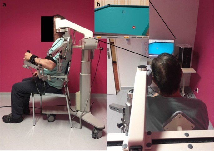
Each patient made 4 visits over 2 days with the same unique rater who was an advanced user of the ArmeoSpring. During the pre-inclusion visit, the patients were informed by the rater about the protocol details, and the inclusion/exclusion criteria meeting was verified. If included, each patient made 3 visits on 3 consecutive half-days. During the first visit, the patient was comfortably seated on the ArmeoSpring, which was adjusted to allow movements of the UE in a large tridimensional workspace required to perform the assessment exercises (Fig. 1). During the second and third visits, the patient was placed on the device in the same way and performed the same series of exercises as during the first visit.

figure1
Experimental setup. a Installation of the patient performing a training exercise of the impaired upper limb with the ArmeoSpring. b Screenshot of the 2D-horizontal catch assessment exercise used in this study

Continue

, , , , , , , , , , , ,

Leave a comment

[BLOG POST] 6 Basic Principles of Neuroplasticity – The Best Brain Possible

6 Basic Principles of Neuroplasticity

Neuroplasticity is an umbrella term referring to the various capabilities of your brain to reorganize itself throughout life due to your environment, behavior, and internal experiences. To ensure the survival of the species, the human nervous system evolved to adapt to its environment — based on learning from past experiences. This is true for all organisms with a nervous system.

According to the book Neuroplasticity from The MIT Press Essential Knowledge Series,  by Moheb Constandi:

But what does ‘rewiring your brain’ actually mean? It refers to the concept of neuroplasticity, a very loosely defined term that simply means some kind of change in the nervous system. Just 50 years ago, the idea that the adult brain can change in any way was heretical. Researchers accepted that the immature brain is malleable, but also believed that it gradually hardens, like clay poured into a mold, into a permanently fixed structure by the time childhood ended. It was also believed that we were born with all the brain cells we will ever have, that the brain is incapable of regenerating itself, and therefore any damage or injuries it sustains cannot be fixed.

In fact, nothing could be further from the truth.”

In the 1980s, researchers at The University of California at San Francisco (UCSF) confirmed that the human brain remodels itself following the “Hebbian rule.” Donald Hebb, a Canadian psychologist, first proposed that “Neurons that fire together, wire together” meaning that the brain continually alters itself physically and operationally based on incoming stimuli.

Types of Neuroplasticity

Although the definition of the word “neuroplasticity” is vague without further qualification, there are basically two types of neuroplasticity:

  • Functional plasticity: The brain’s ability to move functions from one area of the brain to another area.
  • Structural plasticity: The brain’s ability to actually change its physical structure as a result of learning.

Plasticity occurs throughout the brain and can involve many different physical structures, for instance, neurons, synapses, vascular cells, and glial cells.

Your Brain Never Stops Changing

Far from being fixed, your brain is a highly dynamic structure, which undergoes significant change, not only as it develops, but also throughout your entire lifespan. As mentioned above, science used to believe that the brain only changed during certain periods in youth. While it’s true that your brain is much more plastic in the younger years and capacity declines with age, plasticity happens from birth until death.

The human brain reaches about 80 percent of its adult size by two years of age, and growth is nearly complete by age ten. We now know that extensive plastic changes continue to take place in late adolescence and beyond. Harnessing the process of neuroplasticity in adulthood isn’t quite as simple as some of the neuro-hype would have you believe, but it can be accomplished under specific circumstances.

You are not stuck with the brain you were born with or even the one you have right now.

Plasticity Is How All Learning and Memory Happen

Learning and memory are neuroplastic processes in your brain, involving chemical and structural changes. By altering the number or strength of connections between brain cells, information gets written into memory. It’s not really known exactly where or how the recording and recalling of memories happen, but the most popular candidate site for memory storage is the synapse, the space between neurons, where they communicate.

This means that when you repeatedly practice an activity or access a memory, your neural networks are physically shaped accordingly. When you cease a behavior or recalling a specific memory, your brain eventually disconnects the cells no longer in use for that pattern. This can work to your advantage and disadvantage. For example, it’s how bad habits are createdAll addiction happens because of neuroplasticity. However, it’s also how you can weaken painful traumatic memories and reduce anxiety and depression.

6 Basic Principles of Neuroplasticity

Plasticity Allows For Amazing Adaptability

Regions that are normally specialized to perform specific functions can switch roles and process other kinds of information.

Plasticity was first demonstrated in an experiment with ferrets, who have identical wiring to the auditory cortex and visual cortex as humans except for one important factor. The basic human wiring exists at birth while ferrets grow the circuit after birth. Scientist interrupted the pathway in the ferrets so that nerves from the eye grew into the auditory cortex. The ferrets were then trained to respond to sounds and lights. They “heard” the lights with parts of the brain that would normally process sound. 

In a later experiment, sighted adults were blindfolded 24 hours a day for five days. The subjects spent their time learning Braille and performing various tactile and auditory activities. They had their brains scanned before and at the end of the experiment. In the earlier scans, their auditory cortex showed normal activity upon hearing sounds. As expected, their visual cortices lit up when seeing and their somatosensory cortices buzzed when fingering Braille symbols. After five days of being blindfolded, cortical brain regions that had been dedicated to seeing were now hearing and feeling.

You Encourage or Discourage Neuroplasticity With Your Lifestyle Habits

Neuroplastic change occurs in response to stimuli processed in the brain originating either internally or externally.  External stimuli, including things like exercise and cognitive stimulation, enhance the production of neural stem cells and promote the survival of newborn neurons. Internal stimuli originating from your own mind, such as meditation and visualization have also been shown to increase neuroplasticity.  Certain types of neuroinflammationinsufficient sleep, and stress and depression have proven to decrease neuroplasticity.

You can support your brain and encourage neuroplasticity through your lifestyle habits as follows:

  1. Sleep — and lots of it — is absolutely essential for an optimally functioning brain.
  2. Exercise is fertilizer for your brain and promotes the birth and preservation of new brain cells (neurogenesis).
  3. Learn how to feed your brain the nutrients it needs to be in top form.
  4. Make your mental health a priority. Take steps to decrease stress and depression.

As for specific activities you can do, Dr. Michael Merzenich, one of the original researchers confirming plasticity at UCSF and the co-founder of Posit Science Corporation, gives this advice in the article 8 Practical Ways to Keep Your Mind Sharp:

Look for activities that are attentionally demanding and inherently rewarding, and that continuously involve new elements to master. Those types of activities engage brain chemistry that’s beneficial for learning, remembering and mood — by stimulating the production of acetylcholine (when paying attention), norepinephrine (when encountering something new), and dopamine (when feeling rewarded).”

Brain Change Is Specific

The nature of change in your brain is specific to the experience. Experience-dependent changes are usually focal and time-dependant. Plastic change doesn’t typically occur widespread across the brain.

Research has proven that the biggest changes occurring in the brain as a result of learning new skills. For example, in animal studies, research has shown that learning new motor skills yields more dramatic changes in the brain than does the repetition of previously acquired motor movements.

This doesn’t mean that working on existing skills is not beneficial. Repetition is absolutely necessary for neuroplastic alterations to last. Most initial changes are temporary. Your brain first records the change, then determines whether it should make it permanent or not. It only sticks if your brain judges the experience to be novel enough or if the outcome is important enough.

Neuroplastic change is also regionally specific. For example, if you’re learning a skill using your right hand, the changes will be greatest in the areas responsible for that movement. The right and left sides of your body are controlled by the opposite side of your brain. Hence, training with your right hand will make the most changes to the left side of your brain, and vice versa.

Neuroplasticity can help you or hurt you.

6 Basic Principles of Neuroplasticity

Neuroplasticity Is Both Positive and Negative

When you hear about neuroplasticity, it’s usually in conjunction with remarkable, positive brain and life change — almost like science fiction. And like science fiction, it has a dark side. It’s because of neuroplasticity that addictions become ingrained in your brain, valuable skills are lost as your brain ages, and some brain illnesses and conditions show up in humans.

Bad habits and addictions

Forming a habit involves neuroplastic change in your brain. A person desires something because their plastic brain has become sensitized to the substance or experience and craves it. When an urge is satisfied, dopamine, a feel-good neurotransmitter, is released. The same shot of dopamine that gives pleasure is also an essential component of neuroplastic change. Dopamine assists in building neuronal connections that reinforce the habit.

Every time you act in the same way, a specific neuronal pattern is stimulated and strengthened. We know that neurons that fire together wire together. Your brain, wanting to be efficient, takes the path of least resistance each time and a habit — or a full-blown addiction — is born. Fortunately, breaking habits and addictions is accomplished via the same neuroplastic process in reverse

Brain Decline

A lot of the ways in which our brain function degrades that we typically think of as part of “just getting old” is really negative neuroplastic change. As people age, they unknowingly contribute to their brain’s decline by not using and challenging it as much.

You’ve got a “use it or lose it” brain. Information rarely accessed and behaviors seldom practiced cause neural pathways to weaken until connections may be completely lost in a process called “synaptic pruning.” In his book, Soft-Wired: How the New Science of Brain Plasticity Can Change Your Life, Dr. Michael Merzenich calls backward neuroplastic change “negative learning”.

Negative learning can happen at any age — especially with technology doing so much brain work for us these days. Using a GPS consistently, staring straight ahead at a screen for hours a day, texting and not talking to people face-to-face, and many more habits of the modern lifestyle can contribute to undesirable brain changes.

Mental Illness

It’s also because of neuroplasticity that some of the major brain illnesses and conditions show up in humans. Schizophrenia, bipolar disorder, depression, anxiety, obsessive-compulsive and phobic behaviors, epilepsy, and more occur because of neuroplastic change. For example, depression can develop from neuroplastic changes brought about by many things, such as adverse childhood experiences, life circumstances, trauma, lack of emotional support, and stress.

Fortunately for us, neuroplastic change is reversible. You can improve your brain’s function — through the same neuroplastic processes. It’s possible to overcome a mental health condition by driving a brain back towards normal operation through neuroplastic change. Many studies on brain plasticity have demonstrated that many aspects of your brain power, intelligence, or control – in normal and neurologically impaired individuals – can be improved by intense and appropriately targeted behavioral training.

Source: https://thebestbrainpossible.com/6-basic-principles-of-neuroplasticity/?utm_campaign=shareaholic&utm_medium=facebook&utm_source=socialnetwork&fbclid=IwAR1lakYOzQiE_M3wOoa8twmpHMjwrnONeBKRBb83HsBvx4Bf7FXBgvuJrgU

, , , , , ,

Leave a comment

[WEB SITE] 20 Memory Tips to Improve Your Learning

Improving memory

Source: https://custom-writing.org/#improving-memory

,

Leave a comment

[BLOG POST] Stuck at Home? Resources to Stay Active and Engaged – Collection Spotlight from the National Rehabilitation Information Center

Life is looking very different right now, thanks to the coronavirus outbreak. Many people are staying close to home, teleworking or telelearning, and restricting their social interactions significantly. During this unprecedented time, people may want to explore opportunities to learn and interact online, and they may be looking for activities they can participate in while keeping up the recommended social distancing. We’ve gathered some resources from the NIDILRR community and elsewhere which we hope will help you stay engaged, active, and connected to your community.

Keep Learning

Online courses, webinars, and programs can help you stay mentally engaged. Many of these learning tools also offer continuing education credits which can be applied toward certifications, memberships, and professional licensing.

Stay Active and Engaged Close to Home

We may not be able to go to our favorite gym or exercise class, but we can still be active and stay within the recommended guidelines.

Connect to the Community Virtually

Many of us are turning to our social media feeds and our email inboxes to stay connected to friends, family, and coworkers. It can also be useful for researchers who want to get their research results into the community without traveling to conferences and meetings.

Consider Online Participation in Research

From surveys to phone or web interviews, there are many ways to participate in ongoing research that can benefit you and your community without leaving home. We regularly feature these opportunities in our News and Notes from the NIDILRR Community and Beyond weekly newsletter. Here are just a few currently recruiting participants:

In addition to these resources from the NIDILRR grantee community, you might want to explore these websites from other agencies, organizations, and national sites:

  • National Park Service – find and virtually explore national parks nearby and far away, learn about discount programs for seniors and people with disabilities.
  • Smithsonian Institutions – virtually explore the Smithsonian’s collections and exhibits, plan a future trip, visit the Science Education Center for fun games to play online.
  • National Gallery of Art – virtually explore the exhibits, find lessons and online courses for adults and kids.
  • National Center on Health, Physical Activity, and Disability – find articles, videos, and more to keep you healthy, active, and engaged.
  • 211.org – the Information and Referral community is fully engaged in helping people connect to help in their community. Call 211 or visit 211.org to find your local help line, speak with a community resource specialist, and find the support you need.
  • National Library Service for the Blind and Print Disabled – NLS is a free braille and talking book library service for people with temporary or permanent low vision, blindness, or a physical disability that prevents them from reading or holding the printed page.

We hope you and your community remain healthy, active, and connected during this stressful time. Please contact our information specialists if we can be of any assistance!


via Stuck at Home? Resources to Stay Active and Engaged | Collection Spotlight from the National Rehabilitation Information Center

, , , , ,

Leave a comment

[ARTICLE] Changes in actual arm-hand use in stroke patients during and after clinical rehabilitation involving a well-defined arm-hand rehabilitation program: A prospective cohort study – Full Text

Abstract

Introduction

Improvement of arm-hand function and arm-hand skill performance in stroke patients is reported by many authors. However, therapy content often is poorly described, data on actual arm-hand use are scarce, and, as follow-up time often is very short, little information on patients’ mid- and long-term progression is available. Also, outcome data mainly stem from either a general patient group, unstratified for the severity of arm-hand impairment, or a very specific patient group.

Objectives

To investigate to what extent the rate of improvement or deterioration of actual arm-hand use differs between stroke patients with either a severely, moderately or mildly affected arm-hand, during and after rehabilitation involving a well-defined rehabilitation program.

Methods

Design: single–armed prospective cohort study. Outcome measure: affected arm-hand use during daily tasks (accelerometry), expressed as ‘Intensity-of arm-hand-use’ and ‘Duration-of-arm-hand-use’ during waking hours. Measurement dates: at admission, clinical discharge and 3, 6, 9, and 12 months post-discharge. Statistics: Two-way repeated measures ANOVAs.

Results

Seventy-six patients (63 males); mean age: 57.6 years (sd:10.6); post-stroke time: 29.8 days (sd:20.1) participated. Between baseline and 1-year follow-up, Intensity-of-arm-hand-use on the affected side increased by 51%, 114% and 14% (p < .000) in the mildly, moderately and severely affected patients, respectively. Similarly, Duration-of-arm-hand-use increased by 26%, 220% and 161% (p < .000). Regarding bimanual arm-hand use: Intensity-of-arm-hand-use increased by 44%, 74% and 30% (p < .000), whereas Duration-of-arm-hand-use increased by 10%, 22% and 16% (p < .000).

Conclusion

Stroke survivors with a severely, moderately or mildly affected arm-hand showed different, though (clinically) important, improvements in actual arm-hand use during the rehabilitation phase. Intensity-of-arm-hand-use and Duration-of-arm-hand-use significantly improved in both unimanual and bimanual tasks/skills. These improvements were maintained until at least 1 year post-discharge.


Introduction

After stroke, the majority of stroke survivors experiences significant arm-hand impairments [12] and a decreased use of the paretic arm and hand in daily life [3]. The actual use of the affected hand in daily life performance depends on the severity of the arm-hand impairment [46] and is associated with perceived limitations in participation [78]. Severity of arm-hand impairment is also associated with a decrease of health-related quality of life [9], restricted social participation [10], and subjective well-being [1112].

Numerous interventions and arm-hand rehabilitation programs have been developed in order to resolve arm-hand impairments in stroke patients [613]. In the Netherlands, a number of stroke units in rehabilitation centres implemented a well-described ‘therapy-as-usual’ arm-hand rehabilitation program, called CARAS (acronym for: Concise Arm and hand Rehabilitation Approach in Stroke)[14], serving a broad spectrum of stroke patients across the full stroke severity range of arm-hand impairments. The arm-hand rehabilitation program has been developed to guide clinicians in systematically designing arm-hand rehabilitation, tailored towards the individual patient’s characteristics while keeping control over the overall heterogeneity of this population typically seen in stroke rehabilitation centres. A vast majority of stroke patients who participated in CARAS improved on arm-hand function (AHF), on arm-hand skilled performance (AHSP) capacity and on (self-) perceived performance, both during and after clinical rehabilitation [15]. The term ‘arm-hand function’ (AHF) refers to the International Classification of Functioning (ICF) [16] ‘body function and structures level’. The term ‘arm-hand skilled performance’ (AHSP) refers to the ICF ‘activity level’, covering capacity as well as both perceived performance and actual arm-hand use [17].

Improved AHF and/or AHSP capacity do not automatically lead to an increase in actual arm-hand use and do not guarantee an increase of performing functional activities in daily life [1820]. Improvements at function level, i.e. regaining selectivity, (grip) strength and/or grip performance, do not automatically lead to improvements experienced in real life task performance of persons in the post-stroke phase who live at home [1821]. Next to outcome measures regarding AHF, AHSP capacity and (self-) perceived AHSP, which are typically measured in controlled conditions, objective assessment of functional activity and actual arm-hand use outside the testing situation is warranted [2223].

Accelerometry can be used to reliably and objectively assess actual arm-hand use during daily task performance [2432]and has been used in several studies to detect arm-hand movements and evaluate arm-hand use in the post-stroke phase [203335]. Previous studies have demonstrated that, in stroke patients, movement counts, as measured with accelerometers, are associated with the use of the affected arm-hand (Motor Activity Log score) [3637] and, at function level, with the Fugl-Meyer Assessment [38]. Next to quantifying paretic arm-hand use, accelerometers have also been used to provide feedback to further enhance the use of the affected hand in home-based situations [39]. Most studies consist of relatively small [27304044] and highly selected study populations [45] with short time intervals between baseline and follow-up measurements. As to our knowledge, only a few studies monitored arm-hand use in stroke patients for a longer period, i.e. between time of discharge to a home situation or till 6 to 12 months after stroke [194446]. However, they used a relatively small study sample and their intervention aimed at arm-hand rehabilitation was undefined. Both studies of Connell et al. and Uswatte et al. describe a well-defined arm hand intervention where accelerometry data were used as an outcome measure [2747]. However, the study population described by Connell et al. consisted of a relative small and a relative mildly impaired group of chronic stroke survivors. The study population described by Uswatte et al. consisted of a large group of sub-acute stroke patients within strict inclusion criteria ranges [37], who, due to significant spontaneous neurologic recovery within this sub-acute phase, had a mildly impaired arm and hand [4849]. This means that the group lacked persons with a moderately to severely affected arm-hand, who are commonly treated in the daily rehabilitation setting.

The course of AHF and AHSP of a broad range of sub-acute stroke patients during and after rehabilitation involving a well-defined arm-hand rehabilitation program (i.e. CARAS) [14] has been reported by Franck et al. [15]. The present paper provides data concerning actual arm-hand use in the same study population, and focuses on two objectives. The first aim is to investigate changes in actual arm-hand use across time, i.e. during and after clinical rehabilitation, within a stroke patient group typically seen in daily medical rehabilitation practice, i.e. covering a broad spectrum of arm-hand problem severity levels, who followed a well-described arm-hand treatment regime. The second aim is to investigate to what extent improvement (or deterioration) regarding the use of the affected arm-hand in daily life situations differs between patient categories, i.e. patients with either a severely, moderately or mildly impaired arm-hand, during and after their rehabilitation, involving a well-defined arm-hand rehabilitation program.[…]

Continue —->  Changes in actual arm-hand use in stroke patients during and after clinical rehabilitation involving a well-defined arm-hand rehabilitation program: A prospective cohort study

Fig 3. Mean values for Intensity-of-arm-hand-use during uptime for subgroups 1, 2 and 3.
T = time; bl = baseline; cd = clinical discharge; m = month; Solid line = subgroup 1; Dotted line = subgroup 2; Dashed line = subgroup 3.
https://doi.org/10.1371/journal.pone.0214651.g003

, , , , , , , , , , , , ,

Leave a comment

[BLOG POST] Where does the controversial finding that adult human brains don’t grow new neurons leave ongoing research?

Scientists have known for about two decades that some neurons – the fundamental cells in the brain that transmit signals – are generated throughout life. But now a controversial new study from the University of California, San Francisco, casts doubt on whether many neurons are added to the human brain after birth.

As a translational neuroscientist, this work immediately piqued my interest. It has direct implications for the research my lab does: We transplant young neurons into damaged brain areas in mice in an attempt to treat epileptic seizures and the damage they’ve caused. Like many labs, part of our work is based on a foundational belief that the hippocampus is a brain region where new neurons are born throughout life.

If the new study is right, and human brains for the most part don’t add new neurons after infancy, researchers like me need to reconsider the validity of the animal models we use to understand various brain conditions – in my case temporal lobe epilepsy. And I suspect other labs that focus on conditions including drug addiction, depression and post-traumatic stress disorder are thinking about what the UCSF study means for their investigations, too.

In the brain of a baby who died soon after birth, there are many new neurons (green in this image) in the hippocampus. Sorrells et alCC BY-ND

When and where are new neurons born?

No doubt, the adult human brain is able to learn throughout life and to change and adapt – a capability brain scientists call neuroplasticity, the brain’s ability to reorganize itself by rewiring connections. Yet, a central dogma in the field of neuroscience for nearly 100 years had been that a child is born with all the neurons she will ever have because the adult brain cannot regenerate neurons.

Just over half a century ago, researchers devised a way to study proliferation of cells in the mature brain, based on techniques to incorporate a radioactive label into new cells as they divide. This approach led to the startling discovery in the 1960s that rodent brains actually could generate new neurons.

Neurogenesis – the production of new neurons – was previously thought to only occur during embryonic life, a time of extremely rapid brain growth and expansion, and the rodent findings were met with considerable skepticism. Then researchers discovered that new neurons are also born throughout life in the songbird brain, a species scientists use as a model for studying vocal learning. It started to look like neurogenesis plays a key role in learning and neuroplasticity – at least in some brain regions in a few animal species.

Even so, neuroscientists were skeptical that many nerve cells could be renewed in the adult brain; evidence was scant that dividing cells in mammalian brains produced new neurons, as opposed to other cell types. It wasn’t until researchers extracted neural stem cells from adult mouse brains and grew them in cell culture that scientists showed these precursor cells could divide and differentiate into new neurons. Now it is generally well accepted that neurogenesis takes place in two areas of the adult rodent brain: the olfactory bulbs, which process smell information, and the hippocampus, a region characterized by neuroplasticity that is required for forming new declarative memories.

Adult neural stem cells cluster together in what scientists call niches – hotbeds for cultivating the birth and growth of new neurons, recognizable by their distinctive architecture. Despite the mounting evidence for regional growth of new neurons, these studies underscored the point that the adult brain harbors only a few stem cell niches and their capacity to produce neurons is limited to just a few types of cells.

With this knowledge, and new tools for labeling proliferating cells and identifying maturing neurons, scientists began to look for postnatal neurogenesis in primate and human brains.

What’s happening in adult human brains?

Many neuroscientists believe that by understanding the process of adult neurogenesis we’ll gain insights into the causes of some human neurological disorders. Then the next logical step would be trying to develop new treatments harnessing neurogenesis for conditions such as Alzheimer’s disease or trauma-induced epilepsy. And stimulating resident stem cells in the brain to generate new neurons is an exciting prospect for treating neurodegenerative diseases.

Because neurogenesis and learning in rodents increases with voluntary exercise and decreases with age and early life stress, some workers in the field became convinced that older people might be able to enhance their memory as they age by maintaining a program of regular aerobic exercise.

However, obtaining rigorous proof for adult neurogenesis in the human and primate brain has been technically challenging – both due to the limited experimental approaches and the larger sizes of the brains, compared to reptiles, songbirds and rodents.

Researchers injected a compound found in DNA, nicknamed BrdU to identify brand new neurons in human adult hippocampus – but the labeled cells were extremely rare. Other groups demonstrated that adult human brain tissue obtained during neurosurgery contained stem cell niches that housed progenitor cells that could generate new neurons in the lab, showing that these cells had an inborn neurogenic capacity, even in adults.

But even when scientists saw evidence for new neurons in the brain, they tended to be scarce. Some neurogenesis experts were skeptical that evidence based on incorporating BrdU into DNA was a reliable method for proving that new cells were actually being born through cell division, rather than just serving as a marker for other normal cell functions.

Further questions about how long human brains retain the capacity for neurogenesis arose in 2011, with a study that compared numbers of newborn neurons migrating in the olfactory bulbs of infants versus older individuals up to 84 years of age. Strikingly, in the first six months of life, the baby brains contained lots of chains of young neurons migrating into the frontal lobes, regions that guide executive function, long-range planning and social interactions. These areas of the human cortex are hugely increased in size and complexity compared to rodents and other species. But between 6 to 18 months of age, the migrating chains dwindled to a thin stream. Then, a very different pattern emerged: Where the migrating chains of neurons had been in the infant brain, a cell-free gap appeared, suggesting that neural stem cells become depleted during the first six months of life.

Questions still lingered about the human hippocampus and adult neurogenesis as a source for its neuroplasticity. One group came up with a clever approach based on radiocarbon dating. They measured how much atmospheric ¹⁴C – a radioactive isotope derived from nuclear bomb tests – was incorporated into people’s DNA. This method suggested that as many as 700 new cells are added to the adult human hippocampus every day. But these findings were contradicted by a 2016 study that found that the neurogenic cells in the adult hippocampus could only produce non-neuronal brain cells called microglia.

Rethinking neurogenesis research

Now the largest and most comprehensive study conducted to date presents even stronger evidence that robust neurogenesis doesn’t continue throughout adulthood in the human hippocampus – or if it does persist, it is extremely rare. This work is controversial and not universally accepted. Critics have been quick to cast doubt on the results, but the finding isn’t totally out of the blue.

So where does this leave the field of neuroscience? If the UCSF scientists are correct, what does that mean for ongoing research in labs around the world?

Because lots of studies of neurological diseases are done in mice and rats, many scientists are invested in the possibility that adult neurogenesis persists in the human brain, just as it does in rodents. If it doesn’t, how valid is it to think that the mechanisms of learning and neuroplasticity in our model animals are comparable to those in the human brain? How relevant are our models of neurological disorders for understanding how changes in the hippocampus contribute to disorders such as the type of epilepsy I study?

In my lab, we transplant embryonic mouse or human neurons into the adult hippocampus in mice, after damage caused by epileptic seizures. We aim to repair this damage and suppress seizures by seeding the mouse hippocampus with neural stem cells that will mature and form new connections. In temporal lobe epilepsy, studies in adult rodents suggest that naturally occurring hippocampal neurogenesis is problematic. It seems that the newborn hippocampal neurons become highly excitable and contribute to seizures. We’re trying to inhibit these newborn hyperexcitable neurons with the transplants. But if humans don’t generate new hippocampal neurons, then maybe we’re developing a treatment in mice for a problem that has a different mechanism in people.

Perhaps our species has evolved separate mechanisms for neuroplasticity, distinct from those used by species such as rats and mice. One possibility is that there are other sites in the human brain where neurogenesis occurs – its a big structure and more exploration will be necessary. If it turns out to be true that the human brain has a diminished capacity for neurogenesis after birth, the finding will have important implications for how neuroscientists like me think about tackling brain disorders.

Perhaps most importantly, this work underscores how crucial it is to learn how to increase the longevity of the neurons we do have, born early in life, and how we might replace or repair neurons that become damaged.

via Where does the controversial finding that adult human brains don’t grow new neurons leave ongoing research?


, , , , , , , , , , , ,

Leave a comment

[ARTICLE] Reorganization of finger coordination patterns through motor exploration in individuals after stroke – Full Text


Abstract

Background

Impairment of hand and finger function after stroke is common and affects the ability to perform activities of daily living. Even though many of these coordination deficits such as finger individuation have been well characterized, it is critical to understand how stroke survivors learn to explore and reorganize their finger coordination patterns for optimizing rehabilitation. In this study, I examine the use of a body-machine interface to assess how participants explore their movement repertoire, and how this changes with continued practice.

Methods

Ten participants with chronic stroke wore a data glove and the finger joint angles were mapped on to the position of a cursor on a screen. The task of the participants was to move the cursor back and forth between two specified targets on a screen. Critically, the map between the finger movements and cursor motion was altered so that participants sometimes had to generate coordination patterns that required finger individuation. There were two phases to the experiment – an initial assessment phase on day 1, followed by a learning phase (days 2–5) where participants trained to reorganize their coordination patterns.

Results

Participants showed difficulty in performing tasks which had maps that required finger individuation, and the degree to which they explored their movement repertoire was directly related to clinical tests of hand function. However, over four sessions of practice, participants were able to learn to reorganize their finger movement coordination pattern and improve their performance. Moreover, training also resulted in improvements in movement repertoire outside of the context of the specific task during free exploration.

Conclusions

Stroke survivors show deficits in movement repertoire in their paretic hand, but facilitating movement exploration during training can increase the movement repertoire. This suggests that exploration may be an important element of rehabilitation to regain optimal function.

Background

Stroke often results in impairments of upper extremity, including hand and finger function, with 75% of stroke survivors facing difficulties performing activities of daily living [12]. Critically, impairments after stroke not only include muscle- and joint-specific deficits such as weakness, and changes in the kinetic and kinematic workspace of the fingers [34], but also coordination deficits such as reduced independent joint control [5] and impairments in finger individuation and enslaving [6789]. Therefore, understanding how to address these coordination deficits is critical for improving hand rehabilitation.

Typical approaches to hand rehabilitation emphasize repetition [10] and functional practice based on evidence that such experience can cause reorganization in the brain [11]. Although this has proven to be reasonably successful, functional practice (such as repetitive grasping of objects) does not specify the coordination pattern to be used when performing the tasks. As a result, because of the redundancy in the human body, there is a risk that stroke survivors may adopt atypical compensatory movements to perform tasks [12]. These compensatory movements have been mainly identified during reaching [1314], but there is evidence that they are also present in finger coordination patterns during grasping [15]. Although there is still debate over the role of compensatory movements in rehabilitation [16], there is at least some evidence both in animal and humans that continued use of these compensatory patterns may be detrimental to true recovery [171819].

To address this issue, there has been a greater focus on directly facilitating the learning of new coordination patterns. Specifically, in hand rehabilitation, virtual tasks (such as playing a virtual piano) have been examined as a way to train finger individuation [2021]. In these protocols, individuation is encouraged by asking participants to press a particular key with a finger, while keeping other fingers stationary. A similar approach to improve hand dexterity was also adopted by developing a glove that could be used as a controller for a popular guitar-playing video game [22]. However, directly instructing desired coordination patterns to be produced becomes challenging as the number of degrees of freedom involved in the coordination pattern increase. For example, the hand has approximately 20 kinematic degrees of freedom, and providing verbal, visual or auditory feedback for simultaneously controlling all these degrees of freedom would be a major challenge. A potential solution that has been suggested is not to directly instruct the coordination pattern itself, but rather let participants explore different coordination patterns [23]. This idea of motor exploration is based on dynamical systems theory that suggests that variability and exploration may help participants escape sub-optimal pre-existing coordination patterns and potentially settle in more optimal coordination patterns [24252627]. Such exploration has been shown to be important in adapting existing movement repertoire [28], and has also been shown to be associated with faster rates of learning [29].

In order to test the hypothesis that exploration of novel coordination patterns can improve overall movement repertoire, I used a body-machine interface [3031] to examine how stroke survivors explore and reorganize finger coordination patterns with practice. A body-machine interface maps body movements (in this case finger movements) to the control of a real or virtual object (in this case a screen cursor), which can provide a way to elicit different coordination patterns in the context of an intuitive task. Specifically I examined: (i) how stroke survivors reorganize their finger coordination patterns, (ii) how training to explore novel coordination patterns affects their ability to reorganize their coordination pattern, and (iii) if training to explore novel coordination patterns has an effect on their overall movement repertoire. In this context, I use the term “novel” to indicate coordination patterns that require finger individuation. This assumption is motivated by the finding that stroke survivors have difficulty producing finger individuation even under explicit instruction [69], and therefore it is highly likely that they would not use coordination patterns requiring finger individuation frequently in activities of daily living.[…]

Continue —>  Reorganization of finger coordination patterns through motor exploration in individuals after stroke | Journal of NeuroEngineering and Rehabilitation | Full Text

Fig. 1 a Experimental setup – participants wore a data glove and moved their fingers to control a screen cursor b Schematic of task – participants moved a cursor between two targets using movements of four fingers (thumb excluded). c Experimental protocol. Participants came in for 5 total sessions – an initial assessment phase, followed by a learning phase. d Weighting coefficients of the index and middle (blue), and ring and little (red) fingers during the initial assessment phase, and e weighting coefficients during the learning phase

, , , , , , , , , ,

Leave a comment

[Abstract] Effects of tDCS on motor learning and memory formation: a consensus and critical position paper – Clinical Neurophysiology

Highlights

  • We review investigations of whether tDCS can facilitate motor skill learning and adaptation.
  • We identify several caveats in the existing literature and propose solutions for addressing these.
  • Open Science efforts will improve standardization, reproducibility and quality of future research.

Abstract

Motor skills are required for activities of daily living. Transcranial direct current stimulation (tDCS) applied in association with motor skill learning has been investigated as a tool for enhancing training effects in health and disease. Here, we review the published literature investigating whether tDCS can facilitate the acquisition, retention or adaptation of motor skills. Work in multiple laboratories is underway to develop a mechanistic understanding of tDCS effects on different forms of learning and to optimize stimulation protocols. Efforts are required to improve reproducibility and standardization. Overall, reproducibility remains to be fully tested, effect sizes with present techniques vary over a wide range, and the basis of observed inter-individual variability in tDCS effects is incompletely understood. It is recommended that future studies explicitly state in the Methods the exploratory (hypothesis-generating) or hypothesis-driven (confirmatory) nature of the experimental designs. General research practices could be improved with prospective pre-registration of hypothesis-based investigations, more emphasis on the detailed description of methods (including all pertinent details to enable future modeling of induced current and experimental replication), and use of post-publication open data repositories. A checklist is proposed for reporting tDCS investigations in a way that can improve efforts to assess reproducibility.

Source: Effects of tDCS on motor learning and memory formation: a consensus and critical position paper – Clinical Neurophysiology

, , , , , , ,

Leave a comment